. 1997 Aug 25;138(4):731-46.
doi: 10.1083/jcb.138.4.731.

Novel genes involved in endosomal traffic in yeast revealed by suppression of a targeting-defective plasma membrane ATPase mutant

Affiliations

Novel genes involved in endosomal traffic in yeast revealed by suppression of a targeting-defective plasma membrane ATPase mutant

W j Luo et al. J Cell Biol. .

Abstract

A novel genetic selection was used to identify genes regulating traffic in the yeast endosomal system. We took advantage of a temperature-sensitive mutant in PMA1, encoding the plasma membrane ATPase, in which newly synthesized Pma1 is mislocalized to the vacuole via the endosome. Diversion of mutant Pma1 from vacuolar delivery and rerouting to the plasma membrane is a major mechanism of suppression of pma1(ts). 16 independent suppressor of pma1 (sop) mutants were isolated. Identification of the corresponding genes reveals eight that are identical with VPS genes required for delivery of newly synthesized vacuolar proteins. A second group of SOP genes participates in vacuolar delivery of mutant Pma1 but is not essential for delivery of the vacuolar protease carboxypeptidase Y. Because the biosynthetic pathway to the vacuole intersects with the endocytic pathway, internalization of a bulk membrane endocytic marker FM 4-64 was assayed in the sop mutants. By this means, defective endosome-to-vacuole trafficking was revealed in a subset of sop mutants. Another subset of sop mutants displays perturbed trafficking between endosome and Golgi: impaired pro-alpha factor processing in these strains was found to be due to defective recycling of the trans-Golgi protease Kex2. One of these strains defective in Kex2 trafficking carries a mutation in SOP2, encoding a homologue of mammalian synaptojanin (implicated in synaptic vesicle endocytosis and recycling). Thus, cell surface delivery of mutant Pma1 can occur as a consequence of disturbances at several different sites in the endosomal system.


Figures
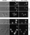
Figure 1
Figure 1
Indirect immunofluorescence localization of mutant Pma1 in a prevacuolar compartment. Localization of Pma1 and V-ATPase in pma1-7 and pma1-7 vps27ts cells. Mid-log cultures were harvested, fixed, and spheroplasted. Cells were permeabilized with SDS before staining with rabbit anti-Pma1 and mouse monoclonal antibody against the 60-kD subunit of V-ATPase, followed by species-specific CY3- and DTAF-conjugated secondary antibodies. (Upper panel) Mutant Pma1 staining is coincident with V-ATPase staining at the vacuolar membrane in pma1-7 cells at 30°C. In these cells, mutant Pma1 is also seen as spots distinct from the vacuole (arrowheads). Nomarski optics show vacuoles as indentations. (Lower panel) pma1-7 vps27ts cells were shifted to 37°C for 1 h. Both mutant Pma1 and the 60-kD V-ATPase subunit are accumulated in the prevacuolar compartment.
Figure 2
Figure 2
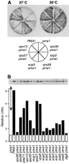
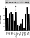
Suppression of temperature-sensitive growth and stabilization of mutant Pma1 protein in sop mutants. (A) Growth at 30 and 37°C on plates (synthetic complete medium). PMA1 (L5488) cells grow at 30 and 37°C, whereas pma1-7 (ACY7) cells grow at 30°C but do not grow at 37°C. sop/vps pma1 cells (WLY2, WLY12, WLY22, WLY 25, WLY33, WLY40) grow at 30 and 37°C. (B) Western blot showing steady-state levels of Pma1 protein. Lysate was prepared from PMA1, pma1-7, and sop pma1-7 cells exponentially growing at 30°C. Samples were normalized to lysate protein. Pma1 protein was detected using anti-Pma1 antibody and 125I–protein A followed by autoradiography. Bar graph shows quantitation of Pma1 levels in wild-type and sop pma1-7 mutants by densitometric scanning of the autoradiogram. Pma1 levels are normalized to that of pma1-7 (arrow), set arbitrarily to 1.0. Western blot is representative of three to five independent measurements in which the standard deviation of the mean is on average 26%.
Figure 3
Figure 3
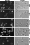
Mutant Pma1 is rerouted to the cell surface in sop mutants as seen by indirect immunofluorescence. Cells exponentially growing at 30°C were fixed, permeabilized, and stained with rabbit anti-Pma1 antibody followed by CY3-conjugated secondary antibody. PMA1 (L5488) cells show cell surface Pma1 localization (top panel) while pma1-7 cells (ACY7) display striking intracellular staining (second panel). Suppressed pma1-7 cells display a predominant plasma membrane distribution of mutant Pma1 (lower three panels). sop pma1 strains shown are WLY2, WLY22, and WLY25.
Figure 4
Figure 4
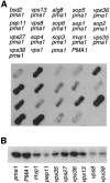
A subset of sop mutants is defective for vacuolar protein sorting. (A) Western blot detecting secretion of CPY. Cells were overlayed with nitrocellulose overnight. Secreted CPY adsorbed to the membrane was visualized with rabbit anti-CPY followed by horseradish peroxidase–conjugated secondary antibody and chemiluminescence detection reagents. PMA1 (L5488) and pma1-7 (ACY7) are shown as non–CPY-secreting controls. vps1Δ (ACX58-3C), which secretes the vast majority of newly synthesized pro-CPY (Raymond et al., 1992), is included as a positive control. (B) Western blot showing intracellular CPY. Protein lysate was prepared from exponentially growing cells, as described in Methods. Samples were normalized to lysate protein. CPY was detected as described above. Strains assayed (left to right) are: ACY7, L3852, WLX17-6D, WLX13-3B, WLX18-6D, ACY33, WLX12-7C, WLX15-4C, WLX16-1A, and WLX14-10A. Mature CPY associated with vps cells is substantially decreased by comparison with PMA1 and pma1-7 cells.
Figure 5
Figure 5
Visualization of endocytosis in sop mutants by FM 4-64 staining. Exponentially growing wild-type cells and sop mutants were stained with FM 4-64 for 5 min at 30°C, washed, and incubated in fresh YPD for 1 h before visualization and photography. Vacuolar membrane staining is seen in wild-type cells (L3852). In vps36 (WLX12-7C), a class E vps mutant, FM 4-64 dye is accumulated in a prevacuolar compartment. A similar accumulation of dye as a spot near the vacuole is seen in vps38 (WLX14-10A) and vps13 (WLX15-4C). Bright punctate staining in the cytoplasm remains in vps8 (WLX16-1A) after 1 h of chase.
Figure 6
Figure 6
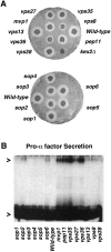
Production of biologically active α factor and secretion of unprocessed pro–α factor by sop mutants. (A) Secretion of biologically active mature α factor was detected by halo assay. Wild-type (L3852) and sop mutants were patched on plates with synthetic complete medium and allowed to grow overnight. The MATα strains were then replica-plated onto a lawn of MATa bar1 cells (LM23-3AZ; Table I). After 1–2 d, halos surrounding patches of α cells were scored. No halo is observed surrounding kex2Δ cells (BFY106-4D), which is included as a control. sop mutant strains are: WLX19-3A, WLX3-2A, WLX9-12C, WLX8-1B, WLX10-2A, WLX11-1C, WLX12-7C, WLX13-3B, WLX14-10A, WLX15-4C, WLX16-1A, WLX17-6D, WLX18-6D, and ACY33. (B) Secretion of pro–α factor by wild-type and sop cells. Exponentially growing cells (0.4 OD600/0.5 ml) were labeled at room temperature with Expre35S35S for 50 min. Culture medium was collected, adjusted to 1% SDS, and boiled. Secreted α factor was immunoprecipitated from the medium, and analyzed by SDS-PAGE (15% polyacrylamide gel) and fluorography. The lower arrowhead indicates mature α factor, a 3.5-kD peptide, and the upper arrowhead indicates precursor α factor with a molecular mass of ∼125 kD. Wild-type cells secrete mature α factor exclusively. A subset of sop cells secrete both unprocessed and mature α factor. Strains assayed are: WLX19-3A, WLX3-2A, WLX9-12C, WLX8-1B, WLX10-2A, WLX11-1C, L3852, WLX17-6D, WLX13-3B, WLX18-6D, ACY33, WLX12-7C, WLX15-4C, WLX16-1A, and WLX14-10A.
Figure 7
Figure 7
Steady-state Kex2 levels in sop mutants. Western blot measuring steady-state level of Kex2p. Lysate (100 μg protein) from wild-type (L3852) and sop mutants (strains listed in Fig. 6 B legend) was resolved by SDS-PAGE and transferred to nitrocellulose. Kex2p was detected by rabbit anti-Kex2 antibody followed by 125Iprotein A and autoradiography. Bar graph shows quantitation of Kex2 levels in sop mutants normalized to that of wild-type (arrow) by densitometric scanning of the autoradiogram. Measurements are representative of two to four experiments.
Figure 8
Figure 8
Sop2 is a member of the inositol 5-phosphatase family and a homologue of synaptojanin. Alignment of Sop2 with rat brain synaptojanin (gi1166575) and two yeast ORFs. OrfN2160 on chromosome XIV is available from GenBank/EMBL/DDBJ under accession number Z50161. Orf P40559, a hypothetical 108.4-kD protein on chromosome IX, is in the SWISS-PROT protein sequence database under accession number P40559. Protein sequences were aligned using the Megalign program (DNAStar, Madison, WI). Identical amino residues are boxed, and hyphens indicate gaps introduced to maximize alignment. Asterisks indicate conserved motifs GDXN(Y/F)R and P(S/ A)W(C/T)DRIL that define inositol 5-phosphatases (Majerus, 1996). Sop2 is 31% identical with synaptojanin along its full length.
Figure 8
Figure 8
Sop2 is a member of the inositol 5-phosphatase family and a homologue of synaptojanin. Alignment of Sop2 with rat brain synaptojanin (gi1166575) and two yeast ORFs. OrfN2160 on chromosome XIV is available from GenBank/EMBL/DDBJ under accession number Z50161. Orf P40559, a hypothetical 108.4-kD protein on chromosome IX, is in the SWISS-PROT protein sequence database under accession number P40559. Protein sequences were aligned using the Megalign program (DNAStar, Madison, WI). Identical amino residues are boxed, and hyphens indicate gaps introduced to maximize alignment. Asterisks indicate conserved motifs GDXN(Y/F)R and P(S/ A)W(C/T)DRIL that define inositol 5-phosphatases (Majerus, 1996). Sop2 is 31% identical with synaptojanin along its full length.

Similar articles

Cited by

References

    1. Altschul SF, Gish W, Miller W, Myers EW, Lipman DJ. Basic local alignment tool. J Mol Biol. 1990;215:403–410. - PubMed
    1. Armstrong J, Patel S, Riddle P. Lysosomal sorting mutants of coronavirus E1 protein, a Golgi membrane protein. J Cell Sci. 1990;95:191–197. - PubMed
    1. Attree O, Olivos IM, Okabe I, Bailey LC, Nelson DL, Lewis RA, McInnes RR, Nussbaum RL. The Lowe's oculocerebrorenal syndrome gene encodes a protein highly homologous to inositol polyphosphate-5-phosphatase. Nature (Lond) 1992;358:239–242. - PubMed
    1. Benito B, Moreno E, Losario R. Half-life of plasma membrane ATPase and its activating system in resting yeast cells. Biochim Biophys Acta. 1991;1063:265–268. - PubMed
    1. Burns N, Grimwade B, Ross-Macdonald PB, Choi E-Y, Finberg K, Roeder GS, Snyder M. Large-scale analysis of gene expression, protein localization, and gene disruption in Saccharomyces cerevisiae. . Genes Dev. 1994;8:1087–1105. - PubMed

Publication types

MeSH terms