. 1997 Aug 11;138(3):629-41.
doi: 10.1083/jcb.138.3.629.

Microtubules orient the mitotic spindle in yeast through dynein-dependent interactions with the cell cortex

Affiliations

Microtubules orient the mitotic spindle in yeast through dynein-dependent interactions with the cell cortex

J L Carminati et al. J Cell Biol. .

Abstract

Proper orientation of the mitotic spindle is critical for successful cell division in budding yeast. To investigate the mechanism of spindle orientation, we used a green fluorescent protein (GFP)-tubulin fusion protein to observe microtubules in living yeast cells. GFP-tubulin is incorporated into microtubules, allowing visualization of both cytoplasmic and spindle microtubules, and does not interfere with normal microtubule function. Microtubules in yeast cells exhibit dynamic instability, although they grow and shrink more slowly than microtubules in animal cells. The dynamic properties of yeast microtubules are modulated during the cell cycle. The behavior of cytoplasmic microtubules revealed distinct interactions with the cell cortex that result in associated spindle movement and orientation. Dynein-mutant cells had defects in these cortical interactions, resulting in misoriented spindles. In addition, microtubule dynamics were altered in the absence of dynein. These results indicate that microtubules and dynein interact to produce dynamic cortical interactions, and that these interactions result in the force driving spindle orientation.


Figures
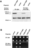
Figure 2
Figure 2
GFP–TUB3 expression and incorporation into functional microtubules. (A) Western blot of crude yeast protein extracts probed with anti–α-tubulin antibody. Strain TSY425, containing the GFP–TUB3 plasmid, and strain TPS510, containing the vector plasmid, were grown in the presence of either 2% galactose, or 2% galactose + 0.5% glucose as the carbon source. Both strains show endogenous total α-tubulin (TUB1p, TUB3p) running as a doublet at 56 kD, while the GFP–TUB3 fusion protein runs at 83 kD. Similar results were obtained with a COOH-terminal fusion protein, TUB3–GFP (data not shown). (B) GFP– TUB3 expression rescues the benomyl supersensitivity of a tub3:: TRP1 haploid strain (DBY2375). (Top) Growth of tub3::TRP1 cells transformed with plasmids as noted above, and a 1:10 dilution of cells (second row), when grown with 2% galactose. (Bottom) Growth of the same strains, and 1:10 dilutions, in the presence of 5 μg/ml benomyl. All cells were incubated at 30°C. tub3:: TRP1 containing either vector alone, TUB3–GFP, or TUB1–GFP remain benomyl supersensitive, while both TUB3 and GFP– TUB3 rescue the benomyl supersensitivity.
Figure 1
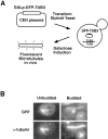
Figure 1
GFP–TUB3 produces fusion protein that incorporates into microtubules. (A) Schematic showing the process by which fluorescent microtubules are generated in vivo. The CEN plasmid carrying GFP–TUB3, pTS417, is transformed into the diploid yeast strain, TPS507, and the resulting strain, TSY425, is then grown in the presence of 2% galactose as the carbon source. After galactose induction, fluorescent microtubules are formed in vivo. (B) Colocalization of GFP–TUB3 with α-tubulin in microtubules. After growth of TSY425 in 2% galactose, cells were fixed and stained for α-tubulin. Fluorescence of rhodamine- labeled α-tubulin was compared with GFP fluorescence; both α-tubulin and GFP colocalized to microtubule structures in unbudded and budded cells. Fluorescent cytoplasmic microtubules and the spindle pole body are seen in unbudded cells (left), whereas a fluorescent mitotic spindle is seen in the budded cell shown (right). The right panel also contains an unbudded cell in which GFP fluorescence of the spindle pole body, but not the microtubules, can be seen.
Figure 3
Figure 3
Yeast microtubules in living yeast cells as visualized by GFP fluorescence. (A) Schematic diagram showing relevant structures in unbudded and budded cells. Arrows on the microtubules denote the dynamic growing and shrinking behavior of microtubules. During mitosis (right), the spindle pole bodies duplicate, and a mitotic spindle is formed, consisting of a bundle of microtubules. Note: although the nuclei are drawn in the schematic, they cannot be seen in fluorescent images of yeast cells. (B) Cytoplasmic microtubules and the spindle pole body are visualized during time-lapse images of a single unbudded cell. The spindle pole body, at the center of the microtubule array, is marked by an arrow. The time, in seconds, is noted above each frame. A faint vacuole, which has lower background staining, can be seen below the spindle pole body. Up to six cytoplasmic microtubules grow and shrink, and come in contact with the cortex during the time sequence. (C) Cytoplasmic microtubules, spindle pole bodies, and a short spindle are visualized in a budded cell. The two spindle pole bodies at each end of the spindle are marked by arrows. Growing and shrinking of cytoplasmic microtubules are shown over the time course, during which the spindle twists slightly out of focus. Vacuoles in both the mother and bud can also be seen. Bars, 2 μm.
Figure 4
Figure 4
Cytoplasmic microtubules interact dynamically with the cell cortex and result in associated movements of the spindle during mitosis. (A) The cytoplasmic microtubule sweeps back and forth along the bud cortex over the course of the time sequence. A short, faint cytoplasmic microtubule can also be seen in the mother cell, as well as the two spindle pole bodies and long spindle that spans the bud neck. In this cell, a bright spot of GFP staining, not corresponding to any microtubule structure, is seen in the mother cell. (B) Sweeping of a cytoplasmic microtubule along the cortex of the mother cell results in the spindle being pulled further into the mother cell. Note how the end of the spindle and the spindle pole body in the bud cell are pulled up into the neck by the third frame. (C) Shrinking of a microtubule at the cortex results in spindle movement toward the direction of the cortical attachment. A cytoplasmic microtubule appears in focus by t = 290 s and remains attached at the mother cortex while shrinking. This shrinking is coupled to a movement of the spindle toward the cortex. By the last frame the left spindle pole body is close to the cortex, and only a short cytoplasmic microtubule remains between them. Bars, 2 μm.
Figure 5
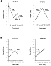
Figure 5
Linear regression analysis of microtubule polymerization and depolymerization rates. (A) Two examples of life-history graphs of wild-type cytoplasmic microtubules, plotting the microtubule length vs time. In the first example, a microtubule in a budded cell shrinks and then undergoes a rescue event. After the next growth phase, a catastrophe event occurs, and the microtubule continues shrinking until the end of the time sequence analyzed. In the second example, one catastrophe occurs between the growing and shrinking phases of a microtubule in a budded cell. The microtubule analyzed in the second graph is shown in Fig. 3 C and is present in the bud from t = 210 to t = 560 s. Rates, as determined by linear regression, are shown over the corresponding growth or shrinkage phases. (B) Examples of life-history graphs of cytoplasmic microtubules in dynein-mutant cells. Note the overall longer length of the microtubules as compared with wild-type microtubules. Both examples are microtubules from budded cells.
Figure 6
Figure 6
Microtubules show aberrant interactions at the cell cortex in dynein-mutant cells. (A) Cytoplasmic microtubules and the spindle pole body are visualized in an unbudded cell. The length of microtubules in unbudded cells remains comparable to that in wild-type unbudded cells. (B) Budded cells contain longer cytoplasmic microtubules that curve around the cell cortex. In this cell, the spindle remains entirely within the mother cell, and cytoplasmic microtubules grow out from both spindle pole bodies. One long microtubule extends into the bud cell and does not show characteristic dynamic interactions with the cell cortex. (C) Three budded cells are shown containing long spindles, all still within the mother cells. While short cytoplasmic microtubules are present, each cell has at least one unusually long microtubule extending into the bud. A knob can be seen on all three long microtubules. The cytoplasmic microtubules have slower dynamics and show aberrant interactions at the cortex. Bars, 2 μm.
Figure 7
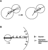
Figure 7
Model of spindle positioning via dynein-dependent interactions at the cell cortex. (A) Sweeping and shrinking interactions between cytoplasmic microtubules and the cortex lead to movement of the elongated spindle across the neck region. A single microtubule extends to the bud cortex, where it sweeps the cortex, as indicated by the dotted lines (left cell). Microtubule shrinking at the cortex results in an associated movement of the spindle toward the attachment site (right cell). (B) Model for how dynein interacts with cytoplasmic microtubules at the cortex. A dynactin attachment complex is localized at the cortex, binds to microtubules, and subsequently binds to a dynein molecule. Dynein remains bound to this complex and either pulls on the microtubule from the cortex or remains attached to the depolymerizing microtubule, resulting in movement of the spindle pole body and spindle. (Small arrow) Direction of dynein's minus end motor activity; (large arrow) direction of subsequent spindle movement. The small boxes at the end of the microtubule denote the depolymerization of the microtubule.

Similar articles

Cited by

References

    1. Belmont LD, Hyman AA, Sawin KE, Mitchison TJ. Real-time visualization of cell cycle-dependent changes in microtubule dynamics in cytoplasmic extracts. Cell. 1990;62:579–589. - PubMed
    1. Burke D, Gasdaska P, Hartwell L. Dominant effects of tubulin overexpression in Saccharomyces cerevisiae. . Mol Cell Biol. 1989;9:1049–1059. - PMC - PubMed
    1. Byers, B. 1981. Cytology of the yeast life cycle. In The Molecular Biology of the Yeast Saccharomyces: Life Cycle and Inheritance. J.N. Strathern, E.W. Jones, and J.R. Broach, editors. Cold Spring Harbor Laboratory, Cold Spring Harbor, NY. 59–96.
    1. Byers B, Goetsch L. Behavior of spindles and spindle plaques in the cell cycle and conjugation of Saccharomyces cerevisiae. . J Bacteriol. 1975;124:511–523. - PMC - PubMed
    1. Cassimeris L, Pryer NK, Salmon ED. Real-time observations of microtubule dynamic instability in living cells. J Cell Biol. 1988;107:2223–2231. - PMC - PubMed

Publication types

MeSH terms