Chondrocytes as a specific target of ectopic Fos expression in early development
- PMID: 9108093
- PMCID: PMC20556
- DOI: 10.1073/pnas.94.8.3994
Chondrocytes as a specific target of ectopic Fos expression in early development
Abstract
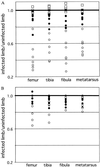
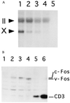
The Finkel-Biskis-Jinkins murine sarcoma virus, which carries v-fos, induces osteosarcomas, whereas high-level expression of exogenous c-fos in transgenic and chimeric mice leads to postnatal development of osteogenic and chondrogenic tumors, respectively. To test whether such target cell specificity of an oncogene can be detected even in early development, we induced ectopic expression of fos in chicken limb buds by microinjecting replication-competent retrovirus into the presumptive leg field of stage 10 embryos. This caused cartilage truncation of all the long bones of the injected leg, which was mainly attributable to chondrodysplasia due to severe retardation of differentiation of the proliferating chondrocytes into mature or hypertrophic chondrocytes, as well as a slight delay in precartilagenous condensation. Expression of genes for all the other known members of chicken AP-1, which include such transforming genes as c-jun and fra-2, however, caused no macroscopic abnormalities in limb formation, indicating a specific function of Fos proteins in embryonic endochondral bone differentiation. The extent of truncation was stronger with v-Fos than with c-Fos, and comparative analysis of these proteins, as well as v-Fos mutants, revealed that strong transforming activity of Fos protein is necessary to cause dysplasia, suggesting that common molecular mechanisms are involved in both embryonic chondrodysplasia and bone tumor formation in postnatal mice.
Figures


Similar articles
-
c-fos-induced osteosarcoma formation in transgenic mice: cooperativity with c-jun and the role of endogenous c-fos.Cancer Res. 1995 Dec 15;55(24):6244-51. Cancer Res. 1995. PMID: 8521421
-
Control of cell cycle gene expression in bone development and during c-Fos-induced osteosarcoma formation.Dev Genet. 1998;22(4):386-97. doi: 10.1002/(SICI)1520-6408(1998)22:4<386::AID-DVG8>3.0.CO;2-2. Dev Genet. 1998. PMID: 9664690
-
Osteoblasts are target cells for transformation in c-fos transgenic mice.J Cell Biol. 1993 Aug;122(3):685-701. doi: 10.1083/jcb.122.3.685. J Cell Biol. 1993. PMID: 8335693 Free PMC article.
-
Retroviruses and oncogenes associated with osteosarcomas.Cancer Treat Res. 1993;62:7-18. doi: 10.1007/978-1-4615-3518-8_2. Cancer Treat Res. 1993. PMID: 8096761 Review. No abstract available.
-
Molecular structure and properties of fos-oncogene.G Batteriol Virol Immunol. 1986 Jan-Jun;79(1-6):11-5. G Batteriol Virol Immunol. 1986. PMID: 3315800 Review.
Cited by
-
c-Fos induces chondrogenic tumor formation in immortalized human mesenchymal progenitor cells.Sci Rep. 2018 Oct 23;8(1):15615. doi: 10.1038/s41598-018-33689-0. Sci Rep. 2018. PMID: 30353072 Free PMC article.
-
Molecular basis for skeletal variation: insights from developmental genetic studies in mice.Birth Defects Res B Dev Reprod Toxicol. 2007 Dec;80(6):425-50. doi: 10.1002/bdrb.20136. Birth Defects Res B Dev Reprod Toxicol. 2007. PMID: 18157899 Free PMC article. Review.
-
C-Fos regulation by the MAPK and PKC pathways in intervertebral disc cells.PLoS One. 2013 Sep 2;8(9):e73210. doi: 10.1371/journal.pone.0073210. eCollection 2013. PLoS One. 2013. PMID: 24023832 Free PMC article.
-
The chromosome-level genome and key genes associated with mud-dwelling behavior and adaptations of hypoxia and noxious environments in loach (Misgurnus anguillicaudatus).BMC Biol. 2023 Feb 1;21(1):18. doi: 10.1186/s12915-023-01517-1. BMC Biol. 2023. PMID: 36726103 Free PMC article.
-
The Role of Activator Protein-1 (AP-1) Family Members in CD30-Positive Lymphomas.Cancers (Basel). 2018 Mar 28;10(4):93. doi: 10.3390/cancers10040093. Cancers (Basel). 2018. PMID: 29597249 Free PMC article. Review.
References
Publication types
MeSH terms
LinkOut - more resources
Full Text Sources
Medical
Miscellaneous

