. 1997 Jan 20;185(2):251-62.
doi: 10.1084/jem.185.2.251.

Distinct costimulatory molecules are required for the induction of effector and memory cytotoxic T lymphocytes

Affiliations

Distinct costimulatory molecules are required for the induction of effector and memory cytotoxic T lymphocytes

Y Liu et al. J Exp Med. .

Abstract

A successful T cell immune response has two major products: effector T cells which directly or indirectly remove the antigens, and memory T cells, which allow a faster and more efficient recall response when challenged by related antigens. An important issue is whether costimulatory molecules on the antigen-presenting cells are involved in determining whether T cells will differentiate into effector or memory cells after antigenic stimulation. To address this issue, we have produced mice with targeted mutations of either the heat-stable antigen (HSA), or both HSA and CD28. We show that CD28/B7 and HSA provide two alternative costimulatory pathways for induction of immunological memory to influenza virus. Furthermore, our results revealed that B7 is essential for the generation of effector T cells from either naive or memory T cells, while HSA is not necessary for the generation of effector T cells. Our results demonstrate that the induction of memory T cells and effector T cells can utilize distinct costimulatory molecules. These results have important implications on lineage relationship between effector and memory T cells.


Figures

Figure 1
Figure 1
Production of mice homozygous for a disrupted HSA gene. (a) The disruption of the HSA gene in C57BL/6 ES line BL/6-III was achieved by replacement of the HSA promotor and the first exon with a neomycin-resistance expression cassette. The coding portions of both HSA exons are depicted by filled boxes. (b) Southern blot analysis of HSA genotypes using a Pvu II fragment located between nucleotides −710, and −1790 relative to the transcriptional start site of the HSA gene as a hybridization probe.
Figure 2
Figure 2
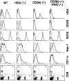
Mice with targeted mutations of HSA and/or CD28 produce normal numbers of T cells, B cells and APC. Mouse spleen cells were analyzed by flow cytometry after staining with either anti-HSA (20C9), anti-CD28 (37N), anti-Mac-1 (TIB128), anti-dendritic cell (HB224), FITC-labeled anti-CD4, and phycoerythrin-labeled anti-CD8 mAbs. CD4, CD8, CD28, B220, and HSA typing was done with freshly isolated spleen cells in pools from three mice. Expression of CD28 on gated T cells is presented. Macrophage and dendritic cells were determined using low-density spleen cells, enriched by centrifugation over a 55% Percoll medium. The genotypes and antibodies used are marked on the panels.
Figure 3
Figure 3
Primary in vivo CTL response against influenza virus in WT mice (a), and mice with a targeted mutation of HSA (b), CD28 (c), or of both HSA and CD28 (d). Spleen cells were harvested from mice on day 7 after A/JAP-infection. The CTL activities were tested on either peptide (AA365-380 of the nucleoprotein from A/JAP virus)- pulsed (EL4-NP), or unpulsed EL4 cells (EL4). Representatives of three independent experiments using pooled spleen cells from 2–4 mice in each group are shown.
Figure 4
Figure 4
Anti-B7 mAbs completely block the production of effector CTL from naive T cells in both WT mice (a) or HSA-KO (b) mice. WT and mutant mice were infected with 1,000 HAU/mouse of influenza virus A/JAP by intraperitoneal injection on day 0. The mice were injected with either a mixture of normal rat and hamster IgG, or anti-B7-1 + anti-B7-2 mAbs (3A12+GL1) on days −1, 0, +1, at a dose of 100 μg/ mouse/injection. Data presented are representative of three experiments using pooled spleen cells from 2–3 mice per group.
Figure 5
Figure 5
Requirement of costimulatory molecules for the generation of primed T cells assayed at 8 d after priming: microplate culture in the absence of exogenous cytokines. (a) Anti-viral CTL of in vitro-stimulated spleen cells from either naive or A/JAPprimed mice. (b) Requirement of B7 family members for the generation of primed T cells. WT and HSA-KO mice were infected with 1,000 HAU/mouse of influenza virus A/JAP by intraperitoneal injection on day 0. On days −1, 0, +1, these mice were injected with either a mixture of normal rat and hamster IgG, or anti-B7-1 + antiB7-2 mAbs (3A12+GL1) at a dose of 100 μg/mouse/injection. Spleen cells were harvested 8 d after viral infection and were restimulated with irradiated, A/JAP-infected syngeneic spleen cells for 6 d in vitro, and CTL activity determined. Responder cells used were from pools of two spleens. Data in b were lysis of NP peptide (AA365-380)-pulsed EL4 target (104/well) only. The lysis of unpulsed EL4 cells is not shown but was always less than 10%. Representative of three experiments, with two mice per group are shown.
Figure 6
Figure 6
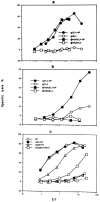
Either HSA or CD28-mediated costimulation is sufficient for generation of primed T cells: bulk cultures. (a and b) Anti-HSA mAb 20C9 blocks T cell memory in CD28KO (b) but not WT mice (a), as measured by recall CTL responses in vitro. CD28-deficient mice and syngeneic WT mice were treated with either normal hamster Ig or anti-HSA mAb (300 μg/mouse/injection) on day −1, day 0, and day 1. On day 0, these mice were injected intraperitoneally with influenza virus A/JAP (1,000 HAU/mouse), spleen cells were harvested on day 7; and were restimulated with A/JAP-infected (1,000 HAU/12 × 106 cells, 37°C,1 h), irradiated syngeneic spleen cells for 5 d (responder: stimulator ratio 5:1, with responder cells at a density of 106/ml). At the end of culturing, viable cells were harvested and the cytotoxicity of these cells was determined by a 6 h 51Cr-release assay. (c) Recall CTL responses in mice with different targeted mutations. The pooled spleen cells from groups of 2–3 mice were harvested on day 7 after i.v. injection of A/JAP (300 HAU), recall CTL activity determined as in a and b. Representatives of 2–3 independent experiments are shown. Note overall CTL responses in c are stronger than in a and b due to a stronger priming via i.v. injection. Dashed lines are the lysis of EL-4 cells in the absence of NP peptide.
Figure 7
Figure 7
Requirement for costimulatory molecules in the generation of memory T cells assayed at 100 d after priming: microplate culture in the absence of exogenous cytokines. Both WT and HSA-KO mice were injected with influenza virus A/JAP (1,000 HAU/mouse) (day 0) and treated with either a mixture of hamster and rat Ig or a mixture of anti-B7-1 plus B7-2 on days −1, 1, 3, 7, 10, 13, 21 (200 μg/ mAb/injection). At 100 d after the viral infection, pooled spleen cells (three mice per group) were stimulated in vitro, CTL activity was determined 5 d after stimulation. Similar results were obtained when memory CTL activity was measured at 30 d after infection.
Figure 8
Figure 8
B7, but not HSA, plays an important role in the induction of effector T cells from memory T cells, regardless of the priming conditions: microplate culture in the absence of exogenous cytokines. Spleen cells from either WT (a and b) or HSA-deficient mice (c and d) pretreated with either normal Ig (a and c) or a mixture of anti-B7-1 and anti-B7-2 (b and d) were infected with A/JAP virus as detailed in the legend to Fig. 3. Increasing numbers of spleen cells were stimulated in vitro for 5 d with A/JAP-infected spleen cells, in the presence of either medium, or a mixture of anti-B7-1 and anti-B7-2, or anti-HSA mAb (final concentration at 5 μg/ml), and CTL activity was determined in a 6-h 51Cr-release assay. Representative of three independent experiments are shown. Similar results were obtained when memory activity is measured in bulk cultures.
Figure 9
Figure 9
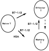
A model for the involvement of costimulatory signals in the generation of effector and memory T cells. Two major products, effector and memory T cells, are produced from naive T cells after viral infection. The production of memory T cells requires costimulation by either HSA or B7, while the production of effector T cells utilizes B7 but not HSA. By definition, memory T cells give rise to effector T cells after further stimulation by antigen. However, effector T cells are unlikely to be mandatory precursors for memory T cells and distinct costimulatory molecules could be used at different phases of the immune response.

Similar articles

Cited by

References

    1. Ada GL, Jones PD. The immune response to influenza infection. Curr Top Microbiol Immunol. 1986;128:1–54. - PubMed
    1. Moskophidis D, Lechner F, Pircher H, Zinkernagel RM. Virus-persistence in acutely infected immunocompetent mice by exhaustion of antiviral cytotoxic effector T cells. Nature (Lond) 1993;362:758–761. - PubMed
    1. Razvi ES, Welsh RM. Programmed cell death of T lymphocytes during acute viral infection: a mechanism for virus-induced immune deficiency. J Virol. 1993;67:5754–5765. - PMC - PubMed
    1. Tripp RA, Lahti JM, Doherty PC. Laser light suicide of proliferating virus-specific CD8 T cells in an in vivoresponse. J Immunol. 1995;155:3719–3721. - PubMed
    1. Müllbacher A. The long-term maintenance of cytotoxic T cell memory does not require persistence of antigen. J Exp Med. 1994;179:317–321. - PMC - PubMed

Publication types