. 1996 Aug 1;16(15):4563-78.
doi: 10.1523/JNEUROSCI.16-15-04563.1996.

Visual motion-detection circuits in flies: small-field retinotopic elements responding to motion are evolutionarily conserved across taxa

Affiliations

Visual motion-detection circuits in flies: small-field retinotopic elements responding to motion are evolutionarily conserved across taxa

E K Buschbeck et al. J Neurosci. .

Abstract

The Hassenstein-Reichardt autocorrelation model for motion computation was derived originally from studies of optomotor turning reactions of beetles and further refined from studies of houseflies. Its applicaton for explaining a variety of optokinetic behaviors in other insects assumes that neural correlates to the model are principally similar across taxa. This account examines whether this assumption is warranted. The results demonstrate that an evolutionarily conserved subset of neurons corresponds to small retinotopic neurons implicated in motion-detecting circuits that link the retina to motion-sensitive neuropils of the lobula plate. The occurrence of these neurons in basal groups suggests that they must have evolved at least 240 million years before the present time. Functional contiguity among the neurons is suggested by their having layer relationships that are independent of taxon-specific neurons, or the absence of orientation-specific motion-sensitive levels in the lobula plate.


Figures

Fig. 1.
Fig. 1.
Schematic organization of the visual system of Diptera. Four retinotopic neuropils (lamina, La; medulla,Me; lobula, Lo; lobula plate, LP) provide outputs to the mid brain (Br), which supply descending neurons (DN) that terminate in motor centers of the thoracic ganglia (Th). Neurons implicated in elementary motion detection are shown in the inset. These are R1–R6 photoreceptors (R), large monopolar cells (LMCs), the transmedullary cells (iTm and Tm1), the bushy T-cells (T4 and T5), and the centrifugal neuron(C2). The lobula plate sends axons of collator neurons(CN) to the brain and contralateral lobula plate.
Fig. 2.
Fig. 2.
Phylogenetic relationships of nematoceran and brachyceran Diptera, based mostly on morphological characters and reconstructed after Wood and Borkent (1989), Woodly (1989), McAlpine (1989), Sinclair et al. (1993), and Cumming et al. (1995). Camera lucida drawings of insects illustrate the morphological variety of investigated taxa. Common names: tipulids, crane flies; culicids, mosquitoes; simuliids, black flies; tabanids, horseflies; bombyliids, bee-flies; dolichopodids, long-legged flies; syrphids, hover flies; glossinids, tsetse flies; and calliphorids, blow flies.
Fig. 7.
Fig. 7.
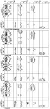
Densitometric analysis of varicose and spiny decorations on medullary neurons, illustrating similarities in profile densities. This method, used for analyzing layer relationships in the calliphorid Sarcophaga bullata (see Strausfeld and Lee, 1993) is used here for four additional taxa. In each case, the first column (A) illustrates medulla inputs; these are the LMCs, L4, L5, and T1 and the photoreceptors R7 and R8. The second and third columns show the densities of varicose and spiny processes of iTm(B) and Tm1 (C). D, The bushy arborization of T4. Comparisons of the output specializations of iTm and Tm1 in the inner medulla demonstrate the relationship of iTm, not Tm1, to T4. Jagged lines indicate changes in density;smooth lines indicate the average.
Fig. 8.
Fig. 8.
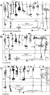
Reconstruction of conserved medulla neurons in three different nematoceran species, based on Golgi impregnation. As in Figure 4, the basic shape and characteristic Gestalt of a subset of medullary neurons remains conserved in these taxa, except that thetipulid iTm has an extra and extremely thin layer of distal processes extending to neighboring columns. However, the basic layer relationships among individual neurons, although a little less precise than in brachyceran species, generally are conserved.
Fig. 3.
Fig. 3.
Reduced silver staining of cross-sections through the medulla of four brachyceran flies. A, Calliphorid;B, syrphid; C, bombyliid; D, tabanid. Images are scaled to equal size for comparison. Differences are in the relative depths of analogous strata. However, as demonstrated in Figures 4, 5, 6, these do not reflect taxonomic differences between cell relationships.
Fig. 4.
Fig. 4.
Layer constancy within a taxon is demonstrated in this tabanid with mass impregnation by the Golgi method. This section shows several LMC terminals (L1, L2), the endings of the lamina monopolar cell L5, and several type iTm(transmedullary neurons). Different individuals of each cell type show the remarkable morphological constancy.
Fig. 5.
Fig. 5.
Reconstructions of conserved medulla neurons in seven different brachyceran species. Although distinct species-specific differences can be observed with respect to features, such as the arrangement of L1 varicosities, the characteristic Gestalten of a subset of medulla elements (L1, L2, L3, L4, L5, Tm1, iTm, C2, and T4) are conserved across taxa. In contrast to the transmedullary cells iTm and Tm1 that are associated with T4 and T5 (see Fig. 9), other retinotopic transmedullary neurons destined for the lobula elements (indicated by asterisks to the right of each figure) have been found in, at most, only two of the investigated taxa and thus seem to be less strongly conserved than neurons serving motion computation.
Fig. 5.
Fig. 5.
Reconstructions of conserved medulla neurons in seven different brachyceran species. Although distinct species-specific differences can be observed with respect to features, such as the arrangement of L1 varicosities, the characteristic Gestalten of a subset of medulla elements (L1, L2, L3, L4, L5, Tm1, iTm, C2, and T4) are conserved across taxa. In contrast to the transmedullary cells iTm and Tm1 that are associated with T4 and T5 (see Fig. 9), other retinotopic transmedullary neurons destined for the lobula elements (indicated by asterisks to the right of each figure) have been found in, at most, only two of the investigated taxa and thus seem to be less strongly conserved than neurons serving motion computation.
Fig. 6.
Fig. 6.
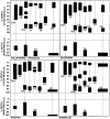
Conservation of depth relationships between specific medulla neurons involved in motion computation. Because of small disparities in the sectioning plane, some of the Golgi reconstructions of Figure 4 show minor discrepancies from their precise absolute depths. Here, depth measurements of several exemplary cells demonstrate the relative depths of specializations for each species of neurons to show that layer relationships are strictly conserved between taxa. For example, in each case the dendritic arborizations ofTm1 correlate in depth with L2 terminals. The bilayered specializations of L1 coincide with those ofL4, L5, C2, and iTm, even in bombyliids, in which in all but C2 an additional layer of specializations can be observed.
Fig. 9.
Fig. 9.
Camera lucida drawings showing coimpregnation of medulla neurons in glossinids (A) and asilids(B). Resolution of neurons in a single column, or direct neighbors, verifies the relative depth relationships of neurons to each other. In both species L1 has been coimpregnated withiTm, illustrating the tight overlap of their neural processes. In asilids, in addition to the iTm neuron restricted to a single column, these taxa possess a neuron(iTm[LF]) with similar terminal arrangements in the inner medulla but having dendrites with a larger field reaching neighboring cartridges.
Fig. 10.
Fig. 10.
Reconstructions and photomicrographs of T5 neurons of seven brachyceran (A–H) and three nematoceran(I–K) species to show the remarkable morphological conservation of this type of nerve cell. A, Calliphorid;B, glossinid; C and D, syrphid;E, asilid; F, bombyliid; G, dolichopodid; H, tabanid; I, simuliid;J, culicid; K, tipulid. Scale in D(for all drawings), E (for all half tones), 20 μm. The phylogenetically basal Nematocera as well as tabanids are characterized by narrower dendritic fields and a relatively thicker lobula stratum than the other species.
Fig. 11.
Fig. 11.
T-cell endings of bombyliids (A)and asilids (B) compared. Except in asilids, brachyceran T5 cells project to four distinct layers of the lobula plate(LP). Their terminals at the two outer layers visit large-diameter, wide-field directional motion-sensitive horizontal neurons (HS). Terminals in two deep layers visit vertical motion-sensitive neurons (VS). Asilids, which among the brachycerans are the closest relatives to bombyliids, lack VS-like elements deep in the lobula plate. All their T-cell terminals project exclusively to HS-like dendrites within the outer strata of the lobula plate.

Similar articles

Cited by

References

    1. Bausenwein B, Fischbach K-F. Activity labeling patterns in the medulla of Drosophila melanogaster caused by motion stimuli. Cell Tissue Res. 1992a;270:25–35. - PubMed
    1. Bausenwein B, Fischbach K-F. Separation of functional pathways in the fly’s medulla: combination of 2-deoxyglucose studies with anatomical fine analysis. Singh RN. Nervous systems principles of design and function 1992b. 223 239 Wiley; New Delhi, India: Eastern.
    1. Bodian D. A new method for staining nerve fibers and nerve endings in mounted paraffin sections. Anat Rec. 1937;69:153–162.
    1. Boschek CB. On the fine structure of the peripheral retina and lamina ganglionaris of the fly, Musca domestica . Z Zellforsch Mikrosk Anat. 1971;118:369–409. - PubMed
    1. Braitenberg V. Patterns and projections in the visual system of the fly. I. Retina–lamina projections. Exp Brain Res. 1967;3:271–298. - PubMed

Publication types

LinkOut - more resources