. 1994 Jun;120(6):1503-15.
doi: 10.1242/dev.120.6.1503.

Developmental control of a G1-S transcriptional program in Drosophila

Affiliations

Developmental control of a G1-S transcriptional program in Drosophila

R J Duronio et al. Development. 1994 Jun.

Abstract

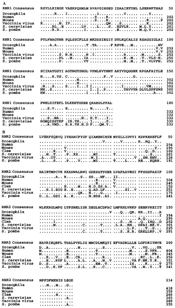
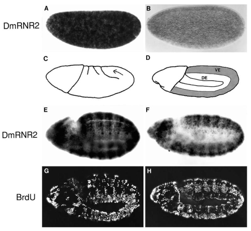
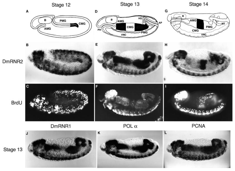
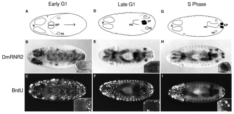
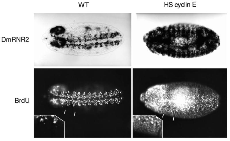
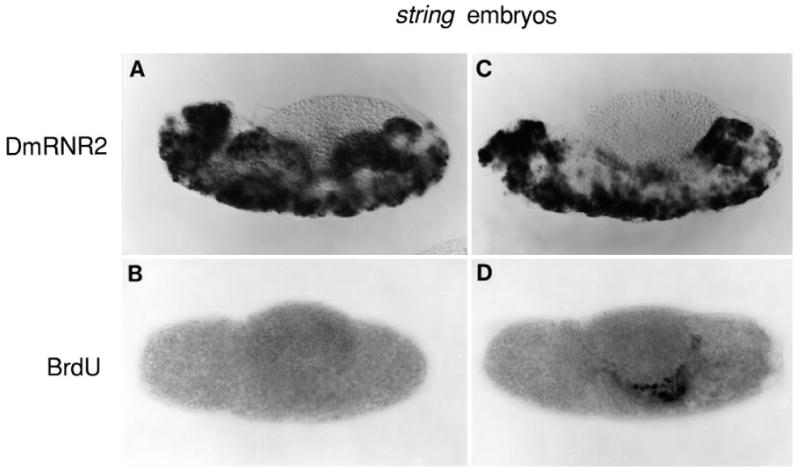
We have defined a coordinate program of transcription of S-phase genes (DNA polymerase alpha, PCNA and the two ribonucleotide reductase subunits) that can be induced by the G1 cyclin, cyclin E. In Drosophila embryos, this program drives an intricate spatial and temporal pattern of gene expression that perfectly parallels the embryonic program of S-phase control. This dynamic pattern of expression is not disrupted by a mutation, string, that blocks the cell cycle. Thus, the transcriptional program is not a secondary consequence of cell cycle progression. We suggest that developmental signals control this transcriptional program and that its activation either directly or indirectly drives transition from G1 to S phase in the stereotyped embryonic pattern.


Figures

Fig. 1
Fig. 1
Predicted amino acid sequence of the DmRNR1 and DmRNR2 cloned PCR products with comparison to other eukaryotic and viral ribonucleotide reductases. Only sequence between and not including the PCR primer sites is shown. The alignments were performed using Geneworks. ‘Consensus’ amino acids appear in ≥2 of the sequences. Dots indicate identities with the consensus. Dashes indicate gaps introduced in a sequence to maximize the alignment. (A) RNR large subunit (RNR1) alignment. The S. cerevisiae genome contains two large subunit genes, RNR1 and RNR3 (Elledge and Davis, 1990), and sequence from the latter is shown in this alignment. (B) RNR small subunit (RNR2) alignment.
Fig. 2
Fig. 2
Embryonic expression of DmRNR2 prior to the appearance of G1-S control. DmRNR2 mRNA was detected by whole embryo in situ hybridizations using a digoxigenin-labeled probe. (A) A preblastoderm embryo with high levels of maternal DmRNR2 message. (B) The maternal message nearly disappears by blastoderm cellularization during early cycle 14. (C) Gastrulation begins shortly after cellularization is complete. Invagination of cells along the ventral midline creates a three-layered germ band. The arrow indicates the movement of cells of the germ band around the posterior tip of the embryo during germ band extension. (D) In a fully germ band extended embryo, the ventral epidermis (VE) extends around to the dorsal side of the embryo as well (shaded region), while the dorsal epidermis (DE) is located laterally. The embryos in E-H are at the germ band extended stage. They have been pulse labeled with BrdU and then hybridized with a DmRNR2 probe prior to immunofluorescent detection of incorporated BrdU (G and H). (E) The constitutive and ubiquitous levels of DmRNR2 that characterize the postblastoderm divisions are beginning to decline in the dorsal epidermis. This same embryo viewed with fluorescent illumination is shown in G. The heavily labeled nuclei in the dorsal epidermis are in early S16, while the unlabeled cells in the ventral epidermis have just completed S15. (F) DmRNR2 expression in a slightly older embryo. Note that DmRNR2 message is significantly reduced in the dorsal epidermis, while the level remains high in ventral epidermis. (H) Fluorescence view of the embryo in F shows the S-phase pattern. The ventral epidermal cells have finished G2 and mitosis of cycle 15 and entered S16 without an intervening G1. Cells in the dorsal epidermis have completed or nearly completed S16 and will enter G1 after progressing through a short G2 and mitosis. In each panel, dorsal is to the top and anterior is to the left. The embryo in E and G is rotated slightly on its side relative to the embryo in F and H such that the ventral midline is apparent.
Fig. 3
Fig. 3
The patterned expression of S-phase genes correlates with the G1-S transition. In each panel, an embryo is displayed in lateral view with dorsal up and anterior to the left. (A) During early stage 12 of embryogenesis, the distinct anterior and posterior invaginations of the midgut primordia (AMG and PMG, respectively) remain separated while germ band retraction initiates. (B) DmRNR2 expression is extinguished in the G1-arrested epidermal cells but persists in neural tissue along the ventral midline and in the brain (B in diagram). At this stage, DmRNR2 expression can be detected in large endodermal cells at the leading edge of the migrating posterior midgut invagination (shaded region in A). These cells will occupy the central portion of the midgut (CMG) after fusion of the midgut primordia. (C) An embryo pulse labeled with BrdU at the same stage as B reveals that the cells of the CMG have entered S17. Replicating nuclei can also be detected in the nervous system. (D) By stage 13, germ band retraction is complete and the midgut primordia have fused on either side of the yolk resulting in three distinct regions of the midgut along the anterior-posterior axis: the AMG, CMG and PMG. (E) In stage 13, DmRNR2 expression has terminated in the CMG and initiated in the AMG and PMG. Several other tissues also accumulate DmRNR2 RNA at this time. Among those visible in this embryo are the hindgut (HG), the anal pads (AP), the pharynx (PH), and the brain (B) and ventral nerve cord (VNC). (F) The S-phase pattern of a stage 13 embryo is identical to the DmRNR2 expression pattern. Clearly visible is S17 in the AMG and PMG. Note that a few CMG cells near the end of S17 have incorporated BrdU (slightly out of focus). (G) The midgut cells begin migrating dorsally and ventrally on either side of a stage 14 embryo to eventually fuse and create a tube that envelopes the yolk. (H) The DmRNR2 expression pattern has changed substantially by stage 14 (this embryo is ∼1 hour older than the embryo in E). Expression has ceased in the AMG, PMG, HG, and PH and RNA reaccumulates in the CMG. Staining is also apparent in the Malpighian tubules (MT) and persists in the VNC. (I) The S-phase pattern at stage 14 changes in concert with the DmRNR2 expression pattern. The CMG has reentered S phase (S18), which is the first endoreduplication S in the midgut. The BrdU incorporation in the Malpighian tubules (MT) at this stage is due to endoreduplication S phases. The pattern of expression of DmRNR1 (J), DNA polymerase α (K), and PCNA (L) during stage 13 is identical to that of DmRNR2 (E).
Fig. 4
Fig. 4
DmRNR2 message accumulation occurs late in G1 prior to the initiation of DNA replication. Embryos were pulse labeled with BrdU and then hybridized with a DmRNR2 probe prior to immunofluorescent detection of incorporated BrdU. All embryos are viewed from the dorsal perspective and anterior is to the left. The schematics indicate the changing position of the anal pads (AP), which occupy the tip of the germ band. In stage 12, the germ band retracts along the same pathway as when it extended. The arrow indicates the direction of cell movement during this process. PS indicates the bilaterally symmetric groups of cells that will form the posterior spiracles, which are already in S phase and express DmRNR2 in the embryos shown. The B in the schematics indicates the two lobes of the brain. In the leftmost three panels (A-C), the cells of the AP are in early G1, and they neither express DmRNR2 (B) nor incorporate BrdU (C). The insets show a close-up view of the right (towards the top) anal pad. In the central panels (D-F), the AP cells are in late G1. At this stage, segmentation is obvious and retraction of the germ band (75%) has revealed the hindgut (HG). The movements of the germ band (arrow in D) are such that the AP will eventually appear more posterior than the PS. The AP now expresses DmRNR2 RNA (E, and indicated by shading in the diagrams), but still fails to incorporate BrdU (F). In the rightmost three panels (G-I), the cells of the AP are in S phase. At this stage, germ band retraction is complete, placing the AP at the end of the hindgut tube. DmRNR2 RNA is still detected (H, and indicated by the shading in G). The fluorescence signal from BrdU labeling of the AP cells is partially quenched by the histochemical staining of the DmRNR2 RNA, but it is nonetheless evident that these cells are in S phase (I, see inset). Note that most of the numerous isolated cells that incorporate BrdU and stain in C, F and I are rapidly dividing cells of the peripheral and central nervous systems.
Fig. 5
Fig. 5
Ectopic production of Drosophila cyclin E induces the G1-S transcriptional program and drives G1 cells into S phase. All panels show a ventral view of a stage 14 embryo either stained for DmRNR2 expression or BrdU incorporation. The VNC is clearly visible in the wild-type (WT) embryos. Note that the G1-arrested epidermal cells on either side of the VNC neither incorporate BrdU nor express DmRNR2. Embryos homozygous for a hsp70-cyclin E transgene (HS cyclin E) were heat shocked at 37°C for 30 minutes. After a subsequent 45 minute recovery period at room temperature, they were either fixed and stained for DmRNR2 expression or pulse labeled with BrdU. The insets represent enlargements of the two segments included between the white markings in the BrdU-labeled embryos.
Fig. 6
Fig. 6
The G1-S transcriptional program operates independently of cell cycle progress. Expression of DmRNR2 (A,C) and BrdU incorporation (B,D) are shown in approximately stage 13 (A,B) and stage 14 (C,D) string mutant embryos. See Fig. 3 panels E, F, H and I for analogous staining of wild-type embryos (note that BrdU incorporation was detected histochemically in this experiment but immunofluorescently in the experiment shown in Fig. 3). In stage 13 string embryos, DmRNR2 is expressed in a near normal pattern (A) despite the lack of detectable DNA replication (B). In stage 14 string embryos, DmRNR2 is also expressed in a near normal pattern (C), and DNA replication is confined to a few cells (note the large nuclei in the center of the midgut in D). These replicating cells are apparently those that would undergo an endoreduplication S phase at this stage (see text).

Similar articles

Cited by

References

    1. Andrews BJ, Herskowitz I. Identification of a DNA binding factor involved in cell-cycle control of the yeast HO gene. Cell. 1989;57:21–29. - PubMed
    1. Andrews BJ, Herskowitz I. Regulation of cell cycle-dependent gene expression in yeast. J Biol Chem. 1990;265:14057–14060. - PubMed
    1. Baldin V, Lukas J, Marcote MJ, Pagano M, Draetta G. Cyclin D1 is a nuclear protein required for cell cycle progression in G1. Genes Dev. 1993;7:812–821. - PubMed
    1. Bjorklund S, Skog S, Tribukait B, Thelander L. S-phase-specific expression of mammalian ribonucleotide reductase R1 and R2 subunit mRNAs. Biochemistry. 1990;29:5452–5458. - PubMed
    1. Blake MC, Azizkhan JC. Transcription factor E2F is required for efficient expression of the hamster dihydrofolate reductase gene in vitro and in vivo. Mol Cell Biol. 1989;9:4994–5002. - PMC - PubMed

Publication types

Associated data