Review
. 1995 Apr 1;30(5):366-80.
doi: 10.1002/jemt.1070300504.

Development and postnatal regulation of adult myoblasts

Affiliations
Review

Development and postnatal regulation of adult myoblasts

Z Yablonka-Reuveni. Microsc Res Tech. .

Abstract

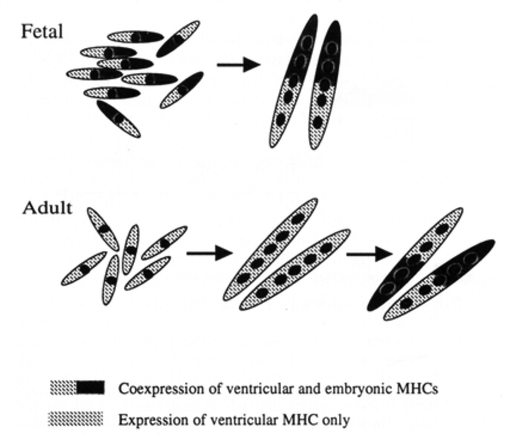
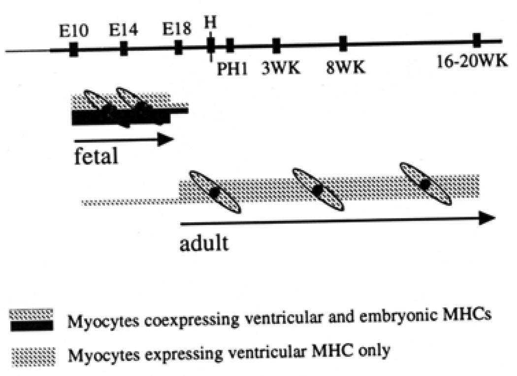
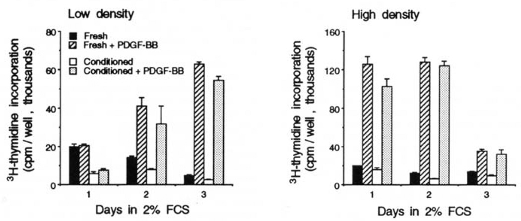
The myogenic precursor cells of postnatal and adult skeletal muscle are situated underneath the basement membrane of the myofibers. It is because of their unique positions that these precursor cells are often referred to as satellite cells. Such defined satellite cells can first be detected following the formation of a distinct basement membrane around the fiber, which takes place in late stages of embryogenesis. Like myoblasts found during development, satellite cells can proliferate, differentiate, and fuse into myofibers. However, in the normal, uninjured adult muscle, satellite cells are mitotically quiescent. In recent years several important questions concerning the biology of satellite cells have been asked. One aspect has been the relationship between satellite cells and myoblasts found in the developing muscle: are these myogenic populations identical or different? Another aspect has been the physiological cues that control the quiescent, proliferative, and differentiative states of these myogenic precursors: what are the growth regulators and how do they function? These issues are discussed, referring to previous work by others and further emphasizing our own studies on avian and rodent satellite cells. Collectively, the studies presented indicate that satellite cells represent a distinct myogenic population that becomes dominant in late stages of embryogenesis. Moreover, although satellite cells are already destined to be myogenic precursors, they do not express any of the four known myogenic regulatory genes unless their activation is induced in the animal or in culture. Furthermore, multiple growth factors are important regulators of satellite cell proliferation and differentiation. Our work on the role of one of these growth factors [platelet-derived growth factor (PDGF)] during proliferation of adult myoblasts is further discussed with greater detail and the possibility that PDGF is involved in the transition from fetal to adult myoblasts in late embryogenesis is brought forward.


Figures

Fig. 1
Fig. 1
Demonstration by indirect immunofluorescence of the laminin component of the basement membrane surrounding muscle fibers in the pectoralis muscle from late embryonic, young, and mature chickens. Unfixed frozen cross sections were prepared from (A) 19-day-old embryo; (B) 19-day-old chicken; and (C) 2-month-old-chicken. Sections were reacted with a monoclonal antibody against chicken laminin (prepared by Bayne et al., 1984) followed by fluorescein-labeled goat anti-mouse IgG; arrows indicate blood vessels that are also positive for laminin (40× objective was used for all micrographs). (Reproduced from Grounds and Yablonka-Reuveni, 1993, with permission of Chapman & Hall.)
Fig. 2
Fig. 2
Electron micrographs of a muscle cross section from the pectoralis muscle of 2-month-old chicken. A: Satellite cell lying between the basement membrane and plasma membrane of muscle fiber. B: Nuclei within the muscle fibers and other cells outside the fiber that can be easily mistaken for satellite cells at the light microscopy level. Adult chicken muscle was immersion fixed in mixed aldehyde, post-fixed in osmium tetroxide, and embedded in epoxy resin. Sections were stained with lead citrate and examined in a Philips 201 TEM operated at 60 KV. (Yablonka-Reuveni and Anderson, unpublished data). A: bar = 1 µm and magnification = ×13,440; B: bar = 1 µm and magnification = ×5,584. Sat, satellite cell nucleus; f, myofiber: fn, nucleus within myofiber; cap, capillary; e, endothelial cell; p, pericyte; black arrows indicate basement membrane: white arrows indicate the plasma membrane of the myofiber and the plasma membrane of the satellite cell.
Fig. 3
Fig. 3
Schematic of embryonic and ventricular MHC expression patterns in myogenic cultures from skeletal muscle of fetal (E10) and adult (2-month-old) chickens detected by monoclonal antibodies against specific MHC. Monoclonal antibodies against specific isoforms of MHC were originally prepared in the laboratory of Dr. E. Bandman (University of California, Davis) and are described in more details in Hartley et al. (1991). This schematic does not exclude the possibility that other sarcomeric MHC are expressed by the cells as well. (Reproduced from Hartley and Yablonka-Reuveni, 1992, with permission of Cordon and Breach. Model is based on data published in Hartley et al., 1991, .)
Fig. 4
Fig. 4
Schematic of the presence of fetal and adult myoblasts during embryogenesis and posthatch stapes of chicken development. E10, E14, and E18 represent days of embryogenesis; PHI represents 1 day after hatching; 3WK, 8WK, and 16–20 WK represent weeks posthatching. H refers to hatching, which occurs on days 20–21. (Reproduced from Hartley and Yablonka-Reuveni, 1992, with permission of Gordon and Breach. Model is based on data published in Hartley el al., 1991, .)
Fig. 5
Fig. 5
Effect of PDGF-BB on proliferation of adult mouse-derived C2 myoblasts. Cells were plated into gelatin-coated, 24-well trays (well diameter = 16 mm) at a density of 1 × 10⁴ cells/well. Cells were maintained for the initial 24 hours in proliferation medium, which consists of MEM fortified with 20% FCS. Medium was then changed to MEM containing 2% FCS in the absence and presence of PDGF-BB (20 ng/ml) and antibodies. Goal anti-PDGF (IgG fraction) and control goat IgG were added to the wells at 50 µg/ml. The 2% FCS medium with or without PDGF and antibodies was replaced every 24 hours to ensure ample supply of additives and cells were harvested following 48 hours in the 2% FCS. Two hours before ceil harvesting, the cultures were exposed to 3H-thymidine at 1 µC/ml. and the extent of 3H-thymidine incorporation into TCA-precipitable material was determined as in Yablonka-Reuveni et al. (1990a). Plotted values represent the average of triplicate wells. FCS used for this study was heat inactivated (56°C, 30 minutes) to avoid possible complement reaction in the presence of the antibodies. Note that our original studies on the response of C2 cells to PDGF were conducted with untreated FCS (Yablonka-Reuveni et al., 1990a); in experiments that are not shown here we demonstrated that 3H-thymidine incorporation by C2 cells maintained in 2% FCS medium alone and its enhancement by PDGF-BB is similar whether untreated or heat-inactivated FCS is used. Antibody against PDGF was purchased from Upstate Biotechnology (Lake Placid, NY); this polyclonal antibody is made in gnat immunized with human PDGF-AB. Using Western blotting we confirmed that this antibody recognizes all three forms of PDGF of human origin as well as rat PDGF-like proteins. Inhibition of PDGF-BB effect in the presence of anti-PDGF was demonstrated by us for C2 cultures initiated at a lower density as well (2.5 × 10³/16-mm well), (Yablonka-Reuveni and Rivera, unpublished results).
Fig. 6
Fig. 6
Saturation binding or PDGF-AB and PDGF-BB to primary myogenic cultures from the pectoral is muscle of 10-day (E10) and 19-day (E19) chicken embryos. Freshly isolated cells were cultured into gelatin-coated 24-well trays (well diameter = 16 mm) using our standard medium for chicken myogenic cultures (MEM containing 10% horse serum and 5% chick embryo extract) for 24 hours. Medium was then replaced with MEM containing 2% FCS for about 18 hours (to reduce any possible exogenous PDGF) and at the end of this period cultures were subjected to a PDGF binding assay. The plotted values represent the average specific binding to triplicate wells, and standard error was less than 5–10%. Cell numbers were determined on parallel triplicate cultures. (Reproduced from Yablonka-Reuveni and Seifert, 1993, with permission of Academic Press.)
Fig. 7
Fig. 7
PDGF-BB and transferrin have a concerted effect on DNA synthesis by chicken myoblasts. Chicken myoblasts isolated by a clonal expansion of a myogenic clone from the pectoralis muscle of a 19-day-old chicken embryo (clone E1915) were cultured at 3 104 cells/16-mm well in MEM-based medium containing 10% horse serum and 5% chicken embryo extract for a period of 24 hours. Medium was than changed to MEM containing 2.5% FCS for 48 hours in the absence or presence of additives. MEM was fortified with iron in the form of FeSO4·7H2O at 0.834 mg/l to provide iron for transferrin. Iron formulation and concentration was based on F-10 medium from Gibco. Highly purified chicken transferrin was from Organon Teknika Corporation (Cappel Research Products, Durham, NO); we used chicken transferrin for this study because species specificity was previously established for the effect of transferrin on chicken myogenesis. Transferrin (Tf) was added to MEM at 20 µg/ml and PDGF-BB was added at 20 ng/ml. The 2.5% FCS-medium and additives were replenished every 24 hours. Prior to cell harvesting, the cultures were exposed for 4 hours to 3H-thymidine at 1 µCi/ml. and the extent of 3H-thymidine incorporation into TCA-precipitable material was determined as for Figure 5. Plotted values represent the average of triplicate wells, (Yablonka-Reuveni, unpublished results.)
Fig. 8
Fig. 8
Effect of PDGF-BB and basic FGF on the proliferation of C2 cultures. Cultures were initiated at low density (1 × 10³ cells/16-mm well) or high density (7.5 × 10³ cells/16-mm well) in proliferation medium (MEM with 20% FCS and 0.5% chicken embryo extract). After 24 hours medium was changed to MEM containing 2% FCS and cultures were maintained in this serum-low medium for various length of time in the absence or presence of growth factors. PDGF-BB was added at 20 ng/ml and bFGF was added at 4 ng/ml. These concentrations of growth factors are maximally effective, as determined in separate experiments. The 2% FCS medium with or without growth factors was replaced every 24 hours. Two hours prior to cell harvesting the cultures were exposed to 3H-thymidine at 1 µC/ml and the extent of 3H-thymidine incorporation into TCA-precipitable material was determined as for Figure 5. Plotted values represent the average of triplicate wells. Highly purified bFGF (human recombinant; produced in yeast) was kindly provided by Dr. S.D. Hauschka (University of Washington, Seattle, WA). (Yablonka-Reuveni and Balestreri unpublished data; study was briefly mentioned in an abstract by Yablonka-Reuveni et al., 1990b).
Fig. 9
Fig. 9
Effect of PDGF-BB and medium conditioned by C2 cells on the proliferation of C2 myoblasts. High-density C2 cultures were prepared as in Figure 5 and maintained in 2% FCS medium for 3 days with daily changes of medium. Medium from the third day lat which time cells are responsive to PDGF, as was shown on parallel cultural was collected, cleared from cell debris by centrifugation (2,000g, 5 minutes), and added to fresh C2 cultures at the Lime of transition from the 20% FCS medium to 2%. Conditioned medium, with or without PDGF-BB (20 ng/ml) was replaced every 24 hours. Incorporation of 3H-thymidine was determined as in Figure 5. Low- and high-density cultures were initiated at 2.5 × 10³ cells/16-mm well and 1 × 10⁴ cells/16-mm well, respectively. Note that proliferation of low-density cultures in conditioned medium is lower than in fresh medium but PDGK effects is similar. Also, the decline in proliferation in high-density cultures after 3 days is due to terminal differentiation of the cells, but more cells ore still proliferating in the presence of PDGF even during the third day. As in Figure 5, FCS used in the 2% FCS medium was heat inactivated, as described in the legend to Figure 5. (Yablonka-Reuveni and Rivera, unpublished data.)

Similar articles

Cited by

References

    1. Allen RE, Boxhorn LK. Inhibition of skeletal muscle satellite cell differentiation by transforming growth factor-beta. J. Cell Physiol. 1987;133:567–572. - PubMed
    1. Allen RE, Boxhorn LK. Regulation of skeletal muscle satellite cell proliferation and differentiation by transforming growth factor-beta, insulin-like growth factor I, and fibroblast growth factor. J. Cell Physiol. 1989;138:311–315. - PubMed
    1. Allen RE, Dodson MV, Luiten LS. Regulation of skeletal muscle satellite cell proliferation by bovine pituitary fibroblast growth factor. Exp. Cell. Res. 1984;152:154–160. - PubMed
    1. Allen RE, Rankin LL, Greene EA, Boxhorn LK, Johnson SE, Taylor RG, Pierce PR. Desmin is present in proliferating rat muscle satellite cells but not in bovine muscle satellite cells. J. Cell. Physiol. 1991;149:525–535. - PubMed
    1. Anderson JE, Liu L, Kardami E. Distinctive patterns of bFGF distribution in degenerating and regenerating areas of dystrophic (mdx) striated muscles. Dev. Biol. 1991;147:96–109. - PubMed

Publication types

LinkOut - more resources