. 1993 Dec 5;268(34):25604-16.

Nascent RNA cleavage by arrested RNA polymerase II does not require upstream translocation of the elongation complex on DNA

Affiliations

Nascent RNA cleavage by arrested RNA polymerase II does not require upstream translocation of the elongation complex on DNA

W Gu et al. J Biol Chem. .

Abstract

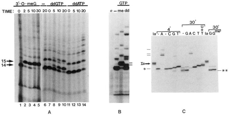
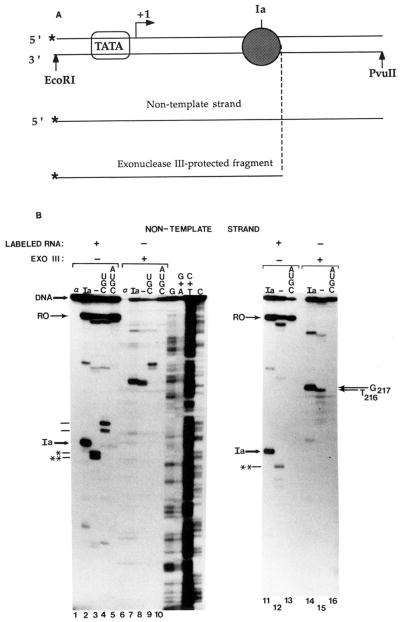
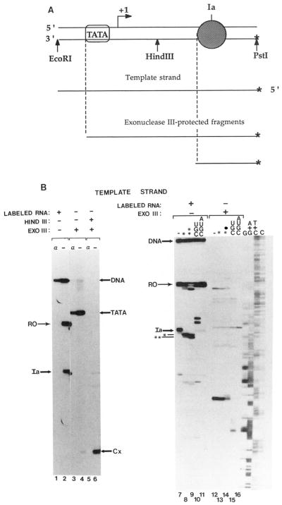
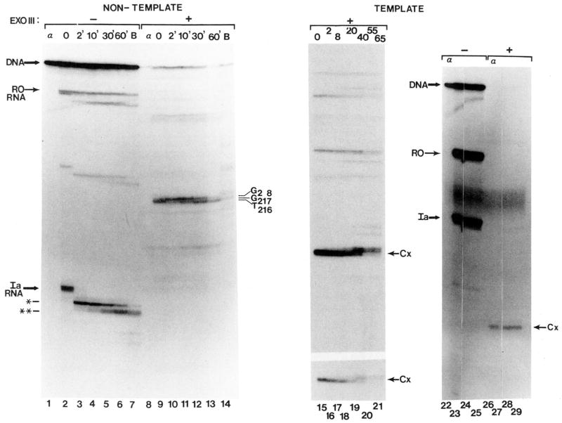
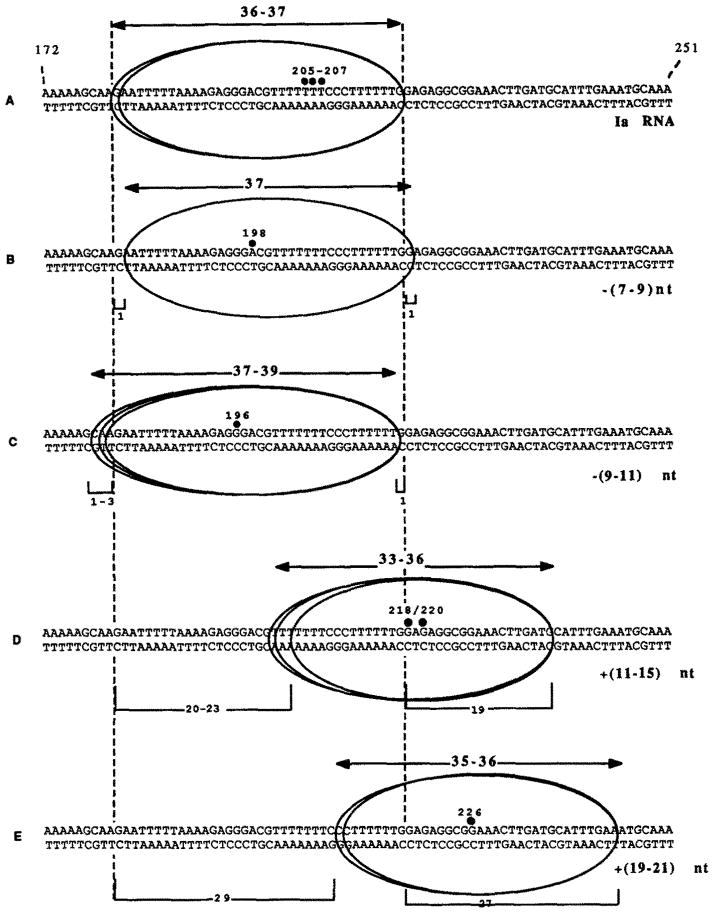
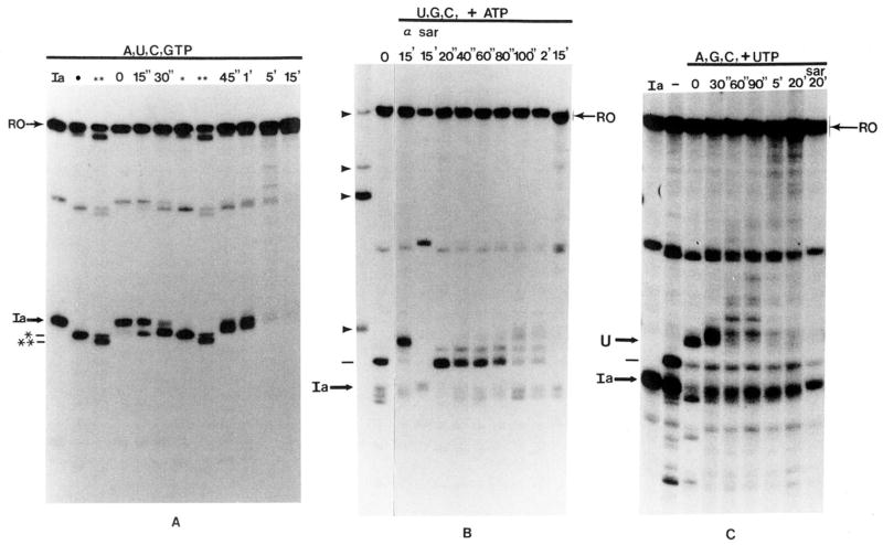
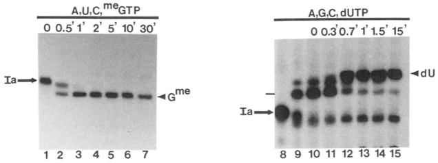
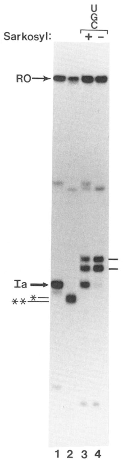
Obstacles incurred by RNA polymerase II during primary transcript synthesis have been identified in vivo and in vitro. Transcription past these impediments requires SII, an RNA polymerase II-binding protein. SII also activates a nuclease in arrested elongation complexes and this nascent RNA shortening precedes transcriptional readthrough. Here we show that in the presence of SII and nucleotides, transcript cleavage is detected during SII-dependent elongation but not during SII-independent transcription. Thus, under typical transcription conditions, SII is necessary but insufficient to activate RNA cleavage. RNA cleavage could serve to move RNA polymerase II away from the transcriptional impediment and/or permit RNA polymerase II multiple attempts at RNA elongation. By mapping the positions of the 3'-ends of RNAs and the elongation complex on DNA, we demonstrate that upstream movement of RNA polymerase II is not required for limited RNA shortening (seven to nine nucleotides) and reactivation of an arrested complex. Arrested complexes become elongation competent after removal of no more than nine nucleotides from the nascent RNA's 3'-end. Further cleavage of nascent RNA, however, does result in "backward" translocation of the enzyme. We also show that one round of RNA cleavage is insufficient for full readthrough at an arrest site, consistent with a previously suggested mechanism of SII action.


Figures

Fig. 1
Fig. 1
A, incorporation of chain-terminating nucleotides into RNA. Initiated complexes were synthesized using pDNAdML as a template. 3′-O-Methyl GTP (1.4 mM, lanes 1–5), water (lane 6), ddGTP (150 μ;M, lanes 7–10), or ddATP (150 pM, lanes 11–14) were added and incubated at 28 °C for the indicated times. B, mobility of RNA chains containing chain terminators. H20 (–), 3′-O-methyl GTP {me, 420 μM), or ddGTP (dd, 170 μM) were incubated with initiated complexes assembled on pDNAdML for 10 min at 28 °C. One reaction (α) received α-amanitin (1 μg/ml) before any nucleotides. The 15-base, ddGMP-containing RNA (lower dash) and that containing 3′-O-methyl GMP are indicated to the right. C, extension of cleaved RNA chains by dideoxynucleotides. Washed elongation complexes arrested at site Ia (lanes Ia) were incubated for 4 or 30 min with SII (rat liver, 140 ng HAP fraction) and 7 mM MgCl2 to generate the first (*) or second (**) cleavage products, respectively. Samples were stopped (lanes –) or incubated with the indicated dideoxynucleotide (G, C, A, or T, 160 μM each) and the other three ribonucleotides (400 μM each) for 15 min at 28 °C. For comparison, one sample received 3′-O-methyl GTP (160 μM) instead of ddGTP. One reaction contained UTP (2 μM, U+T) in addition to ATP, CTP, GTP, and ddTTP to obtain chains terminated at downstream template A residues. *, A198-RNA; **, G196-RNA, dashes at left indicate RNAs terminating at positions 218, 220, and 226 (bottom to top)
Fig. 2
Fig. 2. Exonuclease III mapping of the downstream boundaries of elongation complexes
A, schematic representation of exo III assay of arrested elongation complexes (dark sphere) at site Ia on labeled (*) non-template strand. Bent arrow indicates transcription start site. B, washed complexes arrested at site Ia (Ia) bearing 32P-labeled or unlabeled RNA (as indicated) were assembled on a DNA fragment labeled at the 5′-end of the non-template strand (see “Materials and Methods”). RNA cleavage (lanes 3,8,12, and 15) or elongation (lanes 4, 5, 9, 10, 13, and 16) was carried out by providing bovine brain (lanes 3 and 8) or rat liver (2.4 μg of phosphocellulose fraction) SII, 7 mM MgCl2 and either no nucleotides (–), UTP, CTP, and GTP (UGC) or all four ribonucleotides (AUGC). Complexes were mock treated or digested with exo III as indicated. When present, α-amanitin (1 μg/ml, lanes 1 and 6) was added prior to synthesis of initiated complexes. The major cleavage products are indicated by * and **. RNAs extended to positions G218/220 (lower) and G226 are indicated with dashes to the left. Full-length template strand (DNA) and DNA fragments protected from exo III (G217, 7*16) are indicated. G, G+A, C+T, C, chemical cleavage (Maxam and Gilbert, 1980) at the respective bases of 32P-template DNA; RO, runoff RNA.
Fig. 3
Fig. 3. Exonuclease III mapping of the upstream boundaries of elongation complexes
A, schematic representation of exo III assay of arrested elongation complexes at site Ia on labeled (*) template strand. Encircled TATA indicates TATA-box sequence and associated binding protein(s). Other symbols are as indicated in the legend to Fig. 2A. B, washed complexes arrested at site Ia bearing 32P-labeled or unlabeled RNA were assembled on a DNA fragment 32P-labeled at the 5′-end of the template strand (see “Materials and Methods”). RNA cleavage (lanes 8, 9,13, and 14) or chain extension (lanes 10, 11, 15, and 16) were carried out by adding SII (rat liver, 140 ng; HAP fraction), 7 mM MgCl2, and either no nucleotides (–) or UTP, GTP, and CTP (UGC) or all four ribonucleotides (AUGC) and incubating for 4 (lanes 8 and 13) or 15 (lanes 9–11 and 14–16) min at 28 °C. Complexes were either treated with HindIII (lanes 5–16) or mock treated (lanes 1–4) and then digested with exo III (lanes 3–6 and 12–16) or mock-treated (lanes 1, 2, and 7–11). When present, α-amanitin (1 μg/ml, lanes 1, 3, and 5) was added prior to RNA synthesis. Chemical cleavage of 32P-template DNA at the respective bases is shown in lanes G, A+G, T+C, and C. The positions of DNA template and RNA are indicated as de in the legend to Fig. 2. The DNA fragment protected from exo III by TATA-binding proteins (TATA) and complexes arrested at site Ia (Cx) are indicated.
Fig. 4
Fig. 4. Time course of elongation complex boundary movement
Washed complexes containing labeled (lanes 1–7 and 22–25) or unlabeled RNA (lanes 8–21) were prepared on DNA labeled at the 5′-end of either the non-template (lanes 1–14) or template (lanes 15–29) strands. Complexes were HindIII (lanes 15–21 and 26–29) or mock (lanes 22–25) digested and incubated with SII (180-ng rat liver HAP fraction, lanes 15–20; 240 ng, lane 21; 1 μl of TSK-Φ fraction, lanes 2–6 and 9–13; bovine brain phosphocellulose fraction, lanes 7 and 14) and 7 mM MgCl2. After the indicated time at 28 °C (or 20 min, lanes 22–29) samples were mock or exo III digested as indicated. Control reactions received α-amanitin (lanes α 1 μg/ml) from the start and were stopped before incubation with SII. Some washed complexes at site Ia were incubated with α-amanitin for 20 min at 28 °C before exo III digestion (1 μg/ml, lanes 24, 25, 28, and 29) with (lanes 25 and 29) or without (lanes 24 and 28) SII (60-ng rat liver HAP fraction). DNA fragments protected from exo III digestion are indicated to the right of each panel. The lower panel in lanes 15–21 shows a shorter exposure of the region of the gel containing the exo III protection products.
Fig. 5
Fig. 5. Schematic summary of exonuclease III protection experiments
The upstream and downstream boundaries are displayed along a portion of the template (+172 → +251) for five elongation complexes: A, the complex arrested at site Ia; B, the complex bearing the first cleavage intermediate (*, A198); C, the complex bearing the second cleavage intermediate (**, G196); D, the complex containing RNAs extended to G218 and G2M; and E, the complex containing RNAs extended to G226, Numbered, filled circles depict the positions of RNA 3′-ends, bidirectional arrows show minimum-maximum boundary distances for each complex, vertical dashed lines show boundary positions for complex Ia, numbers in parentheses to the right show number of nucleotides removed (−) or added (+) to transcripts Ia, and numbered brackets show position differences between a boundary (upstream or downstream) determined for complex Ia and that complex.
Fig. 6
Fig. 6
A, time course of SII-dependent elongation from site Ia in the presence of all four nucleotides. Washed complexes (lane Ia) were split into 2 aliquots. One received bovine brain SII and 7 mM MgCl2 and was incubated at 28 °C for 1.5 or 15 min to generate the first (*) and second (**) cleavage intermediates, respectively. The second aliquot of washed complexes received bovine brain SII, MgCl2, and 800 μM each of all four NTPs. Portions of this reaction were stopped after the indicated times at 28 °C and analyzed by electrophoresis with the first and second cleavage intermediates. RO, runoff RNA. B, RNA elongation by an SII-independent elongation complex in the presence of SII. RNA in washed complexes was extended for 10 min at 28 °C to positions G218/G220 (indicated by dash at left, lane 0) in the presence of UTP, CTP, and GTP (800 μM each), bovine brain SII, and 7 mM MgCl2. The reaction was chilled to 4 °C, ATP (800 μM) was added, and samples were stopped at the indicated times after incubation at 28 °C. One sample (sar) was adjusted to 0.25% in Sarkosyl and another (α) to 1 μg/ml in α-amanitin before the addition of ATP and incubation at 28 °C. Arrowheads indicate the position of marker RNAs of 260, 380, 420, and 540 nucleotides (bottom to top). C, RNA elongation by a second SII-independent elongation complex in the presence of SII. Elongation complexes were assembled at site Ia (Ia) and moved to positions G218/G220 (dash to left of figure) as described in the legend to B. These complexes were washed free of nucleotides by centrifugation and resuspension and moved to position C230 (U) after an 8-min incubation at 28 °C in the presence of bovine brain SII, 7 mM MgCl2, and 800 μM each of ATP, GTP, and CTP. The reaction was incubated at 28 °C with UTP (800 μM) for the indicated times. One sample (sar) was made 0.25% in Sarkosyl before the addition of UTP and incubation at 28 °C.
Fig. 7
Fig. 7. Time course of RNA elongation by SII-dependent (lanes 1–7) and SII-independent (lanes 9–15) elongation complexes in the presence of a chain-terminating nucleotide
Washed complexes arrested at site Ia (Ia) were prepared (lanes 1 and 8). Bovine brain SII, MgCl2 (7 mM), ATP, UTP, CTP (800 μM each), and 3′-O-methyl GTP (830 μM, lanes 1–7) were added, and samples were stopped after the indicated times at 28 °C. In lanes 9–15, complexes arrested at site Ia were first moved to G218/G220 as described in the legend to Fig. 6B. These complexes were washed and provided with SII, MgCl2 (7 mM), ATP, GTP, CTP (800 μM each), and 3′-dUTP (800 μM). After the indicated periods of time at 28 °C, samples were stopped and analyzed by electrophoresis. RNAs terminated with 3′-O-methyl GMP and 3′-dUMP are indicated with arrowheads.
Fig. 8
Fig. 8. Identification of a second Sarkosyl-sensitive step during SII-activated elongation by RNA polymerase II
Washed elongation complexes arrested at site Ia were prepared for electrophoresis (lane I) or treated with MgCl2 (7 mM) and bovine brain SII for 30 min at 28 °C (lanes 2–4). One sample was prepared for electrophoresis (lane 2). Reactions were adjusted to 800 μM each in UTP, CTP, and GTP (lanes 3 and 4) and 0.25% Sarkosyl (lane 3) and incubated at 28 °C for 30 min before preparation for electrophoresis.

Similar articles

Cited by

References

    1. Bengal E, Goldring A, Aloni Y. J Biol Chem. 1989;264:18926–18932. - PubMed
    1. Borukhov S, Polyakov A, Nikiforov V, Goldfarb A. Proc Natl Acad Sci U S A. 1992;89:8899–8902. - PMC - PubMed
    1. Borukhov S, Sagitov V, Goldfarb A. Cell. 1993;72:1–20. - PubMed
    1. Cochet-Meilhac M, Chambon P. Biochim Biophys Acta. 1974;353:160–184. - PubMed
    1. Conaway JW, Bond MW, Conaway RC. J Biol Chem. 1987;262:8293–8297. - PubMed

Publication types

Associated data