Somatic expression of herpes thymidine kinase in mice following injection of a fusion gene into eggs
- PMID: 6276022
- PMCID: PMC4883678
- DOI: 10.1016/0092-8674(81)90376-7
Somatic expression of herpes thymidine kinase in mice following injection of a fusion gene into eggs
Abstract
A plasmid, designated pMK, containing the structural gene for thymidine kinase from herpes simplex virus (HSV) fused to the promoter/regulatory region of the mouse metallothionein-I gene, was injected into the pronucleus of fertilized one-cell mouse eggs; the eggs were subsequently reimplanted into the oviducts of pseudopregnant mice. The first experiment produced 19 offspring, one of which expressed high levels of HSV thymidine kinase activity in the liver and kidney. pMK DNA sequences were detected in equal amounts in several tissues of the expressing mouse as well as in three mice that did not express HSV thymidine kinase activity. In all cases, several copies of the pMK plasmid were tandemly duplicated and integrated into mouse DNA. It appears as though multiple copies of the intact plasmid were fused by homologous recombination either before or after integration at a single site in the mouse genome. The overall efficiency of obtaining somatic expression of thymidine kinase in experiments performed to date is about 10% (4/41), and twice this number have integrated pMK DNA. This procedure not only provides a means of introducing new genes into mice, but it will also be a valuable system for studying tissue-specific regulation of gene expression.
Figures


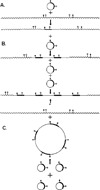
 ). (A) A single plasmid DNA molecule recombines (either by homologous or nonhomologous recombination) with mouse DNA to give a single integrated plasmid. The junction between mouse and plasmid sequences might occur at any site in both plasmid and mouse DNAs. Digestion with either of the two enzymes whose sites are shown would be expected to generate two products, neither of which would be likely to be the same size as linear pMK DNA. (B) After the initial integration event, a number of subsequent events could occur involving homologous recombination of additional copies of pMK into those already integrated, giving rise to a tandem repetition of the integrated pMK sequences. Digestion with either enzyme would generate several copies (three as drawn) of full-length linear pMK molecules, plus single copies of two new junction fragments. (C) Several copies of the plasmid could homologously recombine with one another to generate a tandemly repetitive plasmid, which would then recombine with mouse DNA, again generating a tandemly repeated integrated plasmid. Restriction enzyme analysis would generate the same products as model B.

Similar articles
-
Regulation of alpha genes of herpes simplex virus: expression of chimeric genes produced by fusion of thymidine kinase with alpha gene promoters.Cell. 1981 May;24(2):555-65. doi: 10.1016/0092-8674(81)90346-9. Cell. 1981. PMID: 6263501
-
Differential regulation of metallothionein-thymidine kinase fusion genes in transgenic mice and their offspring.Cell. 1982 Jun;29(2):701-10. doi: 10.1016/0092-8674(82)90186-6. Cell. 1982. PMID: 7116454
-
Herpes simplex virus thymidine kinase activity of thymidine kinase-deficient Escherichia coli K-12 mutant transformed by hybrid plasmids.Proc Natl Acad Sci U S A. 1981 Jan;78(1):582-6. doi: 10.1073/pnas.78.1.582. Proc Natl Acad Sci U S A. 1981. PMID: 6264449 Free PMC article.
-
Regulation of metallothionein--thymidine kinase fusion plasmids injected into mouse eggs.Nature. 1982 Mar 4;296(5852):39-42. doi: 10.1038/296039a0. Nature. 1982. PMID: 6278311
-
Selectable markers for the transfer of genes into mammalian cells.Curr Top Microbiol Immunol. 1982;96:145-57. doi: 10.1007/978-3-642-68315-2_9. Curr Top Microbiol Immunol. 1982. PMID: 6276089 Review. No abstract available.
Cited by
-
Modelling human regulatory variation in mouse: finding the function in genome-wide association studies and whole-genome sequencing.PLoS Genet. 2012;8(3):e1002544. doi: 10.1371/journal.pgen.1002544. Epub 2012 Mar 1. PLoS Genet. 2012. PMID: 22396661 Free PMC article. Review.
-
Completion of the swine genome will simplify the production of swine as a large animal biomedical model.BMC Med Genomics. 2012 Nov 15;5:55. doi: 10.1186/1755-8794-5-55. BMC Med Genomics. 2012. PMID: 23151353 Free PMC article.
-
Engineering Point Mutant and Epitope-Tagged Alleles in Mice Using Cas9 RNA-Guided Nuclease.Curr Protoc Mouse Biol. 2018 Mar;8(1):28-53. doi: 10.1002/cpmo.40. Curr Protoc Mouse Biol. 2018. PMID: 30040228 Free PMC article.
-
CORP: Using transgenic mice to study skeletal muscle physiology.J Appl Physiol (1985). 2020 May 1;128(5):1227-1239. doi: 10.1152/japplphysiol.00021.2020. Epub 2020 Feb 27. J Appl Physiol (1985). 2020. PMID: 32105520 Free PMC article. Review.
-
Dramatic growth of mice that develop from eggs microinjected with metallothionein-growth hormone fusion genes.Nature. 1982 Dec 16;300(5893):611-5. doi: 10.1038/300611a0. Nature. 1982. PMID: 6958982 Free PMC article.
References
-
- Bradford M. A rapid and sensitive method for quantitation of microgram quantities of protein utilizing the principle of protein-dye binding. Analyt. Biochem. 1976;72:248–254. - PubMed
-
- Brinster RL. Cultivation of the mammalian embryo. In: Rothblat G, Cristofala V, editors. Growth, Nutrition and Metabolism of Cells in Culture. Vol. 2. New York: Academic Press; 1972. pp. 251–286.
Publication types
MeSH terms
Substances
Grants and funding
LinkOut - more resources
Full Text Sources
Other Literature Sources
Molecular Biology Databases

