. 2024 Nov 16;25(22):12315.
doi: 10.3390/ijms252212315.

Roscovitine, a CDK Inhibitor, Reduced Neuronal Toxicity of mHTT by Targeting HTT Phosphorylation at S1181 and S1201 In Vitro

Affiliations

Roscovitine, a CDK Inhibitor, Reduced Neuronal Toxicity of mHTT by Targeting HTT Phosphorylation at S1181 and S1201 In Vitro

Hongshuai Liu et al. Int J Mol Sci. .

Abstract

Huntington's disease (HD) is an autosomal dominant neurodegenerative disease caused by a single mutation in the huntingtin gene (HTT). Normal HTT has a CAG trinucleotide repeat at its N-terminal within the range of 36. However, once the CAG repeats exceed 37, the mutant gene (mHTT) will encode mutant HTT protein (mHTT), which results in neurodegeneration in the brain, specifically in the striatum and other brain regions. Since the mutation was discovered, there have been many research efforts to understand the mechanism and develop therapeutic strategies to treat HD. HTT is a large protein with many post-translational modification sites (PTMs) and can be modified by phosphorylation, acetylation, methylation, sumoylation, etc. Some modifications reduced mHTT toxicity both in cell and animal models of HD. We aimed to find the known kinase inhibitors that can modulate the toxicity of mHTT. We performed an in vitro kinase assay using HTT peptides, which bear different PTM sites identified by us previously. A total of 368 kinases were screened. Among those kinases, cyclin-dependent kinases (CDKs) affected the serine phosphorylation on the peptides that contain S1181 and S1201 of HTT. We explored the effect of CDK1 and CDK5 on the phosphorylation of these PTMs of HTT and found that CDK5 modified these two serine sites, while CDK5 knockdown reduced the phosphorylation of S1181 and S1201. Modifying these two serine sites altered the neuronal toxicity induced by mHTT. Roscovitine, a CDK inhibitor, reduced the p-S1181 and p-S1201 and had a protective effect against mHTT toxicity. We further investigated the feasibility of the use of roscovitine in HD mice. We confirmed that roscovitine penetrated the mouse brain by IP injection and inhibited CDK5 activity in the brains of HD mice. It is promising to move this study to in vivo for pre-clinical HD treatment.

Keywords: CDK5; Huntington’s disease; post-translational modification (PTM); roscovitine; toxicity.


Conflict of interest statement

The authors declare no conflicts of interest.

Figures

Figure 1
Figure 1
Kinase assays identified upstream kinases modifying the serine of HTT peptides in vitro. In vitro kinase assay was used to screen the kinase targeting HTT peptide bearing serine 1181, serine 1201, and serine 2653 of HTT. (A) Kinases targeting the serine 1181 of the HTT peptide. (B) Kinases targeting the serine 1201 of the HTT peptide. (C) Kinases targeting the serine 2653 of the HTT peptide. CDK1 is highlighted by blue rectangles and CDK5/p25 by red rectangles. RBC, red blood cell substrate. Dotted lines indicate the 5% cut-off of enzyme activity used for the kinase screening.
Figure 2
Figure 2
HTT peptides as potential as targets for CDKs. (A) The amino acid sequences surrounding serine of 1181, 1201, and 2653 of HTT from different species. S/T-P-x-K/R is a consensus site for both CDK1 and CDK5, which means that the amino acid of S or T followed by P-x-K/R could be phosphorylated by CDK1 and CDK5. (S = serine; T = threonine; P = proline; K = lysine; R = arginine; x is any amino acid). It is well conserved in vertebrates for S1181 and S1201 of the HTT sequence, but not S2653. (B) CDKs target serine 1181 of the HTT peptide. (C) CDKs target serine 1201 of HTT peptide. CDK1 is highlighted by blue rectangles and CDK5/p25 by red rectangles. RBC, red blood cell substrate. Dotted lines indicate the 1% cut-off of enzyme activity used for the kinase screening.
Figure 3
Figure 3
The effect of knocking down CDK5 on the phosphorylation of S1181 and S1201 of mHTT. HEK293 cells were co-transfected with a plasmid expressing full-length mutant huntingtin with 82Q (FL-82Q) and siRNA targeting human CDK5 for 24 h. The expression level of CDK5, phosphorylated S1181-HTT (p-S1181), and phosphorylated S1201-HTT (p-S1201) were detected by western blot. (A) The representative western blot for CDK5. (B) The quantification of CDK5 expression. (C) The representative western blot for p-S1181-HTT. (D) The quantification of p-S1181-HTT expression. (E) The representative western blot for p-S1201-HTT. (F) The quantification of p-S1201-HTT expression. The experiment was repeated by at least two different analysts. Each experiment had n = 3. One representative experiment is presented. 2166 is the anti-HTT antibody (MAB2166, Millipore). RNAi = pooled siRNA targeting human CDK5. A two-tailed Student t-test was used. * p < 0.05; ** p < 0.01.
Figure 4
Figure 4
The effect of CDK5 overexpression on S1181 of mHTT. HEK293 cells were co-transfected with HTT plasmid FL-82Q and a plasmid expressing human CDK5 for 24 h. The expression levels of CDK5, p-S1181, and p-S1201 were detected by western blot. (A) The representative western blot for CDK5, p-S1181, 2166, and MW1. (B) The quantification of CDK5 expression. (C,D) The quantification of p-S1181 expression. (E) The quantification of total huntingtin (MAB2166) expression. (F) The quantification of mutant huntingtin (MW1) expression. The experiment was repeated by at least two different analysts. Each experiment had n = 3. One representative experiment is presented. 2166 is the anti-HTT antibody (MAB2166). MW1 is the anti-HTT antibody that binds to mutant huntingtin (clone MW1). RNAi = siRNA targeting human CDK5. A two-tailed Student t-test was used. ** p < 0.01; *** p < 0.001.
Figure 5
Figure 5
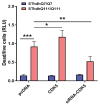
CDK5 level altered cell toxicity induced by mHTT. The effect of CDK5 overexpression or knocking down was evaluated in mouse striatal cells with different poly Q (SThdh Q7/Q7 or SThdh Q111/Q111). Cells were transfected with either a plasmid expressing human CDK5 or pooled siRNA targeting mouse CDK5 for 24 h. Cell death was measured with CytoTox-Glo cytotoxicity assay kit (Promega). The experiment was repeated by at least two different analysts. Each experiment had n = 3. One representative experiment is presented. Two-way ANOVA with Tukey’s multiple comparation was used for analysis. * p < 0.05; ** p < 0.01; *** p < 0.001.
Figure 6
Figure 6
Phospho-null modification of S1181 and S1201 of mHTT altered the mHTT-induced cell toxicity. S1181 and S1201 in the plasmid of FL-82Q were artificially modified to alanine (A). (A) Modified plasmids were transiently transfected into HEK293 cells for 72 h, and cell toxicity was measured by caspase3/7 activity. (B) The modified plasmids were transient transfected in primary cortical neurons, and cell death was analyzed by nuclei condensation assay. The experiment was repeated by at least two different analysts. Each experiment had n = 3. One representative experiment is presented. One-way ANOVA with Dunnett’s multiple comparation was used for analysis. * p < 0.05; ** p < 0.01; *** p < 0.001.
Figure 7
Figure 7
Roscovitine protected cells from mHTT-induced toxicity. Roscovitine was used in different HD cell models to evaluate its effect on mHTT-induced toxicity. (A). The effect of roscovitine on mHTT-induced toxicity in SThdh cells expressing Q111 cells. SThdh Q111/Q111 cells were treated with roscovitine for 24 h under serum withdrawal conditions, and cell toxicity was measured using a CytoTox kit. (B). The effect of roscovitine on mHTT-induced toxicity in primary cortical neuronal HD cell model. Primary cultured cortical neurons were transiently transfected with plasmids expressing either normal Q (23Q) or poly Q (82Q) for 4 h and then treated with or without roscovitine for 48 h. A nuclei condensation assay was used to evaluate cell death. The experiment was repeated by at least two different analysts. Each experiment had n = 3. One representative experiment is presented. W/O = serum withdrawal. One-way ANOVA with Dunnett’s multiple comparation was used for analysis. * p < 0.05; ** p < 0.01.
Figure 8
Figure 8
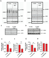
Roscovitine reduced the phosphorylation of S1181 and S1201 of mHTT. Roscovitine reduced p-S1181-HTT and p-S1201-HTT in vitro by inhibiting CDK5 but not CDK1. (A) The representative western blot for p-S1181 in HEK293 cells transfected with a plasmid expressing FL-82Q and treated with or without roscovitine for 48 h. (B) The representative western blot for p-S1201 in HEK293 cells transfected with a plasmid expressing FL-82Q and treated with or without roscovitine for 48 h. (C) The representative western blot for CDK5 in HEK293 cells transfected with a plasmid expressing FL-82Q and treated with or without roscovitine for 48 h. (D) The representative western blot for CDK1 in HEK293 cells transfected with a plasmid expressing FL-82Q and treated with or without roscovitine for 48 h. (E) Quantification of p-S1181 in A. (F) Quantification of p-S1201 in B. (G) Quantification of CDK5 in C. (H) Quantification of CDK1 in D. The experiment was repeated by at least two different analysts. Each experiment had n = 3. One representative experiment is presented. Ros~ = Roscovitine. A two-tailed Student t-test was used. * p < 0.05; ** p < 0.01.
Figure 9
Figure 9
Roscovitine penetrated the mouse brain and inhibited CDK5 activity in the zQ175 HD mouse brain. Variant doses of roscovitine were injected into 4-month-old mice through intraperitoneal injection (IP). Plasma and brain samples were collected at different time points after injection. The concentration of roscovitine was measured by LCMS/MS. (A) Brain concentration of roscovitine at different time points after acute IP injection with variant doses. Each group has 4–5 mice. (B) The CDK5 activity in the brain of mice injected with roscovitine for three weeks. Two doses of roscovitine were injected into 4-month-old zQ175HD mice by IP daily for three weeks. Twenty-four hours after the last injection, the mouse brain samples were collected, and the CDK5 activity was measured using a commercial kit from Promega. Animal number is indicated in the bar graph for each group. R-25 = 25 mg/kg of roscovitine. R-50 = 50 mg/kg of roscovitine. One-way ANOVA with Fish’s LSD was used for analysis. * p < 0.05; ** p < 0.01.
Figure 10
Figure 10
Tolerability of mice injected with roscovitine by IP administration. (A) The body weight loss of zQ175 HD mice injected with roscovitine. (B) The stride length (by CatWalk) in zQ175 HD mice before and after being injected with roscovitine. Roscovitine was injected by IP daily for three weeks. Animal number for each group is indicated in the bar graphs. V = Vehicle, R = roscovitine. R-25 = 25 mg/kg of roscovitine. R-50 = 50 mg/kg of roscovitine. One-way ANOVA with Fish’s LSD was used for analysis.
Figure 11
Figure 11
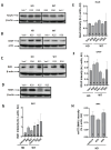
Molecular neurobiological measurement in the brain of zQ175 HD mice injected with roscovitine by IP. Brain samples were collected as previously described. The cortex was dissected, and lysates were made for western blot analysis. (A) The expression of a pan-neuronal marker, NeuN. (B) The expression of astrocyte marker GFAP. (C) The expression of microglial marker IBA1. (D) The expression of mHTT marker MW1. (E) The quantification of NeuN. (F) The quantification of GFAP. (G) The quantification of IBA1. (H) The quantification of MW1. Animal number is indicated in the bar graph for each group. Veh~ = Vehicle, R25 = 25 mg/kg of roscovitine. R50 = 50 mg/kg of roscovitine. One-way ANOVA with Fish’s LSD was used for analysis.

Similar articles

References

    1. MacDonald M.E., Ambrose C.M., Duyao M.P., Myers R.H., Lin C., Srinidhi L., Barnes G., Taylor S.A., James M., Groot N., et al. A novel gene containing a trinucleotide repeat that is expanded and unstable on Huntington’s disease chromosomes. The Huntington’s Disease Collaborative Research Group. Cell. 1993;72:971–983. doi: 10.1016/0092-8674(93)90585-E. - DOI - PubMed
    1. Tabrizi S.J., Flower M.D., Ross C.A., Wild E.J. Huntington disease: New insights into molecular pathogenesis and therapeutic opportunities. Nat. Rev. Neurol. 2020;16:529–546. doi: 10.1038/s41582-020-0389-4. - DOI - PubMed
    1. Bates G.P., Dorsey R., Gusella J.F., Hayden M.R., Kay C., Leavitt B.R., Nance M., Ross C.A., Scahill R.I., Wetzel R., et al. Huntington disease. Nat. Rev. Dis. Primers. 2015;1:15005. doi: 10.1038/nrdp.2015.5. - DOI - PubMed
    1. Saudou F., Humbert S. The Biology of Huntingtin. Neuron. 2016;89:910–926. doi: 10.1016/j.neuron.2016.02.003. - DOI - PubMed
    1. Li P.P., Moulick R., Feng H., Sun X., Arbez N., Jin J., Marque L.O., Hedglen E., Chan H.Y.E., Ross C.A., et al. RNA Toxicity and Perturbation of rRNA Processing in Spinocerebellar Ataxia Type 2. Mov. Disord. 2021;36:2519–2529. doi: 10.1002/mds.28729. - DOI - PMC - PubMed

MeSH terms

LinkOut - more resources