MLKL deficiency elevates testosterone production in male mice independently of necroptotic functions
- PMID: 39572538
- PMCID: PMC11582601
- DOI: 10.1038/s41419-024-07242-z
MLKL deficiency elevates testosterone production in male mice independently of necroptotic functions
Abstract
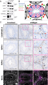
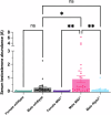
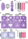
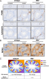
Mixed lineage kinase domain-like (MLKL) is a pseudokinase, best known for its role as the terminal effector of the necroptotic cell death pathway. MLKL-mediated necroptosis has long been linked to various age-related pathologies including neurodegeneration, atherosclerosis and male reproductive decline, however many of these attributions remain controversial. Here, we investigated the role of MLKL and necroptosis in the adult mouse testis: an organ divided into sperm-producing seminiferous tubules and the surrounding testosterone-producing interstitium. We find that sperm-producing cells within seminiferous tubules lack expression of key necroptotic mediators and thus are resistant to a pro-necroptotic challenge. By comparison, coordinated expression of the necroptotic pathway occurs in the testicular interstitium, rendering cells within this compartment, especially the lysozyme-positive macrophages, vulnerable to necroptotic cell death. We also uncover a non-necroptotic role for MLKL in regulating testosterone levels. Thus, MLKL serves two roles in the mouse testes - one involving the canonical response of macrophages to necroptotic insult, and the other a non-canonical function in male reproductive hormone control.
© 2024. The Author(s).
Conflict of interest statement
Competing interests: KMP, JMH, KEL, ALS, and JMM contribute to or have contributed to a project developing necroptosis inhibitors in collaboration with Anaxis Pharma. The other authors declare no competing interests. Ethics declaration: All experiments were approved by the WEHI Animal Ethics Committee following the Prevention of Cruelty to Animals Act (1996) and the Australian National Health and Medical Research Council Code of Practice for the Care and Use of Animals for Scientific Purposes (1997).
Figures


Similar articles
-
Viral MLKL Homologs Subvert Necroptotic Cell Death by Sequestering Cellular RIPK3.Cell Rep. 2019 Sep 24;28(13):3309-3319.e5. doi: 10.1016/j.celrep.2019.08.055. Cell Rep. 2019. PMID: 31553902
-
The pseudokinase MLKL regulates hepatic insulin sensitivity independently of inflammation.Mol Metab. 2019 May;23:14-23. doi: 10.1016/j.molmet.2019.02.003. Epub 2019 Feb 20. Mol Metab. 2019. PMID: 30837196 Free PMC article.
-
Activation of the pseudokinase MLKL unleashes the four-helix bundle domain to induce membrane localization and necroptotic cell death.Proc Natl Acad Sci U S A. 2014 Oct 21;111(42):15072-7. doi: 10.1073/pnas.1408987111. Epub 2014 Oct 6. Proc Natl Acad Sci U S A. 2014. PMID: 25288762 Free PMC article.
-
MLKL: Functions beyond serving as the Executioner of Necroptosis.Theranostics. 2021 Mar 4;11(10):4759-4769. doi: 10.7150/thno.54072. eCollection 2021. Theranostics. 2021. PMID: 33754026 Free PMC article. Review.
-
Necroptotic movers and shakers: cell types, inflammatory drivers and diseases.Curr Opin Immunol. 2021 Feb;68:83-97. doi: 10.1016/j.coi.2020.09.008. Epub 2020 Nov 4. Curr Opin Immunol. 2021. PMID: 33160107 Review.
References
-
- Samson AL, Garnish SE, Hildebrand JM, Murphy JM. Location, location, location: a compartmentalized view of TNF-induced necroptotic signaling. Sci Signal. 2021;14:eabc6178. - PubMed
MeSH terms
Substances
Grants and funding
LinkOut - more resources
Full Text Sources
Miscellaneous

