. 2024 Nov 22;386(6724):eadq8587.
doi: 10.1126/science.adq8587. Epub 2024 Nov 22.

Specific tRNAs promote mRNA decay by recruiting the CCR4-NOT complex to translating ribosomes

Affiliations

Specific tRNAs promote mRNA decay by recruiting the CCR4-NOT complex to translating ribosomes

Xiaoqiang Zhu et al. Science. .

Abstract

The CCR4-NOT complex is a major regulator of eukaryotic messenger RNA (mRNA) stability. Slow decoding during translation promotes association of CCR4-NOT with ribosomes, accelerating mRNA degradation. We applied selective ribosome profiling to further investigate the determinants of CCR4-NOT recruitment to ribosomes in mammalian cells. This revealed that specific arginine codons in the P-site are strong signals for ribosomal recruitment of human CNOT3, a CCR4-NOT subunit. Cryo-electron microscopy and transfer RNA (tRNA) mutagenesis demonstrated that the D-arms of select arginine tRNAs interact with CNOT3 and promote its recruitment whereas other tRNA D-arms sterically clash with CNOT3. These effects link codon content to mRNA stability. Thus, in addition to their canonical decoding function, tRNAs directly engage regulatory complexes during translation, a mechanism we term P-site tRNA-mediated mRNA decay.


Conflict of interest statement

Competing interests: J.T.M is a scientific advisor for Ribometrix, Inc. and owns equity in Orbital Therapeutics, Inc. UT Southwestern Medical Center has filed a provisional patent covering sequence modifications of tRNAs that impact mRNA stability.

Figures

Fig. 1.
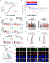
Fig. 1.. Select arginine codons are enriched in the ribosomal P-site of CNOT3-bound ribosomes.
(A and B) Sucrose density gradient profiles of HEK293T cell lysate and western blot analysis of fractions without (A) or with RNase A treatment (B). (C) Western blot analysis of CNOT3 or control immunoprecipitates. Representative results from n=3 biological replicates shown for sucrose density gradient profiles and CNOT3 IP. (D) Schematic of CNOT3-selective ribosome profiling. Figure created with BioRender.com. (E) Meta-codon plots showing the triplet periodicity of ribosome profiling reads. (F) Codon enrichment in ribosomal E, P, and A-sites of CNOT3-bound ribosomes. (G) Sequence logo representation of amino acid enrichment in ribosomal E, P, and A-sites of CNOT3-bound ribosomes.
Fig. 2.
Fig. 2.. mRNAs rich in CGG, CGA, and AGG arginine codons are destabilized by CNOT3.
(A) Cumulative distribution function (CDF) plots showing the fold-change in half-lives of the following sets of mRNAs in CNOT3-depleted HEK293T cells, measured by SLAM-seq: i) mRNAs with high weighted CGG/CGA/AGG scores, calculated as the sum of the enrichment values of each of these codons in the P-site of CNOT3-bound ribosomes, normalized to total codon number, in each mRNA; ii) mRNAs rich in arginine encoded by CGC, AGA, and CGU. mRNAs rich in these codons that also had a high weighted CGG/CGA/AGG score (top 1000) were excluded from this gene set. P values were calculated by one-sided Wilcoxon rank sum test. (B) Stability of a reporter construct encoding 42 arginine-centered tripeptides, with arginine encoded by CGG, CGA, or AGG, or a control reporter with arginine codons replaced with codons that were not enriched in the P-site of CNOT3-bound ribosomes. CNOT3 knockout (sgCNOT3-1) or control (sgNT1) HEK293T cells were treated with 1 μg/mL doxycycline, and reporter mRNA levels relative to GAPDH at each time point were measured by qRT-PCR. n=3 biological replicates (mean ± SD shown). P values were calculated by student’s t test, comparing sgCNOT3-1 to sgNT1 for each reporter. **P<0.01; ***P<0.001; n.s., not significant. (C) GSEA analysis of global mRNA half-life data showing stabilization of mRNAs encoding mitochondrial ribosomal proteins in CNOT3-depleted HEK293T cells and Jurkat cells. Genes are ordered left to right along the x-axes based on fold-change of half-lives in CNOT3-depleted cells compared to control cells (high to low). The rank of genes within each geneset is indicated with vertical black lines. mRNA stability in CNOT3-depleted Jurkat cells was reported previously (33). (D) GSEA showing that human mRNAs encoding mitochondrial ribosomal proteins exhibit a high weighted CGG/CGA/AGG score. Genes are ordered left to right along the x-axes based on weighted CGG/CGA/AGG score (high to low). The rank of genes within each geneset is indicated with vertical black lines. (E and F) qRT-PCR analysis of mitochondrial ribosomal protein mRNAs, normalized to GAPDH, in HEK293T (E) and Jurkat cells (F) infected with lentivirus expressing the indicated sgRNAs. n=3 biological replicates (mean ± SD shown). P values were calculated by student’s t test. *P<0.05; **P<0.01; ***P<0.001. (G and H) Flow cytometry analysis of HEK293T (G) and Jurkat cells (H) expressing the indicated sgRNAs and stained with MitoTracker. Representative data from n=3 biological replicates shown. (I) Schematic of mitochondrial translation assay. Cytosolic translation was inhibited with anisomycin (ANS) and nascent mitochondrial peptides were labeled with L-homopropargylglycine (HPG). Figure created with BioRender.com. (J) Representative images of HPG-labeled Jurkat cells expressing the indicated sgRNAs. TOMM20 is a mitochondrial membrane protein. Representative data from n=3 biological replicates shown.
Fig. 3.
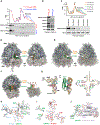
Fig. 3.. Structural analysis of a human CNOT3-ribosome complex.
(A) Sucrose density gradient profiles and western blot analysis of in vitro translation reactions performed with 41×LRCGGD or 41×LKAAGD mRNA. (B) Western blot analysis of CCR4-NOT components in combined polysome fractions from in vitro translation reactions assembled as in (A). (C) Sucrose density gradient profiles and western blot analysis of combined polysome fractions from in vitro translation reactions assembled on the indicated mRNAs. Representative data from n=3 biological replicates shown for panels A-C. (D and E) Cryo-EM density map (D) and cartoon model (E) of the human CNOT3-ribosome complex. The 60S subunit is shown in cyan/grey and the 40S subunit in light blue/grey. CNOT3 is highlighted in green and the P-site tRNA in orange. (F) Clipped density map (left) and atomic model (right) highlighting the ribosomal E, P, and A-sites. CNOT3 and the P-site tRNA are colored as before. (G) Cartoon model and secondary structure representation of CNOT3-arginyl tRNA interactions, highlighting tRNA elements in the D-loop (cyan), D-stem (red), and anticodon stem (purple) contacted by CNOT3 (green). AAS, amino acid acceptor stem; TSL, T stem loop; ASL, anticodon stem loop; DSL, D stem loop. (H-J) Experimental Cryo-EM density (grey surface) of the three CNOT3/tRNA interaction elements: D-loop (H), D-stem (I), and anticodon stem (J). Modeled residues are shown as sticks and colored as in panel G.
Fig. 4.
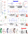
Fig. 4.. A U13:A22:A46 triplet in select arginine tRNAs promotes co-translational CNOT3 recruitment.
(A) Schematic of arginine tRNAs, showing their key distinguishing features and the codons they decode, arranged by their enrichment in the P-site of CNOT3-bound ribosomes. (B) Secondary structure and cartoon depictions of tRNA and CNOT3, highlighting the CNOT3 interaction with nucleotide 22 of the P-site tRNA as well as the 13:22:46 base triplet. (C to G) In vitro translation of 41×LRCGUD mRNA in the presence of in vitro transcribed arginine tRNA variants, followed by western blot analysis of combined polysome fractions to assess CNOT3 recruitment. For panels D-G, 2 μg tRNA per 100 μL reaction volume were used. All experiments were performed with n=3–6 biological replicates (mean ± SD shown). P values were calculated by student’s t test, comparing to m1 (D), WT (E), m10 (G), or as indicated with brackets (F). **P<0.01; ***P<0.001; n.s., not significant. (H to J) Structural models of CNOT3/Not5 interactions with the 13:22:46 base triplet of tRNAArg,CCG (H), tRNAiMet from PDB 6TB3 (14) (I), and the rebuilt tRNALeu,UAA model (PDB 93CI) (19) (J). The inset in (I) shows the tRNAArg,UCU 13:22:46 triplet from PDB 8ISS (45). (K) In vitro translation of the 73×CGG/CGA/AGG mRNA in lysates from cells expressing Flag-tagged wild-type CNOT3 or CNOT3 E95A, followed by western blot analysis of combined polysome fractions. n=3 biological replicates (mean ± SD shown). P values were calculated by student’s t test. ***P<0.001.
Fig. 5.
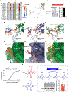
Fig. 5.. An extra nucleotide in the D-loop α element preceding the GG motif blocks CNOT3 recruitment.
(A) Alignment of the D-arms of human arginine tRNAs and tRNAs that have an extra nucleotide in the α element. (B) Secondary structure and cartoon depictions of tRNA and CNOT3, highlighting the CNOT3 interaction with the D-loop α element of the P-site tRNA. (C) In vitro translation of the 73×CGG/CGA/AGG mRNA in lysates from cells expressing Flag-tagged wild-type CNOT3 or CNOT3 R59S, followed by western blot analysis of combined polysome fractions. n=3 biological replicates (mean ± SD shown). P values were calculated by student’s t test. ***P<0.001. (D to I) Molecular models of CNOT3/Not5 interactions with the α element of P-site tRNAArg,CGG (D and G), tRNAiMet from PDB 6TB3 (14) (E and H) and tRNALys,UUU from PDB 6SGC (48) with an aligned, superposed CNOT3 model from tRNAArg,CGG (F and I). (J) CDF plot showing the fold-change in half-lives of mRNAs rich in codons decoded by tRNAs with the α element insertion relative to other transcripts in CNOT3-depleted HEK293T cells, measured by SLAM-seq. P value calculated by one-sided Wilcoxon rank sum test. (K) Comparison of the D-arms of human tRNAiMet and tRNAMet. (L) In vitro translation of 41×LMAUGD mRNA in the presence of in vitro transcribed tRNAMet variants (10 μg tRNA per 100 μL reaction volume), followed by western blot analysis of combined polysome fractions to assess CNOT3 recruitment. All experiments were performed with n=3 biological replicates (mean ± SD shown). P values were calculated by student’s t test, comparing mutants to WT. **P<0.01.
Fig. 6.
Fig. 6.. Slow decoding promotes P-site tRNA-mediated decay (PTMD).
(A to C) Pearson correlation of codon enrichment in the ribosomal A-site of CNOT3-bound ribosomes and A-site dwell time in HEK293T cells when the P-site is occupied by any codon (A), a CGG/CGA/AGG codon (B), or any codon other than CGG/CGA/AGG (C). Note that panel A is also shown in Fig. S1N and duplicated here to facilitate comparison with other panels. (D) Proposed mechanism of PTMD. Slow decoding, resulting in a ribosome with empty A- and E-sites, provides an opportunity for CNOT3 enter the E-site and probe the P-site tRNA. (i) If the P-site tRNA has the U13:A22:A46 triplet and lacks the extended α-element (i.e., tRNAs that decode CGG/CGA/AGG arginine codons), CNOT3 binding will be stabilized and mRNA decay will be favored. (ii) If the P-site tRNA is neutral, lacking both the extended D-loop α element and the U13:A22:A46 triplet, CNOT3 binding may be transient. However, if an extended ribosomal stall occurs due to scarcity of a charged tRNA that can enter the A-site, CCR4-NOT-mediated decay may occur. (iii) If the P-site tRNA has the extended D-loop α element (e.g., tRNAs that decode N, K, I, Y, M, F, and T), CNOT3 accommodation will be sterically blocked, CNOT3 will exit, and translation will resume.

Similar articles

References

    1. Passmore LA, Coller J, Roles of mRNA poly(A) tails in regulation of eukaryotic gene expression. Nat Rev Mol Cell Biol 23, 93–106 (2022). - PMC - PubMed
    1. Gallie DR, The cap and poly(A) tail function synergistically to regulate mRNA translational efficiency. Genes Dev 5, 2108–2116 (1991). - PubMed
    1. Kahvejian A, Svitkin YV, Sukarieh R, M’Boutchou MN, Sonenberg N, Mammalian poly(A)-binding protein is a eukaryotic translation initiation factor, which acts via multiple mechanisms. Genes Dev 19, 104–113 (2005). - PMC - PubMed
    1. Garneau NL, Wilusz J, Wilusz CJ, The highways and byways of mRNA decay. Nat Rev Mol Cell Biol 8, 113–126 (2007). - PubMed
    1. Yi H et al., PABP Cooperates with the CCR4-NOT Complex to Promote mRNA Deadenylation and Block Precocious Decay. Mol Cell 70, 1081–1088 (2018). - PubMed

LinkOut - more resources