. 2024 Nov 6:14:1442743.
doi: 10.3389/fcimb.2024.1442743. eCollection 2024.

Proteomics of circulating extracellular vesicles reveals diverse clinical presentations of COVID-19 but fails to identify viral peptides

Affiliations

Proteomics of circulating extracellular vesicles reveals diverse clinical presentations of COVID-19 but fails to identify viral peptides

Melisa Gualdrón-López et al. Front Cell Infect Microbiol. .

Abstract

Extracellular vesicles (EVs) released by virus-infected cells have the potential to encapsulate viral peptides, a characteristic that could facilitate vaccine development. Furthermore, plasma-derived EVs may elucidate pathological changes occurring in distal tissues during viral infections. We hypothesized that molecular characterization of EVs isolated from COVID-19 patients would reveal peptides suitable for vaccine development. Blood samples were collected from three cohorts: severe COVID-19 patients (G1), mild/asymptomatic cases (G2), and SARS-CoV-2-negative healthcare workers (G3). Samples were obtained at two time points: during the initial phase of the pandemic in early 2020 (m0) and eight months later (m8). Clinical data analysis revealed elevated inflammatory markers in G1. Notably, non-vaccinated individuals in G1 exhibited increased levels of neutralizing antibodies at m8, suggesting prolonged exposure to viral antigens. Proteomic profiling of EVs was performed using three distinct methods: immunocapture (targeting CD9), ganglioside-capture (utilizing Siglec-1) and size-exclusion chromatography (SEC). Contrary to our hypothesis, this analysis failed to identify viral peptides. These findings were subsequently validated through Western blot analysis targeting the RBD of the SARS-CoV-2 Spike protein's and comparative studies using samples from experimentally infected Syrian hamsters. Furthermore, analysis of the EV cargo revealed a diverse molecular profile, including components involved in the regulation of viral replication, systemic inflammation, antigen presentation, and stress responses. These findings underscore the potential significance of EVs in the pathogenesis and progression of COVID-19.

Keywords: COVID-19 patients; SARS-CoV-2; antibody response; extracellular vesicles; ganglioside-capture (CD169/Siglec-1); immunocapture (CD9); proteomics profiling; size-exclusion chromatography (SEC).


Conflict of interest statement

HP, MMo, and LF are shareholders of Innovex Therapeutics. SM-T was a former employee of Innovex Therapeutics. JM-P has received institutional grants and educational/consultancy fees from AbiVax; AstraZeneca; Gilead Sciences; Grifols; Janssen; Merck Sharp & Dohme; and ViiV Healthcare; all outside the submitted work. JC and JB are shareholders of Albajuna Therapeutics SL, NI-U reports institutional grants from Grifols, Dentaid, Hipra and Amassence. The remaining authors declare that the research was conducted in the absence of any commercial or financial relationships that could be construed as a potential conflict of interest. The author(s) declared that they were an editorial board member of Frontiers, at the time of submission. This had no impact on the peer review process and the final decision.

Figures
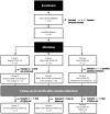
Figure 1
Figure 1
Flowchart of study design. After enrollment of 62 individuals in early 2020, two participants revoked informed consent and six did not meet the criteria of eligibility. Remaining samples were allocated to three different groups: Group 1 (G1, n=19), positive SARS-CoV-2 RT-PCR with severe clinical condition; Group 2 (G2, n=26), positive SARS-COV-2 RT-PCR with mild-symptoms related to SARS-CoV-2 and asymptomatic (ASX) individuals; Group 3 (G3, n=15) negative SARS-CoV-2 RT-PCR healthcare workers. An active follow-up visit was performed eight months after the initial recruitment for most of the individuals.
Figure 2
Figure 2
Humoral immune response of the patient’s cohort. Antibodies profiles at beginning of the study (m0) and eight months later (m8) in the study groups severe (G1), mild and asymptomatic (G2) and non-COVID-19 hospital care workers (G3). (A) Total reactive IgG antibodies against the S protein of the SARS-CoV-2 measured by an in-house ELISA. The lower limit of detection (dashed line) of the assay was 3.2. All Ig levels (UA) without value but measured by ELISA were plotted as 1.6 (average of 0 and 3.2 AU/ml that is lower limit of detection). (B) Neutralization assay antibody titers against the wild-type SARS-CoV-2 pseudoviruses in serum samples. The lower limit of detection (dashed line) of the assay was 60. All nAbs levels with ID50<60 were plotted as 30 (average of 0 and 60 that is lower limit of detection). (C, D) Difference in the log value of anti-S IgG and neutralizing antibodies between the last sampling time (m8) and peak of the infection (m0) depending on the study group (G1/severe, G2/Mild/ASX and G3/Non-COVID-19) and COVID-19 vaccination. Red line represents stable antibody titers throughout the study. The circles represent individual participants, and the bars mean with SD. Individuals from G3 infected between m0 and m8 are denoted with squared symbols. Vaccinated individuals are highlighted with red-filled symbols in (A, B) Longitudinal comparisons for each group were tested using Wilcoxon signed rank test. Comparisons between groups in each time point were assessed by Kruskal-Wallis and Conover’s post-hoc tests. p-values: *p<0.05, ***p<0.001 in (A, B).
Figure 3
Figure 3
Western blot analysis of CD9+/CD63+/CD81+ EVs. Plasma from severe, Mild/ASX COVID-19 patients and non-COVID-19 individuals was used to immunocapture CD9+/CD63+/CD81+ EVs. Immunocaptured EVs were analyzed for the presence of SARS-CoV2 Spike Receptor binding domain (RBD). The membrane was cut at 35kDa and an anti-CD81 antibody was used as a control of quality and quantity of EVs. Recombinant Spike protein (30ng and 5ng) and a cell lysate of Vero cells uninfected and infected with SARS-CoV-2 were used as controls for S-RBD detection.
Figure 4
Figure 4
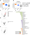
Proteomic analysis of CD9+ EVs from COVID-19 patients isolated by immunocapture at the infection peak. Seven individuals from G1, ten from G2 and 5 from G3 were used for CD9+ plasma-derived EVs isolation by magnetic immunocapture. Immunocaptured EVs were digested and analyzed by LC-MS/MS mass spectrometry. (A) Distribution of total number of proteins identified in each group at the infection peak. (B) Distribution and identity of the EV markers identified in the proteome from each cohort group. Statistical differences were tested using a One-way ANOVA (Kruskal Wallis post-test) with Dunn’s multiple comparisons. (C-E) CD9+ EVs protein abundance from each infected group was compared to abundance in the non-COVID-19 group. Volcano plot representation highlighting proteins up and down-regulated with statistical significance (q-val <0.05) (dotted red line). Proteins listed aside each plot correspond to proteins uniquely identified in each group. (F) Functional enrichment analysis by Gene Ontology (GO) of protein differentially present in CD9+ EVs from Severe COVID-19 patients when compared to non-COVID-19 individuals. Plots show the top enriched terms for Biological processes, Cellular Components and Molecular Functions with an FDR <0.05. *p value <0.05, **p value <0.005, ns, no-significant
Figure 5
Figure 5
Proteomic analysis of plasma-derived EVs from COVID-19 patients isolated by mS1 mediated capture at the infection peak. Plasma from seven individuals from G1 and five from G3 at the peak of infection were used for isolation of EVs using mS1 mediated capture followed by LC-MS/MS. (A) Distribution of total number of proteins identified in isolated EVs. (B) Distribution and identity of the EV markers in the proteome of the analyzed groups. Statistical significance was evaluated by a t-test (Mann Whitney). (C) EVs protein abundance from severe COVID-19 and non-COVID-19 individuals were compared. Volcano plot highlighting proteins found up and down-regulated in severe COVID-19 patients with statistical significance (q-val <0.05). Proteins listed aside correspond to proteins uniquely identified in each group. (D) Functional enrichment analysis by GO of protein differentially present in mS1-captured EVs from severe COVID-19 patients when compared to non-COVID-19 individuals. Plots show the top enriched terms for Biological processes, Cellular Components and Molecular Functions with an FDR <0.05.
Figure 6
Figure 6
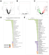
Proteomic analysis of CD9+ plasma-derived EVs from COVID-19 patients isolated by immunocapture at early infection and 8 months thereafter. Samples from 7 individuals from G1, 10 from G2, and 5 from G3 at early infection (m0) and 8 months thereafter (m8) were used for CD9+ plasma- derived EVs isolation via magnetic immunocapture. Immunocaptured EVs were digested and analyzed by LC-MS/MS. (A–C) CD9+-EVs protein abundance from each infected group at early infection was compared to the abundance 8 months later. Volcano plots show proteins significantly upregulated or downregulated (q-val < 0.05): (A) Severe, (B) Mild/ASX, and (C) Non-COVID. (D, E) Functional enrichment analysis by GO of proteins upregulated (D) and downregulated (E) in CD9+ EVs from non-COVID-19 individuals (G3) at the first time point compared to 8 months thereafter. Plots show the top enriched terms for Biological processes, Cellular Components and Molecular Functions with an FDR <0.05.

Similar articles

References

    1. Boechat J. L., Chora I., Morais A., Delgado L. (2021). The immune response to SARS-CoV-2 and COVID-19 immunopathology - Current perspectives. Pulmonology 27, 423–437. doi: 10.1016/j.pulmoe.2021.03.008 - DOI - PMC - PubMed
    1. Brustolin M., Rodon J., Rodríguez de la Concepción M. L., Ávila-Nieto C., Cantero G., Pérez M., et al. . (2021). Protection against reinfection with D614- or G614-SARS-CoV-2 isolates in golden Syrian hamster. Emerg. Microbes Infect. 10, 797–809. doi: 10.1080/22221751.2021.1913974 - DOI - PMC - PubMed
    1. Buzas E. I. (2023). The roles of extracellular vesicles in the immune system. Nat. Rev. Immunol. 23, 236–250. doi: 10.1038/s41577-022-00763-8 - DOI - PMC - PubMed
    1. Chams N., Chams S., Badran R., Shams A., Araji A., Raad M., et al. . (2020). COVID-19: A multidisciplinary review. Front. Public Health 8, 383. doi: 10.3389/fpubh.2020.00383 - DOI - PMC - PubMed
    1. de Gassart A., Geminard C., Fevrier B., Raposo G., Vidal M. (2003). Lipid raft-associated protein sorting in exosomes. Blood 102, 4336–4344. doi: 10.1182/blood-2003-03-0871 - DOI - PubMed

Grants and funding

The author(s) declare financial support was received for the research, authorship, and/or publication of this article. The proteomics of EVs isolated by CD9 was performed at the proteomics facility of Utrecht (Netherlands) as part of an EPIC-XS grant (0000205) to CT and HP. The proteomics analyses of EVs isolated by SEC were performed in the IJC Proteomic unit, which is part of the of Proteored, PRB3 and is supported by grant PT17/0019, of the PE I+D+i 2013-2016, funded by ISCIII and ERDF”. The CRG/UPF Proteomics Unit is part of the Spanish Infrastructure for Omics Technologies (ICTS OmicsTech), and it is supported by “Secretaria d’Universitats i Recerca del Departament d’Economia i Coneixement de la Generalitat de Catalunya” (2017SGR595). We thank Foundation Dormeur for financial support for the acquisition of the QuantStudio-5 real time PCR system. JM-P was supported by the Spanish Ministry of Science,Innovation and Universities (grants PID2022-139271OB-I00 and CB21/13/00063, Spain), and Fundació La Marató de TV3 (grant 202120-30-31-32, Spain). Funded in part by the European Union. The PCR-4-ALL and UNDINE projects have received funding under the Horizon Europe research and innovation programme (grant agreement No 101095606 and No 101057100 respectively). CF-B and HP acknowledge support from the Spanish Ministry of Science and Innovation (MICINN) (PID2022- 142908OB-I00) and support from the grant CEX2023-0001290-S funded by MCIN/AEI/10.13039/501100011033, and support from the Generalitat de Catalunya through the CERCA Program. This research is part of the ISGlobal’s Program on the Molecular Mechanisms of Malaria which is partially supported by the Fundación Ramón Areces. We also acknowledged the internal funding from IrsiCaixa through the crowdfunding initiative YoMeCorono (JM-P) and ISGlobal (HP) to initiate these studies.

LinkOut - more resources