Review
. 2024 Oct 11:15:1462496.
doi: 10.3389/fimmu.2024.1462496. eCollection 2024.

Genomic instability as a driver and suppressor of anti-tumor immunity

Affiliations
Review

Genomic instability as a driver and suppressor of anti-tumor immunity

Marta Requesens et al. Front Immunol. .

Abstract

Genomic instability is a driver and accelerator of tumorigenesis and influences disease outcomes across cancer types. Although genomic instability has been associated with immune evasion and worsened disease prognosis, emerging evidence shows that genomic instability instigates pro-inflammatory signaling and enhances the immunogenicity of tumor cells, making them more susceptible to immune recognition. While this paradoxical role of genomic instability in cancer is complex and likely context-dependent, understanding it is essential for improving the success rates of cancer immunotherapy. In this review, we provide an overview of the underlying mechanisms that link genomic instability to pro-inflammatory signaling and increased immune surveillance in the context of cancer, as well as discuss how genomically unstable tumors evade the immune system. A better understanding of the molecular crosstalk between genomic instability, inflammatory signaling, and immune surveillance could guide the exploitation of immunotherapeutic vulnerabilities in cancer.

Keywords: MMRd; cGAS-STING; chromosomal instability; genomic instability; immune evasion; tumor-infiltrating lymphocytes.


Conflict of interest statement

The authors declare that the research was conducted in the absence of any commercial or financial relationships that could be construed as a potential conflict of interest.

Figures
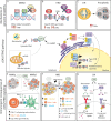
Figure 1
Figure 1
The cGAS/STING pathway and its inflammatory consequences in genomically unstable tumors. (A-C) Genomic instability can lead to the accumulation of dsDNA and other acid nucleic structures in the cytoplasm, which can activate cGAS. (D) Activation of cGAS either by nuclear DNA, micronuclei or other acid nucleic results in inflammatory signalling and expression of type I IFNs, ISG and NF-κB target genes. (E) In MMRd tumor, activation of cGAS results in increased CXCL10 and CCL5 production, which increase the number of tumor-infiltrating CD8+ T and NK cells as well as increased expression of IFN-β, which enhances DC-T cell interactions. (F) In HRD tumors, or tumors treated with DNA damaging agents such as PARP-inhibitors, activation of the cGAS pathways results in expression of the chemokines and cytokines, leading to a more inflamed TME with higher number of cytotoxic immune cells. (G) The cGAS/STING pathway in CIN/aneuploid cells has both pro and anti-tumor effects. The activation of the IRF3-type I IFN signalling axis results in immune surveillance and apoptotic signals, whereas NF-κB signalling mainly promotes IL-6/STAT3 pro-survival signals and enhance metastatic potential. MMRd, mismatch-repair deficient; MMRp, mismatch-repair proficient; MSH, mutS homolog; MLH1, mutL homolog 1; PMS1, PMS1 Homolog 2; SNV, single-nucleotide variations; indels, insertions-deletions; TMB, tumor-mutational burden; HRD, homologous-recombination deficiency; DSB, double-strand break; BRCA, breast cancer gene; CNAs, copy number alterations; CIN, chromosomal instability; mtDNA, mitochondrial DNA; cGAS, cyclic GMP-AMP synthase; STING, stimulator of interferon genes; TBK1, TANK-binding kinase 1; IRF, interferon regulatory factor; NF-κB, nuclear factor κB; ISG, interferon stimulated genes; CCL, C-C motif chemokine ligand; CXCL, C-X-C motif chemokine ligand; CD, cluster of differentiation; NK, natural killer; DC, dendritic cell; TNF, tumor necrosis factor; IL, interleukin; IFN, interferon; IFNR, interferon receptor; JAK1, janus kinase 1; STAT, signal transducer and activator of transcription; P, phosphorylation.
Figure 2
Figure 2
Immunogenicity of MMRd tumors. Frameshifts and SNV eventually result in the expression of neoantigens loaded in the MHC-I complex. Recognition of neoantigens activates CD8+ T cells and subsequent expression of cytotoxic molecules including granzyme B, IFN-γ or perforin, leading to the elimination of tumor cells. Tumor cells can also express neoantigens loaded on the MHC-II, which can be recognized by CD4+ T cells and initiate a cytotoxic response. In parallel, CD4+ T cells can also express pro-inflammatory cytokines and chemokines to recruit myeloid and NK cells that contribute to enhance anti-tumor immunity. Innate γδ T cells can target tumor cells via the NKG2D ligand-receptor interaction. High TMB correlates with the presence of TLSs in the tumor, which are highly organized hubs of immune cells that shape both adaptive and humoral immune responses. In particular, TLSs contain B-cell producing antibodies that may bind tumor antigens and trigger antibody-dependent cytotoxicity (ADCC). MHC, Major histocompatibility complex; TCR, T cell receptor; CD, cluster of differentiation; PD-1, programmed-death 1; TLS, tertiary lymphoid structure; Ig, immunoglobulin; CXCL, C-X-C motif chemokine ligand; CCL, C-C motif chemokine ligand; TNF, tumor necrosis factor; IFN, interferon; M1, macrophage type 1; NK, natural killer; IL, interleukin; NKG2D, natural killer group 2 D.
Figure 3
Figure 3
Immune recognition of CIN/aneuploid cells. CIN/aneuploidy trigger a wide range of cellular stressors resulting in the expression of immune activating ligands at the cell surface. CRT is expressed upon ER stress and facilitates phagocytosis by APCs and cytotoxicity by NK cells via the NKp46 receptor. CRT is also known to increase expression of MHC-I for the recognition of stressed cells by CD8+ T cells. In parallel, both DNA damage and ER-stress can upregulate the expression of NKG2D and DNAM-1-ligands, widely known to potently activate NK cells. Finally, secretion of soluble factors by CIN/Aneuploid cells may contribute to immune infiltration, immune activation and increase expression of MHC-I by tumor cells, altogether enhancing their recognition by the immune system. CIN, chromosomal instability; IFN, interferon; ISG, interferon stimulated genes; CXCL, C-X-C motif chemokine ligand; CCL, C-C motif chemokine ligand; MHC, Major histocompatibility complex; TCR, T cell receptor; CD, cluster of differentiation; M1, macrophage type 1; DC, dendritic cell; NK, natural killer; NKG2D, natural killer group 2 D; DNAM-1, DNAX accessory molecule; CRT, calreticulin; ER, endoplasmic reticulum; cGAS, cyclic GMP-AMP synthase; STING, stimulator of interferon response cGAMP interactor.
Figure 4
Figure 4
Mechanisms of immune evasion in MMRd tumors. Immune evasion occurs from the selection of clones with more immune evasion features upon an immune selective pressure or immunotherapy. Tumor cells with low MHC-I and/or MHC-II are preferentially selected for. Loss or downregulation of antigen presenting complexes can occur via mutations in antigen presentation genes, mutations in regulators of their transcription, or via disruption of the IFN signalling. Disruption of the IFN signalling generally occurs via mutations in JAK1/2 which in turn result in lower MHC-I expression, lower expression of ISG, CXCL9 and CXCL10, and resistance to apoptosis. Next to tumor cell intrinsic mechanisms, an immunosuppressive TME can also drive immune evasion. The TME of MMRd tumors often displays high expression of immune checkpoint ligands that, together with chronic stimulation, result in T cell exhaustion. In addition, the TME may have high infiltration of M2 macrophages, T reg, neutrophils and MDSCs expressing various immunosuppressive factors, which may also contribute to lower DC maturation, lower T cell activation and a microenvironment that suppress cytotoxic responses, ultimately favoring tumor growth. MHC, major histocompatibility complex; IFN, interferons; IFNR, interferon receptor; B2m, B-2-microglobulin; TAP, transporter associated with antigen presenting; JAK, janus-kinase; HLA, human leukocyte antigen; ISG, interferon stimulated gene; CXCL, C-X-C motif chemokine ligand; PD-1, programmed death 1; IL-2, interleukin 2; LAG-3, lymphocyte-activation gene 3; TIM-3, T-cell immunoglobulin and mucin-domain containing-3; PD-L, programmed death ligand; CD, cluster of differentiation; CEACAM, carcinoembryonic antigen-related cell adhesion molecule; CTLA-4, cytotoxic T lymphocyte-associated protein 4; TIGIT,: T cell immunoreceptor with Ig and ITIM domains; LAIR-1, leukocyte associated immunoglobulin like receptor 1; DC, dendritic cells; T reg, regulatory T cell; FOXP3, Forkhead Box P3; M2, macrophage type 2; TGF-β, transforming growth factor β; VEGF, vascular endothelial growth factor; EREG, epiregulin; IDO, Indoleamine 2,3-Dioxygenase; CCL2, C-C motif chemokine Ligand 2; MDSCs, Myeloid-derived suppressor cells.
Figure 5
Figure 5
Mechanisms of immune evasion in CIN/aneuploid tumors. A main mechanism of immune evasion in CIN/aneuploid tumors is dysregulation of the IFN pathway. This can occur in multiple ways including 1) loss of the 9p21 arm, which deletes a IFN gene cluster or loss of 9p24 with deletes JAK2, 2) loss of the 21q arm which deletes the IFN receptors, 3) reduced STAT1 activity, 4) chronic stimulation of STING which consequently eschews inflammatory signalling towards NF-κB, while downregulating IFN-signalling. Loss of IFN signalling may also result in decrease MHC-I expression, decrease expression of ISG, CXCL9/10 and resistance to apoptosis. In parallel, increase NF-κB signalling promotes pro-survival signals and initiation of metastasis. CIN tumors also overexpress ENPP1, which eventually leads to the accumulation of adenosine in the TME and inhibition of T cell activity. ER stress and specific chromosome loses or gains can result in an immunosuppressive TME that leads to the accumulation of FOXP3+ T reg, MDSCs, M2 macrophages and immunosuppressive soluble factors, altogether contributing to a dysfunctional and exhausted T cell state. IFN, interferon; IFNR, interferon receptor; ISG, interferon stimulated gene; cGAs; cyclic GMP-AMP synthase; STING, stimulator of interferon genes; PD-1, programmed death 1; LAG-3, lymphocyte-activation gene 3; TIM-3, T-cell immunoglobulin and mucin-domain containing-3; TOX, thymocyte selection associated high mobility group box; CD, cluster of differentiation; UPR, unfolded protein response; ER, endoplasmic reticulum; NF-κB, nuclear factor κB; ENPP1, ectonucleotide pyrophosphatase/phosphodiesterase 1; AMP: adenosine monophosphate; GMP, guanosine monophosphate; IL, interleukin; PD-L1, programmed death ligand 1; T reg, regulatory T cells; TME, tumor microenvironment.

Similar articles

References

    1. Negrini S, Gorgoulis VG, Halazonetis TD. Genomic instability an evolving hallmark of cancer. Nat Rev Mol Cell Biol. (2010) 11:220–8. doi: 10.1038/nrm2858 - DOI - PubMed
    1. Hanahan D, Weinberg RA. Hallmarks of cancer: The next generation. Cell. (2011) 144(5):646–74. doi: 10.1016/j.cell.2011.02.013 - DOI - PubMed
    1. Jackson SP, Bartek J. The DNA-damage response in human biology and disease. Nature. (2009) 461:1071–8. doi: 10.1038/nature08467 - DOI - PMC - PubMed
    1. Gasser S, Orsulic S, Brown EJ, Raulet DH. The DNA damage pathway regulates innate immune system ligands for the NKG2D receptor. Nature. (2005) 436:1186. doi: 10.1038/nature03884 - DOI - PMC - PubMed
    1. Nastasi C, Mannarino L, D’incalci M. DNA damage response and immune defense. Int J Mol Sci. (2020) 7:1–28. doi: 10.3390/ijms21207504 - DOI - PMC - PubMed

Grants and funding

The author(s) declare financial support was received for the research, authorship, and/or publication of this article. The present study was funded by a Graduate School of Medical Sciences grant of the RUG to MR.

LinkOut - more resources