

The neuronal density in the rostral pole of substantia nigra pars compacta in Wistar Albino rats from Rijswijk rats: A link to spike-wave seizures
- PMID: 39544190
- PMCID: PMC11557302
- DOI: 10.14440/jbm.2024.0027
The neuronal density in the rostral pole of substantia nigra pars compacta in Wistar Albino rats from Rijswijk rats: A link to spike-wave seizures
Abstract
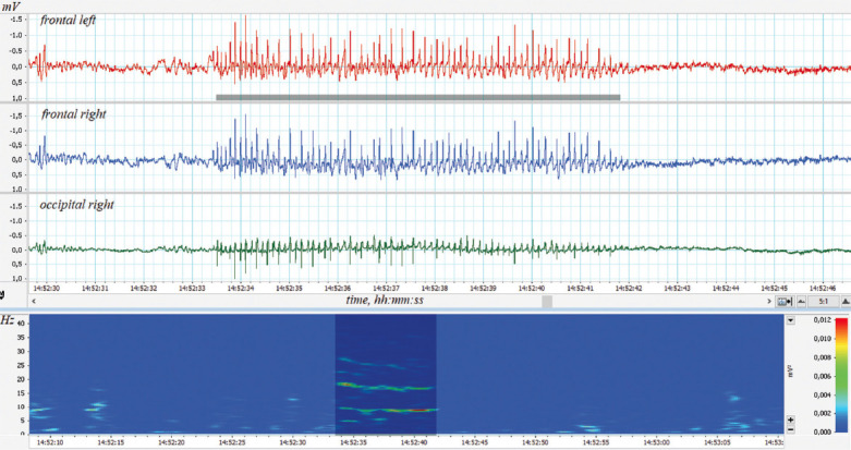
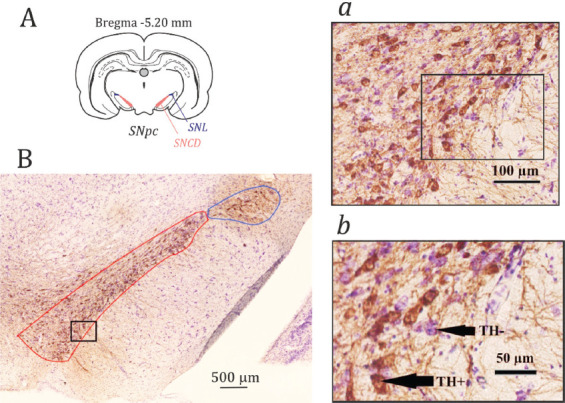
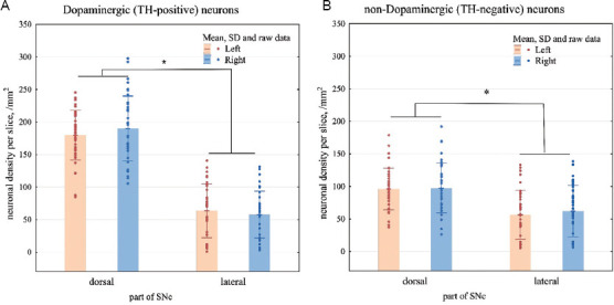
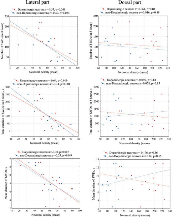
This study aimed to investigate the role of the nigrostriatal dopaminergic system in the modulation of absence epilepsy. Immunochemical analysis of the rostral pole of the substantia nigra pars compacta (SNpc) was conducted on 13 adult male Wistar Albino rats from Rijswijk rats. The rostral pole of the SNpc included the dorsal and lateral parts. The neuronal density in the dorsal part was higher than in the lateral part. The ratio of dopaminergic to non-dopaminergic neurons in the lateral part of the SNpc was 1:1, while in the dorsal part, it was around 1.9:1. All rats exhibited spontaneous spike-wave discharges (SWDs) on their electrocorticograms. SWDs are known to be a hallmark of absence seizures in both human patients and rat models. In this study, we found that the number and duration of SWDs were negatively correlated with dopaminergic and non-dopaminergic neurons only in the lateral part of the SNpc. However, in the dorsal part of the SNpc, no correlations were found between neuronal density and the severity of absence epilepsy. Our findings suggest that the lateral SNpc may be involved in modulating the severity of absence epilepsy in genetically prone subjects. This contributes to a better understanding of the role of the nigrostriatal dopaminergic system in the absence of epilepsy.
Keywords: Absence epilepsy; Electrocorticography; Spike-wave discharges; Substantia nigra pars compacta; Tyrosine hydroxylase; WAG/Rij.
© 2024 The Journal of Biological Methods, All rights reserved.
Conflict of interest statement
The authors declare no conflicts of interest.
Figures
Similar articles
-
Spike-wave seizures, slow-wave sleep EEG and morphology of substantia nigra pars compacta in WAG/Rij rats with genetic predisposition to absence epilepsy.Brain Res Bull. 2021 Sep;174:63-71. doi: 10.1016/j.brainresbull.2021.06.003. Epub 2021 Jun 4. Brain Res Bull. 2021. PMID: 34090934
-
Spike-Wave Seizures, NREM Sleep and Micro-Arousals in WAG/Rij Rats with Genetic Predisposition to Absence Epilepsy: Developmental Aspects.Life (Basel). 2022 Apr 12;12(4):576. doi: 10.3390/life12040576. Life (Basel). 2022. PMID: 35455067 Free PMC article.
-
Prolongation of absence seizures and changes in serotonergic and dopaminergic neurotransmission by nigrostriatal pathway degeneration in genetic absence epilepsy rats.Pharmacol Biochem Behav. 2022 Feb;213:173317. doi: 10.1016/j.pbb.2021.173317. Epub 2021 Dec 30. Pharmacol Biochem Behav. 2022. PMID: 34974062
-
Spike-wave discharges in adult Sprague-Dawley rats and their implications for animal models of temporal lobe epilepsy.Epilepsy Behav. 2014 Mar;32:121-31. doi: 10.1016/j.yebeh.2014.01.004. Epub 2014 Feb 15. Epilepsy Behav. 2014. PMID: 24534480 Free PMC article. Review.
-
Sleep Disturbances in Rats With Genetic Pre-disposition to Spike-Wave Epilepsy (WAG/Rij).Front Neurol. 2021 Nov 5;12:766566. doi: 10.3389/fneur.2021.766566. eCollection 2021. Front Neurol. 2021. PMID: 34803898 Free PMC article. Review.
References
LinkOut - more resources
Full Text Sources
Miscellaneous