. 2024 Oct 27;13(21):1778.
doi: 10.3390/cells13211778.

Targeted Delivery to Dying Cells Through P-Selectin-PSGL-1 Axis: A Promising Strategy for Enhanced Drug Efficacy in Liver Injury Models

Affiliations

Targeted Delivery to Dying Cells Through P-Selectin-PSGL-1 Axis: A Promising Strategy for Enhanced Drug Efficacy in Liver Injury Models

Te-Sheng Lien et al. Cells. .

Abstract

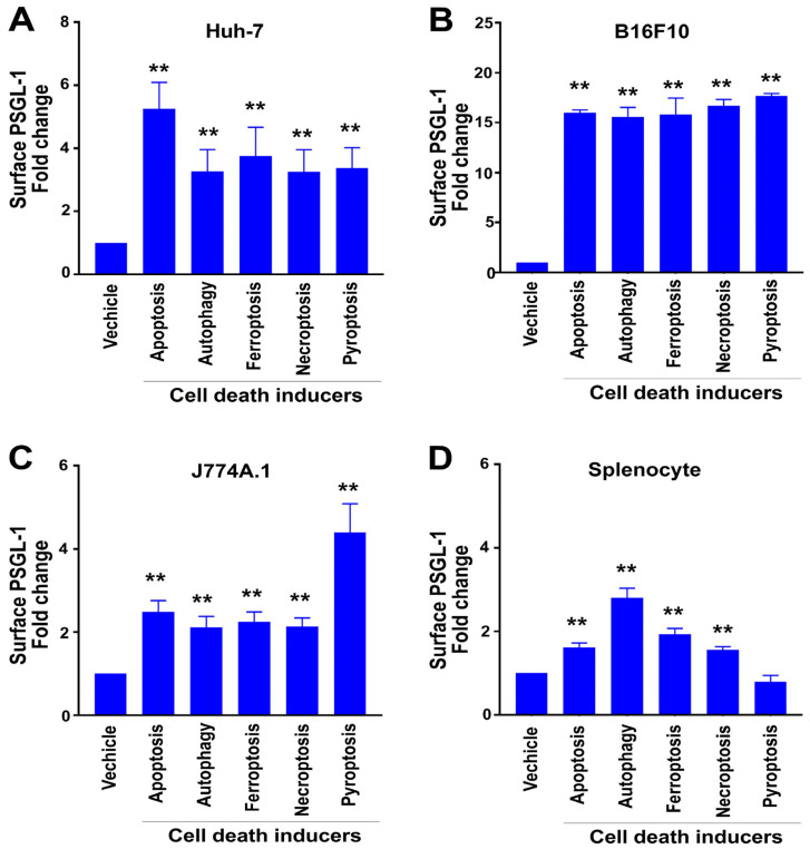
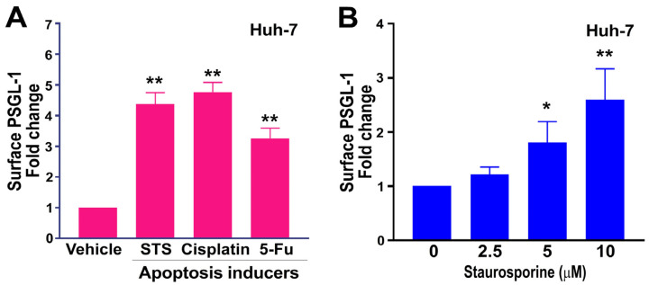
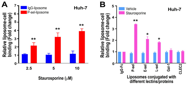
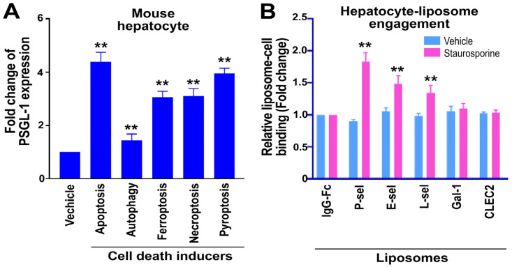
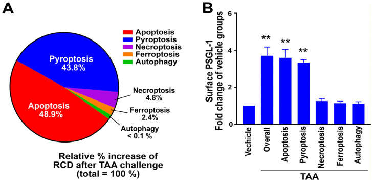
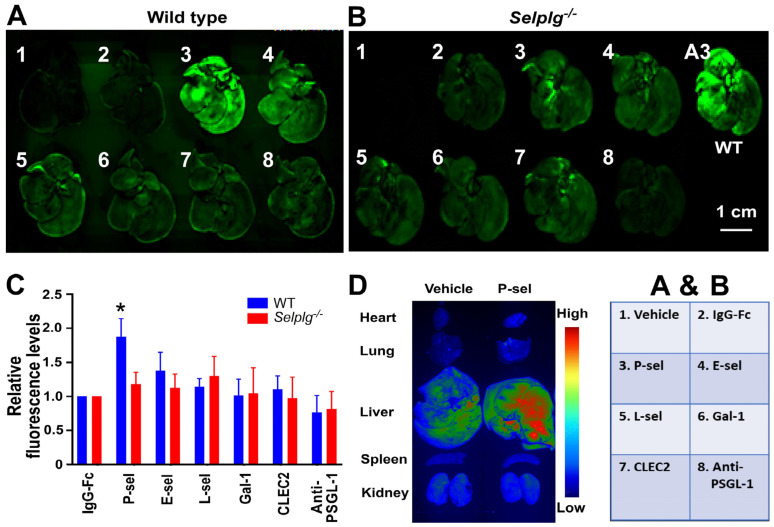
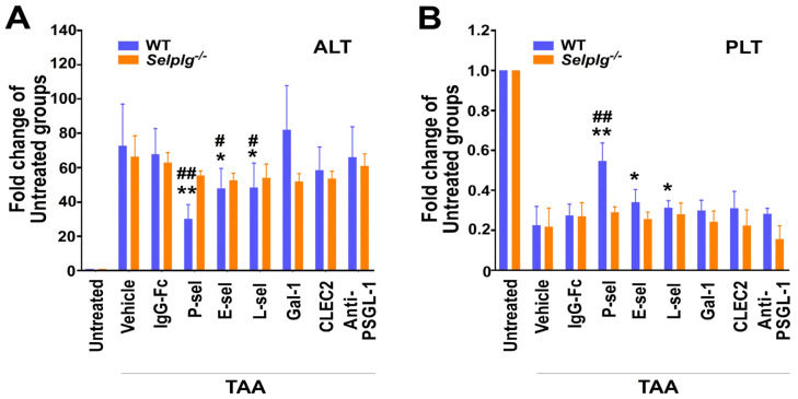
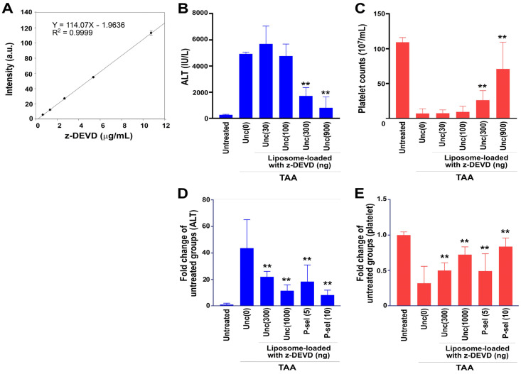
To minimize off-target adverse effects and improve drug efficacy, various tissue-specific drug delivery systems have been developed. However, even in diseased organs, both normal and stressed, dying cells coexist, and a targeted delivery system specifically for dying cells has yet to be explored to mitigate off-target effects within the same organ. This study aimed to establish such a system. By examining the surfaces of dying cells in vitro, we identified P-selectin glycoprotein ligand-1 (PSGL-1) as a universal marker for dying cells, positioning it as a potential target for selective drug delivery. We demonstrated that liposomes conjugated with the PSGL-1 binding protein P-selectin had significantly greater binding efficiency to dying cells compared to control proteins such as E-selectin, L-selectin, galectin-1, and C-type lectin-like receptor 2. Using thioacetamide (TAA) to induce hepatitis and hepatocyte damage in mice, we assessed the effectiveness of our P-selectin-based delivery system. In vivo, P-selectin-conjugated liposomes effectively delivered fluorescent dye and the apoptosis inhibitor z-DEVD to TAA-damaged livers in wild-type mice, but not in PSGL-1 knockout mice. In TAA-treated wild-type mice, unconjugated liposomes required a 100-fold higher z-DEVD dose compared to P-selectin-conjugated liposomes to achieve a comparable, albeit less effective, therapeutic outcome in lowering plasma alanine transaminase levels and alleviating thrombocytopenia. This emphasizes that P-selectin conjugation enhances drug delivery efficiency by approximately 100-fold in mice. These results suggest that P-selectin-based liposomes could be a promising strategy for targeted drug delivery, enabling both diagnosis and treatment by specifically delivering cell-labeling agents and rescue agents to dying cells via the P-selectin-PSGL-1 axis at the individual cell level.

Keywords: apoptosis inhibitor z-DEVD; drug-induced liver damage; dying-cell-specific drug delivery system; fluorescent dye delivery; image tracking and diagnosis; regulated cell death.


Conflict of interest statement

The authors declare no conflicts of interest. The funders had no role in the design of the study; in the collection, analyses, or interpretation of data; in the writing of the manuscript, or in the decision to publish the results.

Figures

Figure 1
Figure 1
Measurement of relative surface PSGL-1 expression after F4/80+ splenocytes, Huh-7, B16F10, and J774A.1 cells were treated with cell death inducers. (A) Human hepatoma cell line Huh-7, (B) mouse melanoma cell line B16F10, (C) mouse macrophage cell line J774A.1, and (D) primary F4/80+ splenocytes were treated with inducers of apoptosis (staurosporine, 10 μM), autophagy (rapamycin, 2 μM), ferroptosis (erastin, 20 μM), necroptosis (TNF-α, 0.2 μM), and pyroptosis (nigericin, 10 μM) for 4 h. Surface PSGL-1 expression levels were then measured by flow cytometry. Vehicle control groups were normalized to 1-fold. ** p < 0.01 indicates markedly increased vs. respective vehicle-treated groups. The study included three experiments with two replicates per group [N = 3 (3 independent experiments), with n = 2 (2 technical replicates per experiment)].
Figure 2
Figure 2
Apoptosis inducers induced surface PSGL-1 expression in Huh-7 cells. (A) Apoptosis inducers like staurosporine (STS; 10 μM), cisplatin (50 μM), and 5-FU (50 μM) increased surface PSGL-1 expression on Huh-7 cells after 4 h. (B) A dose-dependent increase in PSGL-1 expression on the surfaces of Huh-7 cells was observed with STS treatments ranging from 0 to 10 μM over 4 h. ** p < 0.01, * p < 0.05 vs. vehicle-treated groups (A,B). The vehicle groups (A,B) were set to 1-fold. Three experiments were performed with two replicates per group [N = 3 (3 independent experiments), with n = 2 (2 technical replicates per experiment)].
Figure 3
Figure 3
Huh-7 cell binding levels with fluorescent-dye-loaded liposomes were analyzed. Huh-7 cells were treated with or without the apoptosis inducer staurosporine, and their surface PSGL-1 expression (A) and engagement with fluorescein-loaded liposomes conjugated with various lectins and proteins (B) were measured using flow cytometry. Staurosporine treatment induced a dose-dependent increase in P-selectin (P-sel) liposome–Huh-7 cell interaction (A). ** p < 0.01 indicates a significant increase compared to the respective IgG-Fc–liposome groups (A). The IgG–liposome plus 2.5 μM STS-treated group (A) was set to 1-fold. (B) The conjugated lectins and proteins included IgG-Fc, P-selectin (P-sel), E-selectin (E-sel), L-selectin (L-sel), galectin-1 (Gal-1), and CLEC2. As these recombinant proteins contained an IgG-Fc portion, IgG-Fc was used as a control. (B) Both vehicle-treated and staurosporine-treated IgG-Fc groups were normalized to 1-fold. ** p < 0.01, * p < 0.05 indicates a significant increase compared to the respective IgG-Fc groups. The study comprised three experiments, with two replicates per group [N = 3 (3 independent experiments), n = 2 (2 technical replicates per experiment)].
Figure 4
Figure 4
Surface PSGL-1 expression and engagement with liposomes conjugated to various lectins and proteins in primary ASGR1+ mouse hepatocytes. Primary ASGR1+ hepatocytes were treated with or without inducers of apoptosis (staurosporine, 10 μM), autophagy (rapamycin, 2 μM), ferroptosis (erastin, 20 μM), necroptosis (TNF-α, 0.2 μM), and pyroptosis (nigericin, 10 μM) for 4 h in vitro, followed by exposure to fluorescein-loaded liposomes conjugated with different lectins and proteins. Surface PSGL-1 expression levels (A) and cell–liposome engagement levels (B) were analyzed by flow cytometry. The conjugated lectins and proteins included IgG-Fc, P-sel, E-sel, L-sel, Gal-1, and CLEC2. Since these recombinant proteins all contained an IgG-Fc portion, an isotype-matched IgG-Fc was used as a control. Vehicle-treated groups (A) and IgG-Fc-treated hepatocyte groups (B) were normalized to 1-fold. ** p < 0.01 vs. vehicle-treated groups (A), ** p < 0.01 indicated a significant increase vs. respective IgG-Fc groups (B). The study was conducted with three independent experiments, each including two replicates per group [N = 3 (3 independent experiments), with n = 2 (2 technical replicates per experiment)].
Figure 5
Figure 5
RCD profiling and PSGL-1 levels in hepatocytes from TAA-treated mice. (A) The RCD profile in ASGR1+ hepatocytes was assessed following TAA treatment using flow cytometry to evaluate changes in RCD levels. (B) Surface expression levels of PSGL-1 were measured in RCD-marker+ASGR1+ double-positive hepatocytes by flow cytometry. For example, in the apoptosis group, PSGL-1 levels were determined by identifying PSGL-1+ cells within the active caspase-3+ASGR1+ double-positive population. The “overall” groups were analyzed for PSGL-1 expression in ASGR1+ hepatocytes, independent of RCD markers. Vehicle control groups were normalized to a 1-fold change. ** p < 0.01 vs. respective vehicle-treated groups. The study was conducted with three independent experiments, each including two replicates per group [N = 3 (3 independent experiments), with n = 2 (2 technical replicates per experiment)]. A total of 12 mice were used in the study.
Figure 6
Figure 6
Dying-cell-specific targeting via the P-selectin–PSGL-1 axis demonstrated by fluorescent dye delivery to injured mouse liver. Following TAA-induced liver injury, wild-type (WT) (A) and PSGL-1 null (Selplg−/−) (B) mice were injected with fluorescein-loaded liposomes without protein conjugation (vehicle; 1), or conjugated to IgG-Fc (2), P-sel (3), E-sel (4), L-sel (5), Gal-1 (6), CLEC2 (7), and anti-PSGL-1 antibody (8). Fluorescent liver images are shown (A, B), and relative fluorescence intensities were quantified using ImageJ (C), in which the fluorescence levels of mouse liver in the IgG-Fc groups were normalized to 1-fold (C). (D) Pseudo-color imaging revealed that the system selectively delivered fluorescein to injured liver tissue while avoiding non-target organs like the lungs and spleen, where nanoparticles are typically retained. * p < 0.05 vs. IgG-Fc groups. Three experiments with two mice per group (n = 6). A total of 106 (53 WT, 53 Selplg−/−) mice were used in the study.
Figure 7
Figure 7
Rescue of damaged liver through dying-cell-specific targeting via the P-selectin–PSGL-1 axis with delivery of apoptosis inhibitor z-DEVD in mice. After TAA-induced liver injury, wild-type and Selplg−/− mice were injected with caspase-3 inhibitor z-DEVD-loaded liposomes (5 × 10⁷ liposomes, containing 2 μM z-DEVD/mouse) conjugated to various proteins, including IgG-Fc, P-sel, E-sel, L-sel, Gal-1, CLEC2, and anti-PSGL-1 antibody (anti-PSGL-1). Plasma ALT levels (A) and platelet (PLT) counts (B) were measured. ** p < 0.01, * p < 0.05 vs. vehicle groups; ## p < 0.01, # p < 0.05 vs. IgG-Fc groups. Three experiments, with two mice per group (n = 6). A total of 108 (54 WT, 54 Selplg−/−) mice were used in the study.
Figure 8
Figure 8
P-selectin-conjugated liposomes exert a rescue effect that is associated with the suppression of caspase-3 activities in mice. (A) Elevated surface PSGL-1 expression and (B) activated caspase-3 levels in TAA-treated mouse (wild type and Selplg−/−) hepatocytes in vivo were markedly rescued by treatments with P-selectin-conjugated liposomes (P-sel groups) loaded with caspase-3 inhibitor z-DEVD (5 × 10⁷ liposomes, containing 2 μM z-DEVD/mouse). The measurements of untreated groups were normalized to 1-fold. ** p < 0.01, indicated markedly exacerbated vs. respective untreated control groups; # p < 0.05 indicated markedly ameliorated vs. respective IgG-Fc control groups. Three experiments with two mice per group (n = 6) were performed. A total of 48 (24 WT, 24 Selplg−/−) mice were used in the study.
Figure 9
Figure 9
Markedly enhanced rescue efficiency was observed when Red-DEVD was loaded into P-selectin-conjugated liposomes in mice. (A) Quantification of Red-DEVD dosages was performed by measuring the fluorescence intensity of the fluorescent Red-DEVD. (B,C) In vivo testing of unconjugated liposomes (unc) loaded with Red-DEVD, with injection doses ranging from 30 ng to 900 ng per mouse [e.g., unc (30) represents unconjugated liposomes with 30 ng Red-DEVD], was performed to evaluate their ability to reduce TAA-induced increases in circulating ALT levels (B) and thrombocytopenia ((C), reduced platelet counts). The rescue effects on ALT levels (D) and thrombocytopenia (E) in TAA-treated mice, using unconjugated liposomes with Red-DEVD doses of 0–1000 ng per mouse, were compared to those using P-selectin-conjugated liposomes with Red-DEVD doses of 5–10 ng per mouse [e.g., P-sel (5) for P-selectin-conjugated liposomes with 5 ng Red-DEVD]. The untreated groups were normalized to 1-fold. ** p < 0.01 vs. respective TAA-treated unc (0) groups. Three experiments with two replicates per group (n = 6; (AC)), and four experiments with three mice per group (n = 12; (D,E)) were conducted.

Similar articles

References

    1. Nkune N.W., Abrahamse H. Nanoparticle-Based Drug Delivery Systems for Photodynamic Therapy of Metastatic Melanoma: A Review. Int. J. Mol. Sci. 2021;22:12549. doi: 10.3390/ijms222212549. - DOI - PMC - PubMed
    1. Zhang Y., Almazi J.G., Ong H.X., Johansen M.D., Ledger S., Traini D., Hansbro P.M., Kelleher A.D., Ahlenstiel C.L. Nanoparticle Delivery Platforms for RNAi Therapeutics Targeting COVID-19 Disease in the Respiratory Tract. Int. J. Mol. Sci. 2022;23:2408. doi: 10.3390/ijms23052408. - DOI - PMC - PubMed
    1. Hersh A.M., Alomari S., Tyler B.M. Crossing the Blood-Brain Barrier: Advances in Nanoparticle Technology for Drug Delivery in Neuro-Oncology. Int. J. Mol. Sci. 2022;23:4153. doi: 10.3390/ijms23084153. - DOI - PMC - PubMed
    1. Markowski A., Jaromin A., Migdal P., Olczak E., Zygmunt A., Zaremba-Czogalla M., Pawlik K., Gubernator J. Design and Development of a New Type of Hybrid PLGA/Lipid Nanoparticle as an Ursolic Acid Delivery System against Pancreatic Ductal Adenocarcinoma Cells. Int. J. Mol. Sci. 2022;23:5536. doi: 10.3390/ijms23105536. - DOI - PMC - PubMed
    1. Nahrjou N., Ghosh A., Tanasova M. Targeting of GLUT5 for Transporter-Mediated Drug-Delivery Is Contingent upon Substrate Hydrophilicity. Int. J. Mol. Sci. 2021;22:5073. doi: 10.3390/ijms22105073. - DOI - PMC - PubMed

Publication types

LinkOut - more resources