. 2024 Dec;43(24):6383-6409.
doi: 10.1038/s44318-024-00288-x. Epub 2024 Nov 4.

Rab2A-mediated Golgi-lipid droplet interactions support very-low-density lipoprotein secretion in hepatocytes

Affiliations

Rab2A-mediated Golgi-lipid droplet interactions support very-low-density lipoprotein secretion in hepatocytes

Min Xu et al. EMBO J. 2024 Dec.

Abstract

Lipid droplets (LDs) serve as crucial hubs for lipid trafficking and metabolic regulation through their numerous interactions with various organelles. While the interplay between LDs and the Golgi apparatus has been recognized, their roles and underlying mechanisms remain poorly understood. Here, we reveal the role of Ras-related protein Rab-2A (Rab2A) in mediating LD-Golgi interactions, thereby contributing to very-low-density lipoprotein (VLDL) lipidation and secretion in hepatocytes. Mechanistically, our findings identify a selective interaction between Golgi-localized Rab2A and 17-beta-hydroxysteroid dehydrogenase 13 (HSD17B13) protein residing on LDs. This complex facilitates dynamic organelle communication between the Golgi apparatus and LDs, thus contributing to lipid transfer from LDs to the Golgi apparatus for VLDL2 lipidation and secretion. Attenuation of Rab2A activity via AMP-activated protein kinase (AMPK) suppresses the Rab2A-HSD17B13 complex formation, impairing LD-Golgi interactions and subsequent VLDL secretion. Furthermore, genetic inhibition of Rab2A and HSD17B13 in the liver reduces the serum triglyceride and cholesterol levels. Collectively, this study provides a new perspective on the interactions between the Golgi apparatus and LDs.

Keywords: 17-Beta-hydroxysteroid Dehydrogenase 13; AMP-activated Protein Kinase; Organelle Interactions; Ras-related Protein Rab-2A; Very-low-density Lipoprotein.


Conflict of interest statement

Disclosure and competing interests statement. The authors declare no competing interests.

Figures
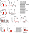
Figure 1
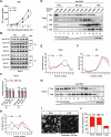
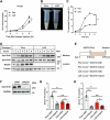
Figure 1. Deletion of Ras-related protein Rab-2A (Rab2A) decreases serum triglyceride and cholesterol levels.
(AF) Serum samples were obtained from Flox and LCK mice under “Random feed” and “Fasted” conditions, followed by subsequent experiments (Male, n = 5 mice per group). Serum triglyceride (TG) levels (A) (Random feed, P = 0.0111; Fasted, P = 0.0089) and their distribution in lipoproteins (C), serum total cholesterol (TC) levels (B) (Random feed, P = 0.0009; Fasted, P = 0.0003) and their distribution in lipoproteins (D) were analyzed. Then, apolipoprotein secretion levels in serum, such as Apo B-100, Apo B-48, Apo-E, Apo-AI, and Apo-CIII were evaluated (E), with grayscale quantification of the respective proteins, where the value in Flox samples was normalized to 1 (Male, n = 3 mice per group) (F). (GL) Flox and LCK mice were fed with a high-fat-high-cholesterol diet (HFHCD) for three months, followed by the collection of serum samples for experiments (Male, n = 6 vs. 5 mice). This encompassed TG levels (G) (P = 0.0302) and detailed distributions in lipoproteins (I), TC levels (H) (P = 0.0033) and detailed distributions in lipoproteins (J), as well as apolipoproteins in serum samples (K). The expression levels of apolipoproteins were quantified, normalizing Flox samples to 1 (Male, n = 3 mice per group) (L) (Apo B-100, p = 0.0053; Apo B-48, P = 0.0407). Data information: Data in (A, B, F, G, H, L) are presented as mean ± SEM. Circles in (A, B, F, G, H, L) correspond to individual mice. P values in (A, B, F, G, H, L) were determined using unpaired two-tailed Student’s t-test. n.s. indicates no significant difference (P > 0.05); *P < 0.05; **P < 0.01. VLDL very low-density lipoprotein, LDL low-density lipoprotein, HDL high-density lipoprotein. Source data are available online for this figure.
Figure 2
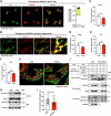
Figure 2. Absence of Rab2A hinders the lipidation process of VLDL2.
(AC) VLDL-TG secretion in male mice were assessed through a tyloxapol injection assay (A) (n = 6 vs. 5 mice) (P = 0.0058). Subsequent analysis and quantification of apolipoprotein secretion levels in serum collected at two hours and four hours following tyloxapol injection (Male, n = 3 mice per group) (B, C). (DH) Quantification of the degree of lipoproteins lipidation within the endoplasmic reticulum (ER), Golgi apparatus (Golgi), and serum of Flox and LCK mice using the sucrose density gradient centrifugation method. Protein imprinting of Apo B-48 (D, G) and subsequent statistical results (E, F, H) elucidated the distribution and proportion of lipoproteins, with fraction 1 representing the top layer of sucrose density and fraction 12 corresponding to the bottom layer. The experiments were replicated twice with similar pattern (Male, n = 1 mouse per group). (I, J) The size of VLDL secreted into the serum of Flox and LCK mice (Male, n = 3 mice per group) was analyzed by negative staining and transmission electron microscope (TEM) imaging. Representative images (I) and statistical data (J) are presented, the number of particles is more than 200. Data information: Data in (A, C) are presented as mean ± SEM. Circles in (C) correspond to individual mice. P value in (A) was determined using two-way ANOVA. P values in (C) were determined using unpaired two-tailed Student’s t test. n.s. indicates no significant difference (P > 0.05); **P < 0.01. Source data are available online for this figure.
Figure 3
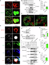
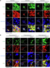
Figure 3. Rab2A orchestrates Golgi-Lipid droplet (LD) interactions.
(A) The subcellular localization of Rab2A was examined in Huh7 cells. Endogenous Rab2A was stained with a primary antibody, and Golgi apparatus was labeled using a primary antibody targeting GM130. Representative images from 5 cells are shown (left), and the percentage of colocalization was quantified with Pearson’s R value (n = 24 cells) (right). (B) The subcellular localization of Rab2A was also confirmed in mouse primary hepatocytes. Endogenous Rab2A was stained with a primary antibody, and Golgi apparatus was labeled with primary antibodies against GM130. Representative images are shown. (C-G) Sucrose density gradient centrifugation facilitated the isolation and purification of Golgi and ER compartments from livers of Flox and LCK mice (Male, n = 3 vs. 5 mice). TG and TC levels were precisely assessed in Golgi fractions (C, D) (TG, P = 0.0445; TC, P = 0.0457) and ER fractions (E, F). Apo B-48 protein levels were also analyzed in Golgi and ER fractions with ADP-ribosylation factor 1 (ARF1) or GRP-94 as the internal control (Male, n = 2 mice per group) (G). (H, I) Mouse primary hepatocytes were isolated, transfected with corresponding Rab2A-siRNA, incubated with 100 μM oleic acid (OA) for 16 h, and then stained with a primary antibody against GM130 (Red). LDs were labeled with Bodipy (Green). Representative images are shown and white arrows indicate potential contacts (H). Statistical data are presented (I) (n = 31 vs. 30 cells) (P = 0.0274). (J) Inhibition of Rab2A attenuated Golgi-LD interfacing, as evidenced by LDs pulldown assay. Purified LDs, isolated from the livers of wild-type mice, were incubated with organelle clusters from primary hepatocytes with or without Rab2A deficiency. The assay was performed twice with similar conclusion and representative results are shown. Data information: Data in (A, CF, I) are presented as mean ± SEM. Circles in (A, I) correspond to individual cell. Circles in (CF) correspond to individual mice. P values in (CF, I) were determined using unpaired two-tailed Student’s t test. n.s. indicates no significant difference (P > 0.05); *P < 0.05. Source data are available online for this figure.
Figure 4
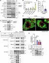
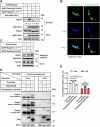
Figure 4. 17-beta-hydroxysteroid dehydrogenase 13 (HSD17B13), an LD-localized protein, binds with Rab2A to mediate Golgi-LD interactions and VLDL secretion.
(A) Mouse primary hepatocytes were isolated, cultured and then stained with a primary antibody against endogenous HSD17B13, with LDs labeled with Bodipy. Representative images are shown. (B) The liver samples from Flox and LCK mice were processed and incubated with Rab2A against primary antibody, facilitating the enrichment of the interaction between Rab2A and HSD17B13 using protein A/G affinity beads, and subsequently visualized via Western blotting. The assay was performed twice with similar results. (C) Mouse primary hepatocytes were isolated, cultured and then stained with primary antibodies against endogenous Rab2A and HSD17B13. The Golgi apparatus was labeled by overexpressing BFP-RCAS1, while LDs were visualized using HCS LipidTOX. Representative images are presented. (DG) Mouse primary hepatocytes were isolated, transfected with corresponding siRNA targeting endogenous HSD17B13. Knockdown efficiency was validated by Western blotting (D). Hepatocytes were incubated with 100 μM oleic acid (OA) for 16 h then fixed and stained for the Golgi apparatus using a primary antibody against GM130 (Red) and LDs using Bodipy (Green). Representative images are shown, with white arrows indicating potential contacts (E). Statistical data are presented (F) (n = 18 cells per group) (P < 0.0001). TG secretion levels were quantified and normalized with cellular total protein content (G) (4 h, P = 0.0209; 8 h, P < 0.0001). (HK) In vivo inhibition of HSD17B13 was achieved in wild-type mouse livers using AAV2/8-shRNA virus (Male, n = 7 vs. 5 mice). Several experiments were conducted to explore the impact of HSD17B13 on organelle communications and VLDL secretion. The effectiveness of knockdown (H), Golgi-LD interfacing (I), TG and TC levels in serum (J, K) (TG, P = 0.0041; TC, P = 0.0267) were systematically assessed. The assay in “I” was performed twice with similar conclusion and representative results are shown. Data information: Data in (F, G, J, K) are presented as mean ± SEM. Circles in (F) correspond to individual cell. Circles in (G) correspond to individual test. Circles in (J, K) correspond to individual mice. P values in (F, G, J, K) were determined using unpaired two-tailed Student’s t test. *P < 0.05; **P < 0.01; ***P < 0.001. Source data are available online for this figure.
Figure 5
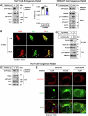
Figure 5. AMPK signaling attenuates Rab2A activity and its role in Golgi-LD interactions and VLDL secretion.
(A, B) Rab2A activity and subcellular distribution in liver samples after overnight fasting were analyzed using the GRASP55 pulldown assay and sucrose density gradient centrifugation assay (Male, n = 3 mice per group) (A), with corresponding statistical analyses presented (B) (Golgi-Rab2A, P = 0.0194; GTP-Rab2A, P = 0.0089; pS231-TBC1D1, P = 0.0454; pT172-AMPKα, P = 0.0006), where levels in “Random feed” state samples were normalized to 1. The assay was performed twice with similar results. (CF) Primary hepatocytes were stimulated with A769662 (100 μM), an AMPK agonist, for 4 h in DMEM medium without fetal bovine serum (FBS), followed by a series of assays. Rab2A activity was primarily evaluated using the GRASP55 pulldown assay (C). Potential Golgi-LDs contact points were then quantified by staining Golgi apparatus with a primary antibody against GM130 (Red) and LDs with Bodipy (Green). Representative images are shown, with white arrows indicating potential contacts (D). Statistical data are presented (E) (n = 22 cells per group) (P < 0.0001). Subsequently, the LDs pulldown assay was performed by incubating purified LDs from the livers of wild-type mice with organelle clusters from primary hepatocytes, with or without A769662 stimulation (F). The assay in “F” was performed twice, yielding similar results, with representative data shown. (G) Primary hepatocytes isolated from Flox and LCK mice were cultured and treated with A769662 (100 μM) for 6 h in DMEM medium without FBS. TG secretion levels were measured and quantified (A769662 stimulation in Flox hepatocytes (0 μM vs. 100 μM), P < 0.0001; Flox vs. LCK in hepatocytes without A769662 stimulation (0 μM), P = 0.0002), and cell lysates were prepared for Western blot analysis (n = 3 per group). Data information: Data in (B, E, G) are presented as mean ± SEM. Circles in (B) correspond to individual mice. Circles in (E) correspond to individual cell. Circles in (G) correspond to individual assay. P values in (B, E, G) were determined using unpaired two-tailed Student’s t test. n.s. indicates no significant difference (P > 0.05); *P < 0.05; **P < 0.01; ***P < 0.001. Source data are available online for this figure.
Figure 6
Figure 6. The graphical abstract describes the pivotal role of Rab2A in mediating Golgi-LD interactions and subsequent VLDL secretion in hepatocytes.
VLDL2, assembled in the ER, is transported to the Golgi via COP-II vesicles for further processing. Our studies reveal that VLDL2 lipidation primarily occurs within the Golgi apparatus, where lipids are transferred from LDs, leading to the formation of mature VLDL1. Both VLDL1 and VLDL2 are secreted from the Golgi apparatus into the serum via vesicular systems. Mechanistically, Golgi-localized Rab2A binds with HSD17B13, an LD-resident protein, orchestrating organelle interactions between the Golgi apparatus and LDs, thereby enhancing lipid transport and VLDL secretion. Additionally, AMPK signaling can potentially attenuate these processes by inhibiting Rab2A activity.
Figure EV1
Figure EV1. Evaluating the rates of lipid secretion in Flox and LCK mice.
(A) Triglyceride (TG) secretion in female mice were assessed through a tyloxapol injection assay (n = 7 vs. 6 mice) (P = 0.0021). (B) A visual examination of blood transparency in Flox and LCK mice was conducted four hours post-tyloxapol injection. (C, D) The evaluation of the TG secretion in primary hepatocytes involved quantifying the secretion level of TG (C) (P < 0.0001) and Apo B-48 (D), with normalization of Flox samples’ secretion level at 1-hour to 1. (EH) The schematic representation outlines the timeline for the Apob knockdown assay (E), followed by the validation of Apo B-48 proteins levels (F) and assessment of serum TG (G) (Flox (shNC vs. shApob), P < 0.0001; shNC (Flox vs. LCK), P < 0.0001; LCK (shNC vs. shApob), P = 0.0079) and TC (H) (Flox (shNC vs. shApob), P < 0.0001; shNC (Flox vs. LCK), p = 0.0009; LCK (shNC vs. shApob), P = 0.0034) levels under “Fasted” condition (Male, n = 10 vs. 5 vs. 7 vs. 8 mice). Data information: Data in (A, C, G, H) are presented as mean ± SEM. Circles in (G, H) correspond to individual mice. P values in (A, C) were determined using two-way ANOVA. P values in (G, H) were determined using unpaired two-tailed Student’s t-test. n.s. indicates no significant difference (P > 0.05), ** indicates P < 0.01; *** indicates P < 0.001.
Figure EV2
Figure EV2. Golgi-localized Ras-related protein Rab-2A (Rab2A) contacts with LDs.
(A, B) Immunofluorescence analysis elucidated Rab2A-mediated organelle contacts between Golgi and LDs in Huh7 cells (A) and primary hepatocytes (B) with overexpression of EGFP-Rab2A plasmids. Golgi was labeled using a primary antibody targeting GM130, while LDs were stained with Bodipy (The white arrow indicates different types of contact points).
Figure EV3
Figure EV3. Confirming the binding efficiency between Rab2A and 17-beta-hydroxysteroid dehydrogenase 13 (HSD17B13) using exogenously expressed Rab2A and HSD17B13.
(A) HEK293T cells were transfected with the BirA*-Rab2A and HSD17B13 plasmids. Rab2A-HSD17B13 binding was evaluated via NeutrAvidin beads pulldown, followed by visualization using Western blotting. (B) HEK293T cells were transfected with Flag-Rab2A and MYC-HSD17B13 plasmids, followed by a Flag-affinity beads pulldown assay to confirm Rab2A and HSD17B13 binding. (C) In vitro binding assay was conducted using purified GST-HSD17B13 and Flag-Rab2A proteins. (D, E) Huh7 cells underwent transfection with HSD17B13-mCherry and EGFP-Rab2A plasmids. Immunofluorescence was employed to observe the binding efficiency between Rab2A and HSD17B13. Representative images are shown (D), and the percentage of HSD17B13 contacting with Rab2A was quantified (E) (n = 19 cells). (F) The regulatory effect of Rab2A activity on the binding between Rab2A and HSD17B13 was examined in HEK293T cells by transfecting with different types of Rab2A plasmids, followed by MYC-affinity beads pulldown assay. Data information: Data in (E) are presented as mean ± SEM. Circles in (E) correspond to individual Huh7 cell. Source data are available online for this figure.
Figure EV4
Figure EV4. Blocking the binding between Rab2A and HSD17B13 using a Rab2A mutant protein suppresses very-low-density lipoprotein (VLDL) secretion in primary hepatocytes.
(AE) Examination of the effects of Rab2A and HSD17B13 binding on TG secretion involving multiple steps. First, vital amino acids for complex binding in Rab2A were identified via Western blotting in HEK293T cells after transfection with relative plasmids (A). Second, the localization of mutant Rab2A protein was assessed using immunofluorescence in Huh7 cells following transfection with EGFP-Rab2A plasmids, with Golgi labeling accomplished using RCAS1-BFP (B). Third, the activity of both wild-type and mutant Rab2A proteins was detected using a GST-GRASP55 pulldown assay to assess influence of specific mutations on Rab2A’s activity (C). Finally, the impact of deficient complex binding on TG secretion was evaluated in Flox and LCK primary hepatocytes after protein overexpression with the AAV-virus system (D). The medium was collected for testing, with values in Flox samples with Free-Flag overexpression normalized to 1 (E) (Flox hepatocytes (Free-Flag vs. Flag-Rab2A), P = 0.0025; Flox hepatocytes (Free-Flag vs. Flag-Rab2A (Δ032-042)), P = 0.0101; Flox hepatocytes (Flag-Rab2A vs. Flag-Rab2A (Δ032-042)), P = 0.0005; Free-Flag (Flox vs. LCK), P < 0.0001; LCK hepatocytes (Free-Flag vs. Flag-Rab2A), P = 0.0030; LCK hepatocytes (Flag-Rab2A vs. Flag-Rab2A (Δ032-042)), P = 0.0063). Data information: Data in (E) are presented as mean ± SEM. Circles in (E) correspond to individual test. P values in (E) were determined using unpaired two-tailed Student’s t-test. n.s. indicates no significant difference (P > 0.05), * indicates P < 0.05; ** indicates P < 0.01; *** indicates P < 0.001.
Figure EV5
Figure EV5. A769662 stimulation attenuates Rab2A activity, Golgi-localization and Rab2A-HSD17B13 binding.
(AC) Huh7 cells were stimulated with A769662 (100 μM), an AMPK agonist, for 4 h in DMEM medium without fetal bovine serum (FBS). Rab2A activity was evaluated using the GRASP55 pulldown assay (A). Its subcellular localization was assessed with immunofluorescence (B). Endogenous Rab2A was stained with a primary antibody, and the Golgi apparatus was labeled with a primary antibody against GM130. The dispersion of Rab2A signaling was quantified via Li’s ICQ value (Li’s colocalization value) in Image J (n = 44 vs. 44 cells), with white arrows indicating potential signaling away from the Golgi (C) (P < 0.0001). (D, E) Huh7 cells, transfected with exogenously expressing Rab2A, were stimulated with A769662 (100 μM) for 4 h in DMEM medium without FBS. The activity of Rab2A was assessed using a GRASP55 pulldown assay and analyzed by Western blotting (D). The subcellular localization of Rab2A was evaluated with immunofluorescence, with lysosomes stained using LysoTracker (E). (F, G) HEK293T cells were transfected with exogenously expressing Rab2A plasmids, with or without co-transfection of HSD17B13 plasmids, and subsequently stimulated with A769662 (100 μM) for 4 h in DMEM medium without FBS. Rab2A activity was evaluated using the GRASP55 pulldown assay (F). The binding efficiency of Rab2A-HSD17B13 complex was assessed by an EGFP-affinity beads pulldown assay (G). Data information: Data in (C) are presented as mean ± SEM. Circles in (C) correspond to individual cell. P value in (C) was determined using unpaired two-tailed Student’s t-test. *** indicates P < 0.001.

Similar articles

References

    1. Abul-Husn NS, Cheng X, Li AH, Xin Y, Schurmann C, Stevis P, Liu Y, Kozlitina J, Stender S, Wood GC et al (2018) A protein-truncating HSD17B13 variant and protection from chronic liver disease. N. Engl J Med 378:1096–1106 - PMC - PubMed
    1. Adam M, Heikelä H, Sobolewski C, Portius D, Mäki-Jouppila J, Mehmood A, Adhikari P, Esposito I, Elo LL, Zhang F-P et al (2018) Hydroxysteroid (17β) dehydrogenase 13 deficiency triggers hepatic steatosis and inflammation in mice. FASEB J 32:3434–3447 - PubMed
    1. Aizawa M, Fukuda M (2015) Small GTPase Rab2B and its specific binding protein Golgi-associated Rab2B interactor-like 4 (GARI-L4) regulate Golgi morphology. J Biol Chem 290:22250–22261 - PMC - PubMed
    1. Barbosa AD, Savage DB, Siniossoglou S (2015) Lipid droplet-organelle interactions: emerging roles in lipid metabolism. Curr Opin Cell Biol 35:91–97 - PubMed
    1. Boren J, Adiels M, Bjornson E, Matikainen N, Soderlund S, Ramo J, Stahlman M, Ripatti P, Ripatti S, Palotie A et al (2020) Effects of TM6SF2 E167K on hepatic lipid and very low-density lipoprotein metabolism in humans. JCI Insight 5:e144079 - PMC - PubMed

Substances

LinkOut - more resources