. 2024 Oct;14(10):100977.
doi: 10.1016/j.jpha.2024.100977. Epub 2024 Apr 10.

Xianling Lianxia formula improves the efficacy of trastuzumab by enhancing NK cell-mediated ADCC in HER2-positive BC

Affiliations

Xianling Lianxia formula improves the efficacy of trastuzumab by enhancing NK cell-mediated ADCC in HER2-positive BC

Feifei Li et al. J Pharm Anal. 2024 Oct.

Abstract

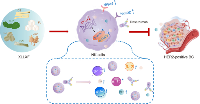
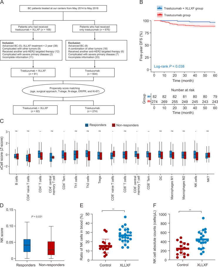
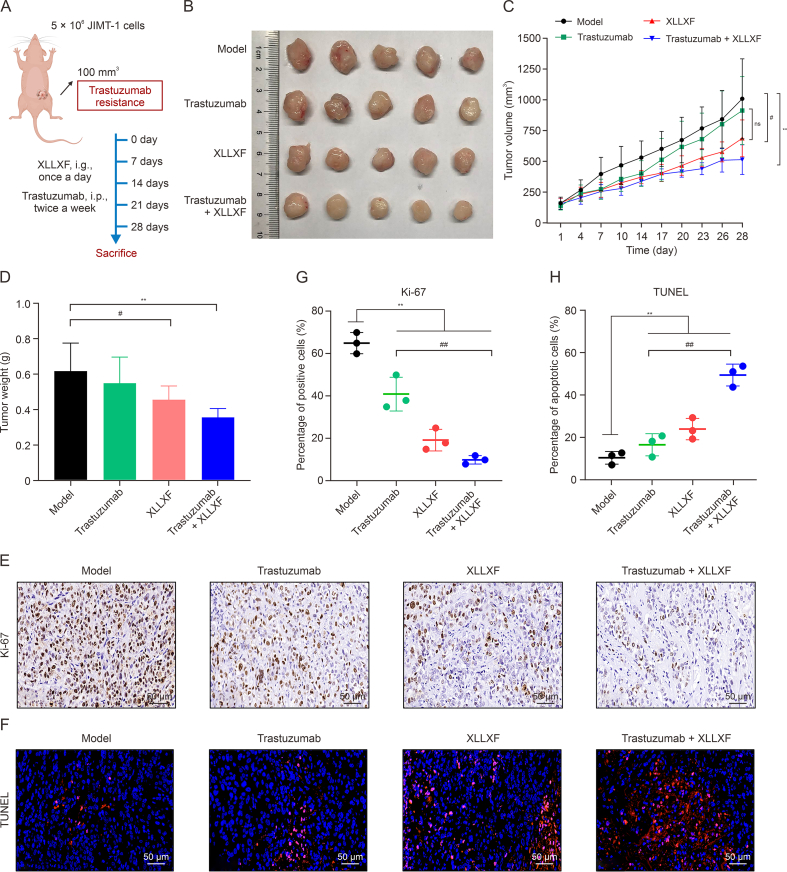
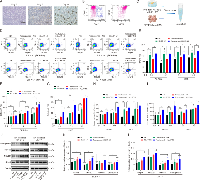
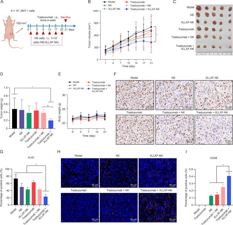
Trastuzumab has improved survival rates in human epidermal growth factor receptor 2 (HER2)-positive breast cancer (BC), but drug resistance leads to treatment failure. Natural killer (NK) cell-mediated antibody-dependent cell cytotoxicity (ADCC) represents an essential antitumor immune mechanism of trastuzumab. Traditional Chinese medicine (TCM) has been used for centuries to treat diseases because of its capacity to improve immune responses. Xianling Lianxia formula (XLLXF), based on the principle of "strengthening body and eliminating toxin", exhibits a synergistic effect in the trastuzumab treatment of patients with HER2-positive BC. Notably, this synergistic effect of XLLXF was executed by enhancing NK cells and ADCC, as demonstrated through in vitro co-culture of NK cells and BC cells and in vivo intervention experiments. Mechanistically, the augmented impact of XLLXF on NK cells is linked to a decrease in cytokine inducible Src homology 2 (SH2) containing protein (CISH) expression, which in turn activates the Janus kinase 1 (JAK1)/signal transducer and activator of transcription 5 (STAT5) pathway. Collectively, these findings suggested that XLLXF holds promise for enhancing NK cell function and sensitizing patients with HER2-positive BC to trastuzumab.

Keywords: ADCC effect; HER2-positive breast cancer; NK cell; Traditional chinese medicine; Trastuzumab.


Conflict of interest statement

The authors declare that there are no conflicts of interest.

Figures

Image 1
Graphical abstract
Fig. 1
Fig. 1
Xianling Lianxia formula (XLLXF) synergizes with trastuzumab to improve the five-year disease-free survival (DFS) in patients with human epidermal growth factor receptor 2 (HER2)-positive breast cancer (BC). (A) Patients recruitment flowchart of cohort study. (B) Kaplan-Meier plots for five-year DFS between the trastuzumab plus XLLXF group (n = 82) and the trastuzumab group (n = 274). (C, D) Bioinformatics analysis: tumor gene expression data obtained from Gene Expression Omnibus (GEO) database demonstrated the degree of immune cell infiltration (C), and further analysis showed that trastuzumab responders exhibited substantially higher levels of natural killer (NK) cell infiltration compared with the non-responders (D). in trastuzumab responders and non-responders groups. (E, F) NK cells proportion (E) and absolute counts (F) in peripheral blood samples of patients with HER2-positive BC. Their peripheral blood samples were collected before (control) and six months after taking XLLXF (XLLXF). P < 0.05, and ∗∗P < 0.01. T-stage: tumor stage; N-stage: node stage; ER: estrogen receptor; PR: progesterone receptor; Tem: effector memory T cell; Tregs: regulatory T cells; DC: dendritic cell; NKT: natural killer T cell.
Fig. 2
Fig. 2
Xianling Lianxia formula (XLLXF) synergizes with trastuzumab to inhibit the tumor growth in human epidermal growth factor receptor 2 (HER2)-positive breast cancer (BC) bearing mice. (A) Schematic diagram of animal model construction and drug administration by Figdraw. (B) Representative images of tumors with the indicated treatment. (C) Tumor size of each group was measured every three days after treatments started. The tumor size data are plotted and shown as mean ± standard deviation (SD). (D) tumor weight of the dissected tumors at the end point was presented. (E) The proliferative cells in tumor tissues were determined by immunohistochemical staining of Ki-67. (F) The apoptotic cells in tumor tissues of each group were determined by terminal dexynucleotidyl transferase-mediated dUTP nick-end labeling (TUNEL) assay. (G, H) The percentage of proliferative cells (G) and apoptotic cells (H) were quantified using lmageJ. Model group: physiological saline, i.g., once a day; trastuzumab group: trastuzumab, i.p., 5 mg/kg, twice a week; XLLXF group: XLLXF, i.g., 5.26 mg/kg, once a day; trastuzumab + XLLXF group: trastuzumab, i.p., 5 mg/kg, twice a week and XLLXF, i.g., 5.26 mg/kg, once a day. ∗∗P < 0.01, #P < 0.05, and ##P < 0.01. ns: not significant.
Fig. 3
Fig. 3
Xianling Lianxia formula (XLLXF) synergizes trastuzumab to increase natural killer (NK) cell activity and function in human epidermal growth factor receptor 2 (HER2)-positive breast cancer (BC)-bearing mice. (A, B) The infiltration of NK cells in tumor tissues was detected by immunofluorescence staining of CD49b (A), and the percentage was quantified using ImageJ (B). Similar to NK1.1 in other mouse strains, the marker CD49b was used to label NK cells in BALB/c mice. (C, D) The protein expressions of NKp46, NK group 2 member D (NKG2D), NK group 2 member A (NKG2A), killer cell immunoglobulin-like receptor 2DL4 (KIR2DL4), and human leucocyte antigen-G (HLA-G) were performed by Western blot (C), and quantified analysis was performed by ImageJ (D). (E, F) The protein expressions of factor-associated suicide (FAS), factor-associated suicide ligand (FASL), perforin, and granzyme B in mouse tumor tissues from different treatment groups were performed by Western blot (E), and quantified analysis was performed by ImageJ (F). Data presented as mean ± standard deviation (SD) normalized to glyceraldehyde-3-phosphate dehydrogenase (GAPDH) levels. (G–H) NK cells (CD49b+ populations) isolated from the spleens of tumor-bearing mice and NKG2D (G) and NKp46, CD107a, granzyme B, and perforin (H) in the NK cells were analyzed by flow cytometry. (I, J) Statistical analysis of the percentages of CD49b+ NK cells in spleen (I), and CD107a, granzyme B, perforin, NKp46, and NKG2D (J). (K) The serum interleukin (IL)-2, interferon-gamma (IFN-γ), and tumor necrosis factor-alpha (TNF-α) levels were quantified by enzyme-linked immunosorbent assay (ELISA). Data are presented as mean ± standard deviation (SD). Model group: physiological saline, i.g., once a day; trastuzumab group: trastuzumab, i.p., 5 mg/kg, twice a week; XLLXF group: XLLXF, i.g., 5.26 mg/kg, once a day; trastuzumab + XLLXF group: trastuzumab, i.p., 5 mg/kg, twice a week and XLLXF, i.g., 5.26 mg/kg, once a day. P < 0.05, ∗∗P < 0.01, and #P < 0.05. SSC-A: side scatter-area.
Fig. 4
Fig. 4
Depletion of natural killer (NK) cells attenuates the potentiating effect of Xianling Lianxia formula (XLLXF) on trastuzumab in human epidermal growth factor receptor 2 (HER2)-positive breast cancer (BC)-bearing mice. (A) Schematic diagram of the model construction and intervention process by Figdraw. (B) Tumor size of each group was measured after treatments started. The tumor size data are plotted and shown as mean ± standard deviation (SD). (C) Representative images of tumors with the indicated treatment. (D) Tumor weight of the dissected tumors at the end point was presented. (E, F) The flow charts (E) and percentages (F) of NK cells (CD49b+ populations) isolated from the spleens of tumor-bearing mice after continuously interventions for four weeks were performed by flow cytometry. (G) The proliferative and apoptotic cells in tumor tissues were determined by immunohistochemical staining of Ki-67 and dexynucleotidyl transferase-mediated dUTP nick end labeling (TUNEL). (H, I) The percentages of proliferative cells (H) and apoptotic cells (I) were quantified using lmageJ. Model group: physiological saline, i.g., once a day; trastuzumab + XLLXF + IgG group: trastuzumab, i.p., 5 mg/kg, twice a day, XLLXF, i.g., 5.26 mg/kg, once a day, and IgG, i.p., 15 mg/kg, every three days; and trastuzumab + XLLXF + anti-asialo GM-1 (anti-ASGM-1) group: trastuzumab, i.p., 5 mg/kg, twice a day, XLLXF, i.g., 5.26 mg/kg, once a day, and anti-ASGM1, i.p., 25 μL, every three days. P < 0.05, ∗∗P < 0.01, #P < 0.05, and ##P < 0.01. UL: upper left; UR: upper right; LR: lower right; LL: lower left.
Fig. 5
Fig. 5
Xianling Lianxia formula (XLLXF) synergizes trastuzumab to enhance the antibody-dependent cell cytotoxicity effect. (A) The expansion of human peripheral blood mononuclear cell (PBMC)-derived primary natural killer (NK) cells in vitro. From left to right, PBMCs were cultured for day 0, 7, and 14. (B) Flow cytometry analysis of the human PBMC-derived primary NK cells (CD3CD56+CD16+ cells). (C) Schematic diagram of the construction of the co-culture system of NK cells and breast cancer (BC) cells. NK cells pretreated with XLLXF or vehicle control were subjected to co-culture with carboxyfluorescein succinimidyl ester (CFSE)-labeled SK-BR-3 and JIMT-1 cells under the indicated ratios of effector (NK cells)-to-target (SK-BR-3/JIMT-1 cells). (D, E) The flow charts (D) and percentages (F) of NKp46+ in NK cells from different treatment groups were performed by flow cytometry. (F, G) Cytotoxicity was quantified by calculating the percentage of allophycocyanin (APC)-positive cells in all target cells by flow cytometry analysis: CFSE-labeled SK-BR-3 (F) and JIMT-1 cells (G) were stained for dead cells with APC-live/dead fixable aqua dead cell stain kit for 30 min on ice. (H, I) The interleukin (IL)-15 (H) and interferon-gamma (IFN-γ) (I) levels in the co-cultured supernatant were quantified by enzyme-linked immunosorbent assay (ELISA). (J–L) The protein expressions of granzyme B, perforin, NK group 2 member D (NKG2D), and NKp46 (J) in NK cells after co-culturing with SK-BR-3 cells (K) or JIMT-1 cells (L) were performed by Western blot. All band intensities were quantified by ImageJ and data presented as mean ± standard deviation (SD) normalized to glyceraldehyde-3-phosphate dehydrogenase (GAPDH) levels. NK group: NK cells co-cultured with breast cancer; XLLXF-NK group: NK cells co-cultured with breast cancer cells after 24 h of XLLXF pre-treatment; trastuzumab + NK group: trastuzumab added at the same time as NK cells co-cultured with breast cancer; and trastuzumab + XLLXF + NK group: trastuzumab added at the same time as NK cells co-cultured with breast cancer cells after XLLXF pre-treatment for 24 h. P < 0.05, ∗∗P < 0.01, ##P < 0.01, and &&P < 0.01; ns: not significant. UL: upper left; UR: upper right; LR: lower right; LL: lower left; E:T: effector-to-target.
Fig. 6
Fig. 6
Xianling Lianxia formula (XLLXF) pre-treatment on natural killer (NK) cells significantly increases their tumor-killing capacity. (A) Schematic diagram of the model construction and intervention process by Figdraw. (B) Tumor size of each group was measured after treatments started. The tumor size data are plotted and shown as mean ± standard deviation (SD). (C) Representative images of tumors with the indicated treatment. (D) Tumor weight of the dissected tumors at the end point was presented. (E) Statistical diagram of body weight of the nude mice with indicated treatment. (F, G) The percentage of proliferative cells in tumor tissues was determined by the immunohistochemical staining of Ki-67 (F), and the percentage was quantified using ImageJ (G). (H, I) The percentage of NK cells in tumor tissues was determined by the immunofluorescent staining of CD56 (H), and the percentage was quantified using ImageJ (I) (n = 5). Model group: physiological saline, i.v., twice a week; NK group: 1 × 107 NK cells, i.v., twice a week; XLLXF-NK group: 1 × 107 NK cells pretreated with 100 μg/mL XLLXF for 48 h, i.v., twice a week; trastuzumab group: trastuzumab, i.p., 5 mg/kg, twice a week; trastuzumab + NK group: trastuzumab, i.p., 5 mg/kg, twice a week, and 1 × 107 NK cells, i.v., twice a week; and trastuzumab + XLLXF-NK group: trastuzumab, i.p., 5 mg/kg, twice a week, and 1 × 107 NK cells pretreated with 100 μg/mL XLLXF for 48 h, i.v., twice a week. P < 0.05, ∗∗P < 0.01, and #P < 0.05.
Fig. 7
Fig. 7
RNA sequencing (RNA-seq) revealed the targets and signaling pathways of natural killer (NK) cells regulated by Xianling Lianxia formula (XLLXF). (A) Representative images of tumors from JIMT-1 xenografted mice with the indicated treatment. (B) Tumor size of each group was measured after treatments started. The tumor size data are plotted and shown as mean ± standard deviation (SD) (n = 6). (C) Tumor weight of the dissected tumors at the end point was presented. (D) NK cells of the spleen were purified by negative magnetic selection using an NK Cell Isolation Kit. The purity of isolated NK cells (CD49b+ populations) was determined by flow cytometry analysis. (E–G) Volcano plot (E), heatmap (F), and Kyoto Encyclopedia of Genes and Genomes (KEGG) enrichment analysis (G) of differentially expressed genes (DEGs) in spleen-derived NK cells from the model and XLLXF groups. (H) A bar graph showing key genes regulated by XLLXF, which are associated with NK cell function. (I) A bar graph showing Janus kinase 1 (JAK1)/signal transducer and activator of transcription 5 (STAT5) signaling pathway key genes differentially expressed in the two groups. Among them, cytokine inducible Src homology 2 (SH2) containing protein (CISH) was highlighted. (J–N) The protein expressions of CISH, NKp46, and NK group 2 member D (NKG2D) in the spleen NK cells and human NK cells (J), and p-JAK1, JAK1, STAT5, and p-STAT5 in human NK cells (K) from different treatment groups were performed by Western blot. Quantified analyses of CISH, NKp46, and NKG2D in the spleen NK cells (L) and human NK cells (M), and JAK1, p-JAK1, STAT5, and p-STAT5 in human NK cells (N) were performed by ImageJ. Data presented as mean ± standard deviation (SD) normalized to β-actin levels. Model group: physiological saline, i.g., once a day; XLLXF: XLLXF, i.g., 5.26 mg/kg, once a day. P < 0.05 and ∗∗P < 0.01; ns: not significant. SSC-A: side scatter-area; FC: fold change; cAMP: cyclic adenosine monophosphate; TNF: tumor necrosis factor; RIG-I: retinoic acid-inducible gene I; NOD: nucleotide-binding oligomerization domain; MAPK: mitogen-activated protein kinase; GPI: glycosylphosphatidylinositol; RI: receptor I; ECM: extracellular matrix; FASL: factor-associated suicide ligand; XCL1: X–C motif chemokine ligand 1; IL2RB: interleukin (IL)-2 receptor subunit beta; IFN: interferon; BCL2L1: B-cell Llymphoma-2 like protein 1; CDKNLA: cyclin-dependent kinase inhibitor 1A; MYC: myelocytomatosis viral oncogene homolog; OSM: oncostatin M; LIFR: leukemia inhibitory factor receptor; JAK: Janus kinase.
Fig. 8
Fig. 8
Xianling Lianxia formula (XLLXF) enhances natural killer (NK) cell activity and antibody-dependent cell cytotoxicity effect by inhibiting cytokine inducible Src homology 2 (SH2) containing protein (CISH) expression and enhancing Janus kinase 1 (JAK1)/signal transducer and activator of transcription 5 (STAT5) signaling. (A, B) Protein (A) and messenger RNA (mRNA) (B) levels of CISH in NK cells by Western blot analysis and reverse transcription-polymerase chain reaction (RT-PCR), respectively. The cells were collected 72 and 48 h after transfection, respectively. (C) The normal control (NC) and small interfering RNA (siRNA)-CISH (knockdown) transfected NK cells pretreated with XLLXF or vehicle control were subjected to co-culture with carboxyfluorescein succinimidyl ester (CFSE)-labeled human epidermal growth factor receptor 2 (HER2)-positive breast cancer (BC) JIMT-1 cells, at the presence of trastuzumab (2.7 μM). After 4 h, cytotoxicity was quantified by calculating the percentage of dead cells in all target cells by flow cytometry analysis. (D) In the effector-to-target cells (E:T) ratios of 5:1 and 10:1, cell viability of JIMT-1 cells was determined by Cell Counting Kit-8 (CCK-8) assay after 24 h and data presented as mean ± standard deviation (SD) normalized to NC. (E, F) Flow cytometry analysis (E) was performed to determine the percentage of NKp46+ in NK cells (F) from the same co-culture system with both JIMT-1 and SK-BR-3 cells at an E:T ratio of 3:1, at the presence of trastuzumab. (G, H) The protein expressions of NKp46, NK group 2 member D (NKG2D), and CISH (G) extracted from NK cells after 24 h co-culturing were performed by Western blot, and quantified analysis was performed by ImageJ (H). (I, J) The protein expressions of p-JAK1, JAK1, p-STAT5, and STAT5 were performed by Western blot (I), and the quantified analysis was performed by ImageJ (J). Data presented as mean ± (SD) normalized to β-actin levels. P < 0.05, ∗∗P < 0.01, and #P < 0.05. ns: not significant.
Fig. 9
Fig. 9
Effect of active ingredients in Xianling Lianxia formula (XLLXF) on natural killer (NK) cells. (A–H) Potential active ingredients of XLLXF on NK cells: at the indicated concentrations, the effect of scutellarein after intervening for 24 h (A) and 48 h (B), the effect of lobetyolin after intervening for 24 h (C) and 48 h (D), the effect of icariin after intervening for 24 h (E) and 48 h (F), and the effect of curcumenol after intervening for 24 h (G) and 48 h (H) on the proliferative capacity of NK cells were determined by Cell Counting Kit-8 (CCK-8) assay. (I–L) After intervening for 24 h of scutellarein (I), lobetyolin (J), icariin (K), and curcumenol (L), flow cytometry analysis was performed to detected the percentage of NKp46+ in NK cells. P < 0.05 and ∗∗P < 0.01. ns: not significancet. SSC-A: side scatter-area.

Similar articles

References

    1. Siegel R.L., Miller K.D., Wagle N.S., et al. Cancer statistics, 2023. CA Cancer J. Clin. 2023;73:17–48. - PubMed
    1. Loibl S., Gianni L. HER2-positive breast cancer. Lancet. 2017;389:2415–2429. - PubMed
    1. Slamon D.J., Leyland-Jones B., Shak S., et al. Use of chemotherapy plus a monoclonal antibody against HER2 for metastatic breast cancer that overexpresses HER2. N. Engl. J. Med. 2001;344:783–792. - PubMed
    1. Wynn C.S., Tang S.C. Anti-HER2 therapy in metastatic breast cancer: Many choices and future directions. Cancer Metastasis Rev. 2022;41:193–209. - PMC - PubMed
    1. Vega Cano K.S., Marmolejo Castañeda D.H., Escrivá-de-Romaní S., et al. Systemic therapy for HER2-positive metastatic breast cancer: Current and future trends. Cancers. 2022;15 - PMC - PubMed

LinkOut - more resources