. 2024 Oct 31;20(10):e1012666.
doi: 10.1371/journal.ppat.1012666. eCollection 2024 Oct.

Dynamic composition of stress granules in Trypanosoma brucei

Affiliations

Dynamic composition of stress granules in Trypanosoma brucei

Htay Mon Aye et al. PLoS Pathog. .

Abstract

Stress granules (SGs) are stress-induced RNA condensates consisting of stalled initiation complexes resulting from translational inhibition. The biochemical composition and function of SGs are highly diverse, and this diversity has been attributed to different stress conditions, signalling pathways involved and specific cell types. Interestingly, mRNA decay components, which are found in ubiquitous cytoplasmic foci known as processing bodies (PB), have also been identified in SG proteomes. A major challenge in current SG studies is to understand the cause of SG diversity, as well as the function of SG under different stress conditions. Trypanosoma brucei is a single-cellular parasite that causes Human African Trypanosomiasis (sleeping sickness). In this study, we showed that by varying the supply of extracellular carbon sources during starvation, cellular ATP levels changed rapidly, resulting in SGs of different compositions and dynamics. We identified a subset of SG components, which dissociated from the SGs in response to cellular ATP depletion. Using expansion microscopy, we observed sub-granular compartmentalization of PB- and SG-components within the stress granules. Our results highlight the importance of cellular ATP in SG composition and dynamics, providing functional insight to SGs formed under different stress conditions.


Conflict of interest statement

The authors have declared that no competing interests exist.

Figures
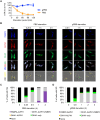
Fig 1
Fig 1. SGs are formed in both PBS and gPBS.
Cells stably expressing PABP2-HA, ALPH1-mScarlet and mNeonGreen-TY-DHH1 were starved in PBS and gPBS for 2 and 3 h, respectively. Cells were analysed for cellular ATP (A) and SG markers (B) at indicated times. The cells were classified based on the presence of SG and SG markers and the quantitation results are shown in (C) and (D). None, cells that did not contain detectable SGs; DHH1-only, cells containing granules marked by DHH1 only; PABP2-ALPH1, DHH1-ALPH1, and PABP2-ALPH1-DHH1, cells containing SGs marked by indicated markers. ALPH1-mScarlet and mNeonGreen-TY-DHH1 were visualized by direct mScarlet and mNeonGreen fluorescence. PABP2-HA was detected by immunofluorescence with anti-HA. Arrows mark the posterior puncta containing ALPH1 only. At least 200 cells were counted at each time point. Scale bar: 5 μm.
Fig 2
Fig 2. Cellular ATP modulates DHH1 dissociation from SGs.
A-B) Proportion of cells with PABP2-, ALPH1- or DHH1-containing SGs were plotted against cellular ATP, using the same data from experiments shown in Fig 1. C-D) Cells were starved in PBS for 30 mins followed by incubation in PBS or gPBS for an additional 90 mins. E-F) Cells expressing PABP2-mScarlet and mNeonGreen-DHH1 were starved in gPBS for 2 h followed by incubation in gPBS or PBS2DG for an additional 1 h. Cellular ATP was measured at specified time points for each starvation condition. Cellular ATP was measured at specified time points for each starvation condition (C) and (E). Percentage of cells containing specified SG profile was quantified at the end of the starvation schemes (D) and (F). None, cells that did not contain detectable SGs; DHH1-containing, cells containing SGs marked by DHH1; DHH1-free, cells containing SGs marked by PABP2 and ALPH1 only. ALPH1-mScarlet and mNeonGreen-TY-DHH1 were visualized by direct mScarlet and mNeonGreen fluorescence, respectively. At least 200 cells were counted at each time point.
Fig 3
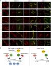
Fig 3. A subset of SG proteins dissociates from SGs in response to cellular ATP depletion.
A) Selected SG proteins were endogenously tagged with mScarlet and expressed in cells stably expressing mNeonGreen-TY-DHH1 or PABP2-TY-GFP. SG localization of the mScarlet-labelled proteins was monitored under specified stress conditions. B) Schematics showing components found in gPBS- or PBS-induced SGs 2 h post starvation. Scale bar: 5 μm.
Fig 4
Fig 4. gPBS, but not PBS, supports dynamic exchange of mRNPs between SGs and polysomes.
A) Experimental setups for co-starvation treatment to monitor mRNP recruitment to SGs during SG formation, and post-starvation treatment to monitor dynamic out-flow of SG materials. B-C) During co-starvation treatments, cells expressing PABP2-mScarlet and mNeonGreen-DHH1 were starved in PBS or gPBS for 2 h in the presence or absence of cycloheximide (50 μg/mL) or puromycin (200 μg/mL). D-E) During post-starvation treatments, cells expressing PABP2-mScarlet and mNeonGreen-DHH1 were starved in PBS or gPBS for 2 h, followed by the addition of cycloheximide or puromycin. Percentage of cells containing SGs was quantified in (C) and (E). Results shown are from 3 independent experiments. Scale bar: 5 μm F) Newly synthesized proteins were labelled with L-Homopropargylglycine (HPG), in cells cultivated in medium (fed), and starved in gPBS or PBS. HPG-labelled nascent proteins were fractionated by SDS-PAGE and visualized by click reaction with Alexa Fluor 488 azide. Equal loading was shown by Coomassie blue staining.
Fig 5
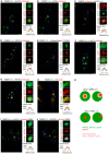
Fig 5. ExM reveals SG and PB components in different compartments.
Cells stably expressing PABP2 and other SG components fused to indicated reporter tags were starved with gPBS (A-H) or PBS (I, J) for 2 h and processed for ExM. The SG components were visualized using antibodies specific to the reporter tags using anti-HA, anti-TY, anti-RFP (for mScarlet), or oligo d(T) for poly A-mRNA. Cell outline is marked in blue. Individual granules are selected for zoomed display to the right of each panel. Scale bar in the zoomed images: 1 μm. Fluorescence intensity profiles of PABP2 and proteins of interest across the granules (white dotted lines) are shown at bottom right of each panel. K) Schematic presentation of the sub-organellar compartmentalization of SG and PB components in stress granules formed in gPBS and PBS (2 h), respectively.
Fig 6
Fig 6. Proline and glucose reduces SG formation in PBS.
Cells expressing PABP2-mScarlet and mNeonGreen-DHH1 were incubated for 2 h in PBS containing 1 g/L proline (proPBS) (A) or glucose at 1 g/L or 2.3 g/L (B). Cells were analysed for cellular ATP levels and presence of SG. Scale bar: 5 μm. At least 200 cells were counted for each starvation condition.

Similar articles

References

    1. Aulas A, Fay MM, Lyons SM, Achorn CA, Kedersha N, Anderson P, et al.. Stress-specific differences in assembly and composition of stress granules and related foci. Journal of Cell Science. 2017. Jan 1;jcs.199240. doi: 10.1242/jcs.199240 - DOI - PMC - PubMed
    1. Kedersha N, Anderson P. Stress granules: sites of mRNA triage that regulate mRNA stability and translatability. Biochemical Society Transactions. 2002. Nov 1;30(6):963–9. doi: 10.1042/bst0300963 - DOI - PubMed
    1. Mazroui R, Di Marco S, Kaufman RJ, Gallouzi IE. Inhibition of the Ubiquitin-Proteasome System Induces Stress Granule Formation. Matera AG, editor. MBoC. 2007. Jul;18(7):2603–18. doi: 10.1091/mbc.e06-12-1079 - DOI - PMC - PubMed
    1. Merrick WC. Cap-dependent and cap-independent translation in eukaryotic systems. Gene. 2004. May;332:1–11. doi: 10.1016/j.gene.2004.02.051 - DOI - PubMed
    1. Hofmann S, Kedersha N, Anderson P, Ivanov P. Molecular mechanisms of stress granule assembly and disassembly. Biochimica et Biophysica Acta (BBA)—Molecular Cell Research. 2021. Jan;1868(1):118876. doi: 10.1016/j.bbamcr.2020.118876 - DOI - PMC - PubMed

MeSH terms

Grants and funding

This work was supported by Singapore Ministry of Education (MOE-T2EP30221-0002 to CYH and A-0008403-00-00 to CYH). The funder had no role in study design, data collection and analysis, decision to publish, or preparation of the manuscript.

LinkOut - more resources