. 2024 Dec 11;15(12):e0202824.
doi: 10.1128/mbio.02028-24. Epub 2024 Oct 29.

Helicase protein DDX11 as a novel antiviral factor promoting RIG-I-MAVS-mediated signaling pathway

Affiliations

Helicase protein DDX11 as a novel antiviral factor promoting RIG-I-MAVS-mediated signaling pathway

Jiyu Zhang et al. mBio. .

Abstract

Type Ι interferon (IFN) production mediated by retinoic acid-inducible gene 1 (RIG-I) and mitochondrial antiviral signaling protein (MAVS) is essential for antiviral innate immune responses. Here, we report the identification of a novel co-sensor for cytosolic nucleic acids: DEAD/H-box helicase 11 (DDX11), a member of the DExD/H (Asp-Glu-x-Asp/His)-box helicase family. Knockdown or knockout of DDX11 attenuated the ability of cells to increase IFN-β, IFN-stimulated gene 56, and C-X-C motif chemokine ligand 10 in response to SeV and poly (I:C) by blocking the activation of TANK-binding kinase 1 and IFN regulatory factor 3. Nucleic acid sensing by DDX11 was independent of the stimulator of IFN genes but was dependent on RIG-I and MAVS. DDX11 regulated RIG-I-MAVS-mediated IFN signaling by specifically interacting with nucleic acid, RIG-I, and MAVS to enhance RIG-I-double-strand RNA and RIG-I-MAVS binding affinity. Overall, our results identified a critical role for DDX11 in the innate immune response and provided molecular insights into the mechanisms by which DDX11 recognized cytosolic nucleic acid and interacted with RIG-Ι and MAVS for potent IFN signaling and antiviral immunity.

Importance: Innate immunity is the first and most rapid host defense against virus infection. Recognition of viral RNA by the retinoic acid-inducible gene 1 (RIG-I)-like receptors (RLRs) initiates innate antiviral immune responses. How the binding of viral RNA to and activation of the RLRs are regulated remains enigmatic. In this study, we identified DEAD/H-box helicase 11 (DDX11) as a positive regulator of the RIG-I-mitochondrial antiviral signaling protein (MAVS)-mediated signaling pathways. Mechanistically, we demonstrated that DDX11 bound to viral RNA, interacted with RIG-I, and promoted their binding to viral RNA. DDX11 also promoted the interaction between RIG-I and MAVS and activation of RIG-I-MAVS signaling. Overall, our results elucidate the role of DDX11 in RIG-I-MAVS-dependent signaling pathways and may shed light on innate immune gene regulation.

Keywords: DDX11; IFN; MAVS; RIG-I.


Conflict of interest statement

The authors declare no conflict of interest.

Figures
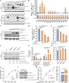
Fig 1
Fig 1
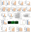
Screening for DExD/H-box helicases that regulate SADS-CoV infection. (A and B) IPI-2I cells were transfected with eukaryotic expression plasmids encoding various DExD/H-box helicases or empty vector for 24 h and then infected with SADS-CoV at a multiplicity of infection (MOI) of 0.1 for 24 h. (A) Protein expression of different DExD/H-box helicases detected by western blotting. (B) The mRNA levels of SADS-CoV N protein verified by qRT-PCR. (C–E) Overexpression of DDX11 inhibits SADS-CoV replication. IPI-2I cells were transfected with HA-tagged swine DDX11 at concentrations of 0.5, 1, and 2 µg or with a control empty vector for 24 h and then infected with SADS-CoV at an MOI of 0.1 for 24 h. (C) Protein expression levels of both DDX11 and SADS-CoV N analyzed by western blotting. Calculated band density values for SADS-CoV N/GAPDH; the values of the empty vector-transfected group are standardized to one. (D) The mRNA levels of the SADS-CoV N protein validated by qRT-PCR. (E) The SADS-CoV TCID50 in the supernatants was titrated on Vero E6 cells. (F–H) Knockdown of DDX11 enhances SADS-CoV replication. IPI-2I cells were transfected with siDDX11-1, siDDX11-2, siDDX11-3, or siNC (negative control) at 50 nM for 36 h and then infected with SADS-CoV at an MOI of 0.1 for 24 h. (F) Protein expression levels of both DDX11 and SADS-CoV N analyzed by western blotting. Calculated band density values for DDX11/GAPDH and SADS-CoV N/GAPDH; the values of the siNC group are standardized to one. (G) The mRNA levels of DDX11 and SADS-CoV N detected by qRT-PCR. (H) The SADS-CoV TCID50 in the supernatants was titrated on Vero E6 cells. (I–K) Knockout of DDX11 enhances SADS-CoV replication. WT HEK293T and DDX11−/− HEK293T cells were infected with SADS-CoV at an MOI of 1 for 24 h. (I) Cellular proliferation was detected in WT or DDX11−/− HEK293T cells, respectively, using CellTiter-Glo at the indicated time points. (J) Protein expression levels of both DDX11 and SADS-CoV N determined by western blotting. Calculated band density values for SADS-CoV N/GAPDH; the values of WT HEK293T cells group are standardized to one. (K) The viral RNA copy number was tested by qRT-PCR at 2, 12, 18, and 24 h post-infection. (L) The SADS-CoV TCID50 in the supernatants was titrated on Vero E6 cells. Means and SD (error bars) of three independent experiments are indicated (*P < 0.05; **P < 0.01; ***P < 0.001; ****P < 0.0001; ns, not significant).
Fig 2
Fig 2
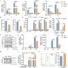
DDX11 positively regulates SeV and poly(I:C)-induced IFN response. (A and B) Overexpression of DDX11 potentiates the activity of the IFN-β promoter in response to stimulation by SeV and poly(I:C). HEK293T cells were transfected with empty vector or HA-DDX11, along with IFN-β-Luc and pRL-TK for 24 h. Cells were infected with SeV (A) or stimulated with poly(I:C) (B) for 12 h. Luciferase activity was quantified utilizing a dual luciferase reporter assay system. (C–E) Overexpression of DDX11 increases the transcription of IFN-β, ISG56, and CXCL10. HEK293T cells were transfected with HA-DDX11 for 24 h and then stimulated with SeV (C), poly(I:C) (D), or VSV-GFP (E) for 12 h. The transcription levels of IFN-β, ISG56, and CXCL10 were evaluated by qRT-PCR. (F and G) Overexpression of DDX24 inhibits the mRNA expression levels of IFN-β. HEK293T were transfected with empty vector, HA-DDX11, or HA-DDX24 for 24 h and then infected with SeV (F) or stimulated with poly(I:C) (G) for 12 h. The cells were harvested for qRT-PCR to detect the mRNA levels of IFN-β. (H) Overexpression of DDX11 increases the phosphorylation of IRF3 and TBK1 stimulated by SeV. HEK293T cells were transfected with empty vector or HA-DDX11 for 24 h and then stimulated with SeV for 12 h. The cell lysates were analyzed by western blotting. The band density values for p-IRF3/IRF3 and p-TBK1/TBK1 were calculated, with the values of the empty vector-treated group being standardized to one. (I–L) Overexpression of DDX11 inhibits VSV-GFP replication. HEK293T were transfected with empty vector or HA-DDX11 for 24 h and then infected with VSV-GFP for 12 h. (I) Protein expression levels of both DDX11 and GFP were detected by western blotting. The band density values for GFP/GAPDH were calculated, with the values of the empty vector-treated group being standardized to one. (J) The mRNA levels of VSV-GFP were verified by qRT-PCR. (K) The propagation of VSV-GFP was monitored and characterized by fluorescence microscopy (bar: 400 µm). (L) Fluorescence was analyzed by flow cytometry. (M–P) Exogenous addition of recombinant IFN-β increases mRNA levels of ISG56, but not DDX11. HEK293T cells were treated with recombinant IFN-β (100 ng/µL) for 0, 2, 4, 6, and 8 h. The mRNA levels of ISG56 (M) and DDX11 (O) were analyzed by qRT-PCR. HEK293T cells were treated with recombinant IFN-β (0, 10, 100, and 1,000 ng/µL) for 8 h, and the mRNA levels of ISG56 (N) and DDX11 (P) were analyzed by qRT-PCR. Means and SD (error bars) of three independent experiments are indicated (*P < 0.05; **P < 0.01; ***P < 0.001; ****P < 0.0001; ns, not significant).
Fig 3
Fig 3
DDX11 knockout reduces SeV- and poly(I:C)-induced IFN response. (A) Knockout of DDX11 and MAVS reduces the transcription of IFN-β after SeV or VSV-GFP infection and poly(I:C) transfection. WT HEK293T, DDX11−/− HEK293T, and MAVS−/− HEK293T cells were infected with SeV and VSV-GFP or stimulated with poly(I:C) for 4 and 8 h. The mRNA levels of IFN-β were analyzed by qRT-PCR. (B and C) Knockout of DDX11 diminishes the activity of the IFN-β promoter in response to SeV infection (B) and poly(I:C) (C) stimulation in HEK293T cells. Luciferase activity was quantified utilizing a dual luciferase reporter assay system. (D) Knockout of DDX11 and MAVS reduces IFN-β secretion after SeV infection. The IFN-β protein in the supernatants of SeV-infected WT HEK293T, DDX11−/− HEK293T, and MAVS−/− HEK293T cells was detected with the human IFN-β quantikine ELISA kit. (E) Overexpression of DDX11 in DDX11−/− HEK293T cells restores the inhibitory effect of knockout of DDX11 on IFN-β. WT HEK293T and DDX11−/− HEK293T cells were transfected with an empty vector or HA-DDX11 and then infected with SeV for 12 h. The mRNA levels of IFN-β were analyzed by qRT-PCR. (F) Knockout of DDX11 reduces phosphorylation of IRF3 and TBK1 stimulated by SeV. WT HEK293T and DDX11−/− HEK293T cells were stimulated with SeV for 12 h and then analyzed by western blotting. The band density values for p-IRF3/IRF3 and p-TBK1/TBK1 were calculated, with the values of SeV-infected WT HEK293T cell group being standardized to one. (G) Knockout of DDX11 inhibits the transcription of ISG56 and CXCL10 after SeV infection. WT HEK293T and DDX11−/− HEK293T cells were stimulated with SeV for 8 and 12 h, and the relative mRNA levels of ISG56 and CXCL10 were evaluated by qRT-PCR. (H–J) Knockout of DDX11 promotes VSV-GFP replication. WT HEK293T and DDX11−/− HEK293T cells were infected with VSV-GFP. (H) Protein expression levels of DDX11 and GFP were detected by western blotting. The band density values for GFP/GAPDH were calculated, with the values of VSV-GFP-infected WT HEK293T cell group being standardized to one. (I) VSV-GFP RNA transcripts were analyzed by qRT-PCR. (J) Fluorescence was analyzed by flow cytometry. Means and SD (error bars) of three independent experiments are indicated (*P < 0.05; **P < 0.01; ***P < 0.001; ****P < 0.0001; ns, not significant).
Fig 4
Fig 4
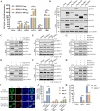
DDX11 targets RIG-I and MAVS. (A) Overexpression of DDX11 increases the IFN-β promoter activation mediated by RIG-I and MAVS. HEK293T cells were transfected with IFN-β-Luc (50 ng), pRL-TK (5 ng), and 100 ng of each plasmid expressing RIG-I, MDA5, MAVS, IRF3, or TBK1, along with an empty vector or a plasmid expressing DDX11 (0, 50, and 100 ng) for 24 h. Luciferase activity was quantified utilizing a dual luciferase reporter assay system. (B) Screening for key molecules in the RLRs signaling pathway that interact with DDX11. HEK293T cells were transfected with plasmids expressing RIG-I, MDA5, MAVS, IRF3, TBK1, or empty vector, together with HA-DDX11. Cellular extracts were harvested and subsequently subjected to immunoprecipitation using an anti-Flag antibody. The immunoprecipitants were analyzed by western blotting using relevant specific antibodies. (C) DDX11 interacts with RIG-I. HEK293T cells were co-transfected with Flag-RIG-I and HA-DDX11 for 24 h. Co-IP and immunoblot analyses of the interaction of DDX11 with RIG-I. (D) Co-IP and immunoblot analyses of the interaction of DDX11 with MAVS. (E and F) Endogenous DDX11 interacts with RIG-I and MAVS. HEK293T cells were infected with SeV for 12 h. The cells were harvested, and Co-IP was performed with anti-DDX11 antibody. IgG was used as a negative control. The immunoprecipitants were analyzed by western blotting using relevant specific antibodies. The band density values for interacting protein/IP protein in the IP samples are presented below the corresponding figure. (G and H) Co-IP and immunoblot analyses of the interaction of DDX11 with RIG-I (G) and MAVS (H) in the presence of RNase A (100 mg/mL). (I) Interaction of DDX11 with RIG-I and MAVS assessed by confocal microscopy (Bar: 5 µm). (J) WT HEK293T, MAVS−/− HEK293T, and RIG-I−/− HEK293T cells were transfected with HA-DDX11 and then infected with SeV for 12 h. The mRNA levels of IFN-β were analyzed by qRT-PCR. (K) WT HEK293T and DDX11−/− HEK293T cells were transfected with plasmid expressing RIG-I and MAVS or empty vector, together with IFN-β-Luc and pRL-TK for 24 h. Luciferase activity was quantified utilizing a dual luciferase reporter assay system. Means and SD (error bars) of three independent experiments were indicated (*P < 0.05; **P < 0.01; ***P < 0.001; ****P < 0.0001; ns, not significant).
Fig 5
Fig 5
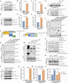
DDX11 acts as an RNA co-sensor to facilitate RIG-I recognition of viral RNA. (A–C) Exogenously expressed DDX11 binds to VSV-GFP-RNA and SADS-CoV-RNA. HEK293T cells were transfected with HA-DDX11 for 24 h and then infected with VSV-GFP for 12 h or SADS-CoV for 24 h. The cells were lysed, and immunoprecipitation was performed with anti-HA or anti-IgG antibody. (A) Detection of the immunoprecipitants by western blotting with the indicated antibodies. (B and C) The immunoprecipitants were analyzed by qRT-PCR to detect bound VSV-GFP-RNA (B) and SADS-CoV-RNA (C). (D–F) Endogenous DDX11 binds to VSV-GFP-RNA and SADS-CoV-RNA. HEK293T cells were infected with VSV-GFP for 12 h or SADS-CoV for 24 h. The cells were lysed, and immunoprecipitation was performed with anti-DDX11 or anti-IgG antibody. (D) The immunoprecipitants were detected by western blotting with indicated antibodies. (E and F) The immunoprecipitants were analyzed by qRT-PCR to detect bound VSV-GFP-RNA (E) and SADS-CoV-RNA (F). (G) DDX11 binds both the 5′ UTR and 3′ UTR transcripts of SADS-CoV. The 5′-UTR or 3′-UTR transcripts of SADS-CoV were labeled and then captured with streptavidin beads. The labeled RNA-bound beads were incubated with HEK293T cell lysates overexpressing HA-DDX11. The bound proteins were analyzed by western blotting with anti-HA antibody. (H) DDX11 binds to poly(I:C). HEK293T cells were transfected with HA-DDX11 for 24 h. The cell lysates were incubated with biotin-labeled poly(I:C) for 4 h at 4°C, followed by the addition of NeutrAvidin beads for 4 h. The proteins bound to the beads were analyzed by western blotting. (I) Amino acids 445–665 of DDX11 are required for the binding of DDX11 to poly(I:C). HEK293T cells were transfected with full-length HA-DDX11 or truncation of DDX11 for 24 h. Cell lysates were incubated with biotin-labeled poly(I:C) for 4 h at 4°C, followed by the addition of NeutrAvidin beads for 4 h. The proteins bound to beads were analyzed by western blotting. (J) Schematics of full-length DDX11 and its serial truncated mutants. (K) Identification of the domain of DDX11 required for the interaction with RIG-I. HEK293T cells were transfected with Flag-RIG-I and DDX11 truncations. Cell lysates were collected for Co-IP and immunoblot analyses. (L) Schematics of full-length RIG-I and its serial truncated mutants. (M) Identification of the domain of RIG-I required for the interaction with DDX11. (N) Overexpression of DDX11 increases RIG-I recognition of poly(I:C). HEK293T cells were transfected with Flag-RIG-I, together with HA-DDX11 or empty vector. Cell lysates were incubated with biotin-labeled poly(I:C) for 4 h. Immunoprecipitation was performed with NeutrAvidin beads, followed by immunoblotting with anti-HA or anti-Flag antibody. Relative quantification of the IP-RIG-I/Input-RIG-I is shown below. (O) Knockout of DDX11 decreases RIG-I recognition of poly(I:C). WT HEK293T cells and DDX11−/− HEK293T cells were transfected with Flag-RIG-I for 24 h. Cell lysates were incubated with biotin-labeled poly(I:C) for 4 h. Immunoprecipitation was performed with NeutrAvidin beads, followed by immunoblotting with anti-Flag antibody. Relative quantification of the IP-RIG-I/Input-RIG-I is shown below. (P and Q) RIG-I binds to SADS-CoV-RNA. HEK293T cells were transfected with Flag-RIG-I for 24 h and then infected with SADS-CoV for 24 h. The cells were lysed, and immunoprecipitation was performed with an anti-Flag or anti-IgG antibody. (P) Detection of the immunoprecipitants by western blotting with the indicated antibodies. (Q) The immunoprecipitants were analyzed by qRT-PCR to detect bound SADS-CoV-RNA. (R) Overexpression of DDX11 increases RIG-I recognition of SADS-CoV-RNA. HEK293T cells were transfected with HA-DDX11 or empty vector for 24 h and then infected with SADS-CoV for 24 h. Immunoprecipitation was performed with anti-RIG-I antibody. The immunoprecipitants were analyzed by qRT-PCR to detect bound SADS-CoV-RNA. (S) Knockout of DDX11 decreases RIG-I recognition of SADS-CoV-RNA. WT HEK293T and DDX11−/− HEK293T cells were infected with SADS-CoV for 24 h. The cells were lysed, and immunoprecipitation was performed with anti-RIG-I or anti-IgG antibody. The immunoprecipitants were analyzed by qRT-PCR to detect bound SADS-CoV-RNA. The mean and SD (error bars) of three independent experiments are indicated (*P < 0.05; **P < 0.01; ***P < 0.001; ****P < 0.0001; ns, not significant).
Fig 6
Fig 6
DDX11 promotes RIG-I and MAVS interaction. (A) Schematics of full-length DDX11 and its serial-truncated mutants. (B) Identification of the domain of DDX11 required for interaction with MAVS. HEK293T cells were transfected with Flag-MAVS and serial-truncated DDX11. Cell lysates were collected for Co-IP and immunoblot analyses. (C) Schematics of full-length MAVS and its serial-truncated mutants. (D) Identification of the domain of MAVS required for interaction with DDX11. (E) The protein levels of DDX11 increase in mitochondria after SeV infection. HEK293T cells were infected SeV for 12 h, and cells were collected for mitochondrial and cytoplasmic extraction. Proteins obtained from the mitochondrial and cytoplasmic fractions were analyzed by western blotting with the indicated antibodies. Calculated band density values for DDX11/GAPDH and DDX11/COX IV; the values of the control group are standardized to one. (F) Localization of DDX11 and mitochondria using confocal microscopy in WT HEK293T and MAVS−/− HEK293T cells. WT HEK293T and MAVS−/− HEK293T cells were transfected with HA-DDX11 for 24 h and then infected with SeV. The mitochondria are colored red and DDX11 protein is colored green. The position of the nucleus was visualized by staining with DAPI (blue); merged images are also presented (Bar: 2 µm). Co-localization of DDX11 and mitochondria was performed using the Pearson correlation coefficient. (G) WT HEK293T and MAVS−/− HEK293T cells were infected with SeV. The cells were collected for mitochondrial and cytoplasmic extraction. Protein from these fractions was analyzed by western blotting with the indicated antibodies. Calculated band densities for DDX11/GAPDH and DDX11/COX IV; the values of the control group are standardized to one. (H and I) DDX11 enhances the interaction of RIG-I with MAVS. (H) Co-IP and western blotting analyses of HEK293T cells transfected with Flag-RIG-I, HA-MAVS, and Myc-DDX11 or empty vector are shown. The band density values for interacting protein/IP protein in the IP samples are presented below the corresponding figure. (I) WT HEK293T and DDX11−/− HEK293T cells were transfected with Flag-RIG-I and Myc-MAVS and then infected with SeV for 12 h. The interaction of RIG-I with MAVS was analyzed by Co-IP and western blotting. The band density values for interacting protein/IP protein in the IP samples are presented below the corresponding figure.
Fig 7
Fig 7
Amino acids 445–665 of DDX11 inhibit RNA virus replication. (A) Screening for key domains of DDX11 that inhibit SADS-CoV replication. HEK293T cells were transfected with an empty vector or plasmids expressing the DDX11 truncations for 24 h and then infected with SADS-CoV for 24 h. Cellular RNA was extracted and analyzed by qRT-PCR to detect mRNA levels of SADS-CoV N. (B and C) Amino acids 445–665 of DDX11 increase the transcription of IFN-β after SeV infection and poly(I:C) transfection in HEK293T cells. The transcription levels of IFN-β in HEK293T cells that transfected with empty vector or DDX11 (aa 1–945), DDX11 (aa 445–665), or DDX11 (aa Δ445–665) expression plasmids and stimulated with SeV (B) or poly(I:C) (C) were evaluated by qRT-PCR. (D) Amino acids 445–665 of DDX11 increase the SeV-mediated IFN-β promoter activity in HEK293T cells. HEK293T cells were transfected with IFN-β-Luc, pRL-TK, and empty vector or DDX11 (aa 1–945), DDX11 (aa 445–665), or DDX11 (aa Δ445–665) expression plasmids for 24 h. Cells were infected with SeV for 12 h, and luciferase activity was quantified utilizing a dual luciferase reporter assay system. (E) Amino acids 445–665 of DDX11 increase the activation of IFN-β luciferase reporters mediated by RIG-I. HEK293T cells were co-transfected with Flag-RIG-I, IFN-β-Luc, pRL-TK plasmid, and empty vector or DDX11 (aa 1–945), DDX11 (aa 445–665), or DDX11 (aa Δ445–665) expression plasmids for 24 h. Luciferase activity was quantified utilizing a dual luciferase reporter assay system. (F and G) Amino acids 445–665 of DDX11 inhibit VSV-GFP replication. HEK293T cells were transfected with plasmids expressing DDX11 (aa 1–945), DDX11 (aa 445–665), or DDX11 (aa Δ445–665) for 24 h and then infected with VSV-GFP for 12 h. (F) Fluorescence was analyzed by flow cytometry. (G) Protein expression of full-length DDX11, DDX11 truncations, and GFP detected by western blotting. Calculated band densities for GFP/GAPDH; the values of the empty vector-transfected group are standardized to one. (H–M) Amino acids 445–665 of DDX11 inhibit SADS-CoV replication. HEK293T or IPI-2I cells were transfected with plasmids expressing DDX11 (aa 1–945), DDX11 (aa 445–665), or DDX11 (aa Δ445–665) for 24 h and then infected with SADS-CoV for 24 h. (H and K) Protein expression of full-length DDX11, DDX11 truncations, and SADS-CoV N detected by western blotting. Calculated band densities for SADS-CoV N/GAPDH; the values of the empty vector-transfected group are standardized to one. (I and L) qRT-PCR assessed transcriptional levels of SADS-CoV N protein. (J and M) The SADS-CoV TCID50 in the supernatants was titrated on Vero E6 cells. Means and SD (error bars) of three independent experiments are indicated (*P < 0.05; **P < 0.01; ***P < 0.001; ****P < 0.0001; ns, not significant).
Fig 8
Fig 8
Schematic depiction of the proposed role of DDX11 in antiviral immunity against RNA virus infection. Upon SADS-CoV or VSV-GFP infection, DDX11 functions as an RNA co-sensor to facilitate RIG-I recognition of viral RNA and subsequently relies on MAVS localization in mitochondria to increase RIG-I-MAVS interactions. This activation cascade induces the phosphorylation of TBK1 and IRF3, which subsequently promotes their nuclear translocation and induces IFN production.

Similar articles

References

    1. Akira S, Uematsu S, Takeuchi O. 2006. Pathogen recognition and innate immunity. Cell 124:783–801. doi:10.1016/j.cell.2006.02.015 - DOI - PubMed
    1. Gürtler C, Bowie AG. 2013. Innate immune detection of microbial nucleic acids. Trends Microbiol 21:413–420. doi:10.1016/j.tim.2013.04.004 - DOI - PMC - PubMed
    1. Hiscott J. 2007. Convergence of the NF-kappaB and IRF pathways in the regulation of the innate antiviral response. Cytokine Growth Factor Rev 18:483–490. doi:10.1016/j.cytogfr.2007.06.002 - DOI - PubMed
    1. Medzhitov R. 2007. Recognition of microorganisms and activation of the immune response. Nature 449:819–826. doi:10.1038/nature06246 - DOI - PubMed
    1. Kato H, Takeuchi O, Sato S, Yoneyama M, Yamamoto M, Matsui K, Uematsu S, Jung A, Kawai T, Ishii KJ, Yamaguchi O, Otsu K, Tsujimura T, Koh C-S, Reis e Sousa C, Matsuura Y, Fujita T, Akira S. 2006. Differential roles of MDA5 and RIG-I helicases in the recognition of RNA viruses. Nature 441:101–105. doi:10.1038/nature04734 - DOI - PubMed

MeSH terms

LinkOut - more resources