. 2024 Oct 28;25(1):388.
doi: 10.1186/s12931-024-03012-9.

GSK3179106 ameliorates lipopolysaccharide-induced inflammation and acute lung injury by targeting P38 MAPK

Affiliations

GSK3179106 ameliorates lipopolysaccharide-induced inflammation and acute lung injury by targeting P38 MAPK

Bin Zheng et al. Respir Res. .

Abstract

Acute lung injury (ALI) is a serious acute respiratory disease that can cause alveolar-capillary barrier disruption and pulmonary edema, respiratory failure and multiple organ dysfunction syndrome. However, there is no effective drugs in clinic until now. GSK3179106 has been reported can alleviate intestinal stress syndrome, but the protective effect of GSK3179106 on ALI has not been elucidated. The present study will evaluate the pharmacological activity of GSK3179106 on lipopolysaccharide (LPS)-induced inflammation and lung injury and clarify its underlying mechanism. We found that GSK3179106 significantly attenuated LPS-induced lung injury in vivo, accompanied by inhibited infiltration of inflammatory cells and reduced expression of inflammatory cytokines. Meanwhile, GSK3179106 dose-dependently reduced the LPS-induced IL-6 expression both in protein and gene levels in macrophages. Mechanistically, GSK3179106 could inhibited the phosphorylation of P38 MAPK induced by LPS. Importantly, results showed that there is a direct combination between GSK3179106 and P38 MAPK. Together, our findings not only clarified the anti-inflammatory activity of GSK3179106 but also discovered its new clinical indications. Therefore, compound GSK3179106 may be a potential candidate for the treatment of acute inflammatory diseases.

Keywords: Acute lung injury; GSK3179106; Lipopolysaccharide; P38 MAPK kinase; Sepsis.


Conflict of interest statement

The authors declare no competing interests.

Figures
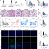
Fig. 1
Fig. 1
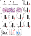
GSK3179106 attenuates septic lung injury in mice. C57BL/6 mice received GSK3179106 (3 and 6 mg/kg) intraperitoneally at 12 h and 1 h prior to LPS (10 mg/kg) administration via the tail vein. The animals were subsequently euthanized under anesthesia 12 h after LPS injection, and lung tissue specimens were harvested. Schematic diagram of the sepsis models induced by LPS. A Lung injury score as assessed by histological analysis of lung tissues. B Representative H&E staining of the mouse lung. The blue arrow highlights the thickening of the alveolar wall. C Total protein concentration in BALF. D Total cells in BALF. E Representative plots of CD11b+LY6G+ cells as a percentage of the total CD45+ cell population. F Statistics of CD11b + LY6G + cells. G Representative LY6G staining images of the septic lung injury. H Statistical plot of MFI. I MPO activity in lung tissue. Data were reported as mean ± SEM. *P < 0.05, **P < 0.01, ***P < 0.001 compared with LPS group; #P < 0.05, ##P < 0.01, ###P < 0.001, ns: no significance compared with CON group n ≥ 5
Fig. 2
Fig. 2
GSK3179106 inhibits sepsis-induced inflammation in mouse lung tissue. C57BL/6 mice received GSK3179106 (3 and 6 mg/kg) intraperitoneally at 12 h and 1 h prior to LPS (10 mg/kg) administration via the tail vein. The animals were subsequently euthanized under anesthesia 12 h after LPS injection, and lung tissue specimens were harvested. AC IL-6 and TNF-α in BALF and IL-6 in serum from the experimental mice were determined with ELISA. D-F The mRNA levels of inflammatory cytokines IL-6, TNF-α, IL-1β in the lung tissues were determined by RT-qPCR after LPS treatment. Cytokine gene expression was normalized to β-actin. Data were reported as mean ± SEM. *P < 0.05, **P < 0.01, ***P < 0.001 compared with LPS group; ##P < 0.01, ###P < 0.001 compared with CON group n ≥ 5
Fig. 3
Fig. 3
GSK3179106 alleviates LPS-induced ALI. C57BL/6 mice were administered GSK3179106 (3 mg/kg) intraperitoneally 6 h prior to an intratracheal injection of LPS (5 mg/kg). The animals were subsequently euthanized under anesthesia 6 h after LPS administration, and lung tissue specimens were collected. Schematic diagram of the ALI models induced by LPS. A Total cells in BALF. B Representative H&E staining images of the mouse lung. The blue arrow highlights the thickening of the alveolar wall. C Lung injury score as assessed by histological analysis of lung tissues. E–H IL-6 and TNF-α in BALF and serum from the experimental mice were determined with ELISA. IJ The mRNA levels of inflammatory cytokines IL-6 and TNF-α in the lung tissues were determined by RT-qPCR after LPS treatment. Cytokine gene expression was normalized to β-actin. K Statistics of CD11b+LY6G+ cells. L MPO activity in lung tissue. M Representative plots of CD11b+LY6G+ cells as a percentage of the total CD45+ cell population and corresponding quantification results in all cells. Data were reported as mean ± SEM. *P < 0.05, **P < 0.01, ***P < 0.001 compared with LPS group; ##P < 0.01, ###P < 0.001 compared with CON group n ≥ 5
Fig. 4
Fig. 4
Effect of GSK3179106 on LPS-inflammation in macrophages. A After RAW264.7 cells were treated with various concentrations of GSK3179106 for 24 h, the cell viability was analyzed via MTT assay. B After RAW264.7 cells were pre-treated with various concentrations of GSK3179106 (0.625, 1.25, 2.5, 5 μM) for 30 min and the supernatant was collected and analyzed for IL-6 after LPS (0.5 μg/mL) stimulation for 24 h by ELISA. DMSO was used as vehicle control. Data were normalized to total protein concentration from the same plate. C For the detection of mRNA levels, RAW264.7 cells were pre-treated with vehicle control (DMSO), various concentrations of GSK3179106 (0.625, 1.25, 2.5, 5 μM) for 30 min followed by incubation with LPS (0.5 μg/mL) for 6 h. The mRNA levels of inflammatory cytokines IL-6 were determined by RT-qPCR and normalized by β-actin mRNA level. D After J774A.1 cells were treated with various concentrations of GSK3179106 for 24 h, the cell viability was analyzed via MTT assay. E After J774A.1 cells were pre-treated with various concentrations of GSK3179106 (0.625, 1.25, 2.5, 5 μM) for 30 min and the supernatant was collected and analyzed for IL-6 after LPS (0.5 μg/mL) stimulation for 24 h by ELISA. DMSO was used as vehicle control. Data were normalized to total protein concentration from the same plate. F For the detection of mRNA levels, J774A.1 cells were pre-treated with vehicle control (DMSO), various concentrations of GSK3179106 (0.625, 1.25, 2.5, 5 μM) for 30 min followed by incubation with LPS (0.5 μg/mL) for 6 h. The mRNA levels of inflammatory cytokines IL-6 were determined by RT-qPCR and normalized by β-actin mRNA level. Data were mean ± SEM of 3 independent experiments. *P < 0.05, **P < 0.01, ***P < 0.001 compared with LPS group; ##P < 0.01, ###P < 0.001 compared with CON
Fig. 5
Fig. 5
GSK3179106 inhibits the transduction of macrophage P38/STAT3 pathway. A RAW264.7 cells were pretreated with different concentrations of GSK3179106 (0.625, 1.25, 2.5, 5 μM) for 30 min followed by incubation with LPS (0.5 μg/mL) for 15 min. The levels of p-P38 were measured by Western blot. The corresponding total protein was the loading control. B J774A.1 cells were pretreated with different concentrations of GSK3179106 (0.625, 1.25, 2.5, 5 μM) for 30 min followed by incubation with LPS (0.5 μg/mL) for 15 min. The levels of p-P38 were measured by Western blot. The corresponding total protein was the loading control. C RAW264.7 cells were stimulated by LPS (0.5 μg/mL) for 5 h. Total protein was extracted and incubated with GSK (5 μM) 4℃ overnight. Then add Pronase (25 ng/μg total protein), that is, add 400 ng Pronase into 16ug total cell protein, incubate at room temperature for 5 min. The protein level of P38 was detected by western blot. The Tubulin was the loading control. D Molecular dockings of GSK3179106 with P38 (6OHD) were analyzed with the Schrödinger software. E RAW264.7 cells were treated with GSK3179106 (5 Μm) for 6 h, and total protein extracted was heated at 44, 48, 52, 56, 60 and 64 ℃ for 5 min, respectively, and placed at room temperature for 3 min. The protein level of P38 was detected by western blot. The Tubulin was the loading control. FG 10–20 mg of lung tissue was weighed in 1.5 mL EP tubes, 500 μL of RIPA was added to each tube, homogenized, and total protein was extracted. The levels of p-P38 were measured by Western blot. The corresponding total protein was the loading control. Data were mean ± SEM of 3 independent experiments. *P < 0.05, **P < 0.01, ***P < 0.001 compared with LPS group; #P < 0.05, ###P < 0.001 compared with CON

Similar articles

References

    1. Liu C, Xiao K, Xie L. Advances in the use of exosomes for the treatment of ALI/ARDS. Front Immunol. 2022;13: 971189. - PMC - PubMed
    1. Kumar V. Pulmonary innate immune response determines the outcome of inflammation during pneumonia and sepsis-associated acute lung injury. Front Immunol. 2020;11:1722. - PMC - PubMed
    1. Dellinger RP, et al. Effects of inhaled nitric oxide in patients with acute respiratory distress syndrome: results of a randomized phase II trial. Inhaled nitric oxide in ARDS study group. Critic Care Med. 1998;26(1):15–23. - PubMed
    1. Luh SP, Chiang CH. Acute lung injury/acute respiratory distress syndrome (ALI/ARDS): the mechanism, present strategies and future perspectives of therapies. J Zhejiang Univ Sci B. 2007;8(1):60–9. - PMC - PubMed
    1. Liu C, Xiao K, Xie L. Progress in preclinical studies of macrophage autophagy in the regulation of ALI/ARDS. Front Immunol. 2022;13: 922702. - PMC - PubMed

MeSH terms

Substances

LinkOut - more resources