. 2024 Sep 17;10(10):1871-1884.
doi: 10.1021/acscentsci.4c00722. eCollection 2024 Oct 23.

Engineering a SARS-CoV-2 Vaccine Targeting the Receptor-Binding Domain Cryptic-Face via Immunofocusing

Affiliations

Engineering a SARS-CoV-2 Vaccine Targeting the Receptor-Binding Domain Cryptic-Face via Immunofocusing

Theodora U J Bruun et al. ACS Cent Sci. .

Abstract

The receptor-binding domain (RBD) of the SARS-CoV-2 spike protein is the main target of neutralizing antibodies. Although they are infrequently elicited during infection or vaccination, antibodies that bind to the conformation-specific cryptic face of the RBD display remarkable breadth of binding and neutralization across Sarbecoviruses. Here, we employed the immunofocusing technique PMD (protect, modify, deprotect) to create RBD immunogens (PMD-RBD) specifically designed to focus the antibody response toward the cryptic-face epitope recognized by the broadly neutralizing antibody S2X259. Immunization with PMD-RBD antigens induced robust binding titers and broad neutralizing activity against homologous and heterologous Sarbecovirus strains. A serum-depletion assay provided direct evidence that PMD successfully skewed the polyclonal antibody response toward the cryptic face of the RBD. Our work demonstrates the ability of PMD to overcome immunodominance and refocus humoral immunity, with implications for the development of broader and more resilient vaccines against current and emerging viruses with pandemic potential.


Conflict of interest statement

The authors declare the following competing financial interest(s): P.A.-B.W. and P.S.K. are named as inventors on patent applications applied for by Stanford University and the Chan Zuckerberg Biohub on epitope restriction for antibody selection.

Figures

Figure 1
Figure 1
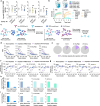
Engineering the RBD of SARS-CoV-2 for PMD. (a) Structure of SARS-CoV-2 RBD (PDB ID:7M7W) showing the binding footprint of one representative antibody from each of the four classes of RBD-binding antibodies; class 1 (Ab CB6, orange), class 2 (Ab C002, light pink), class 3 (S309, yellow), and class 4 (S2X259, blue). (b) BLI competition binding assay of RBD-directed antibodies binding to RBD. Loaded antibodies are displayed in rows, and competing antibodies are displayed in columns. White color indicates no binding of the tested antibody, indicating that the antibodies compete for binding (all competing antibodies are enclosed within dotted lines; unique competition groups are enclosed within solid lines). (c) Screening and identification of permissive lysine installations on RBD. In addition to 10 natural lysines (pink), 11 single lysine substitutions (green) were introduced by site-directed mutagenesis and all 12 proteins were transiently expressed. (d) Expression of lysine-substituted RBDs in Expi-293F cells quantified by dot-blot and normalized to the expression level of wild-type RBD (RWT). Data are presented as mean ± standard deviation (n = 2 biological replicates). Lysines with >40% expression compared to RWT are colored purple in (c). (e) Thermal melting profile of RWT and R7K (RWT with additional R346K, V367K, N440K, R466K, T478K, F490K, L518K substitutions) measured by differential scanning fluorimetry. (f) Expression level of single lysine substitutions cloned into the R7K protein. Expression level normalized to that of R7K. Data are presented as mean ± standard deviation (n = 2). (g) Thermal melting profile of RWT, R11K (R7K plus L452K, V483K, Q498K, and A522K), and R12K (R7K plus N334K, L452K, V483K, Q498K, and A522K), measured by differential scanning fluorimetry. R11K and R12K also contain the repackaging substitutions Y365F, F392W, and V395I to improve stability. (h) BLI binding curves of RWT and R11K with the fragment antigen-binding (Fab) of S2X259. Association was monitored for 5 min (dotted lines), after which dissociation was monitored for 5 min.
Figure 2
Figure 2
Using PMD to create immunofocused antigens. (a) Schematic depicting the PMD strategy. First, RBD is bound to S2X259-conjugated resin (protect). Then, the surface of the protein-mAb complex is rendered non-immunogenic through chemical conjugation (modify). In the last step, the S2X259 epitope is exposed (deprotected) by dissociating it from the mAb resin. (b) Illustration of the modification of two surface exposed lysines on R11K with NHS-PEG4 as in PMD (top) or the cross-linking of two surface exposed lysines with BIS-NHS-PEG5 as in PMD-CL (bottom). (c) SDS-PAGE analysis of the four antigens after expression and purification. (d) Normalized size-exclusion chromatographic traces of purified proteins. (e) Thermal melting temperature and melting temperature onset for RWT, R11K, R11K-PMD, and R11K-CL measured by differential scanning fluorimetry. Data are presented as mean ± standard deviation (n = 3 replicates). (f) BLI binding curves of RBD-directed antibodies from all four classes and ACE2 (expressed as a dimer due to fusion to a human Fc domain) to RWT, R11K, R11K-PMD, and R11K-CL. Association was monitored for 2 min (dotted lines), after which dissociation was monitored for 1 min. (g) BLI association amplitude of each antibody binding to the four antigens (corresponding to curves shown in (f)). (h) Epitope footprints of S2X259-like class 4 antibodies shown in color on the light blue SARS-CoV-2 RBD structure (PDB ID: 7M7W). (i) BLI binding curves of S2X259-like antibodies shown in (h) binding to RWT, R11K, R11K-PMD, and R11K-CL.
Figure 3
Figure 3
Immunogenicity of wild-type RBD and immunofocused RBD antigens. (a) Schematic of the mouse immunization with a four-dose regimen occurring on days 0, 35, 70, and 105. Mice were immunized with 5 μg of antigen adjuvanted with alum/CpG (500 μg/20 μg) via intramuscular injection (n = 5 per group). (b) Serum IgG titers on day 77 against biotinylated-RBDs from SARS-CoV-2 as well as more distantly related Sarbecoviruses including bio-BtKY72, bio-WIV-1, bio-SARS-CoV-1, and bio-BM-4831. (c) Neutralization titers (NT50 – the serum dilution required to neutralize 50% of a given pseudotyped lentivirus) of day 112 serum against wild-type SARS-CoV-2 (D614G), the Beta and Gamma variants of SARS-CoV-2, and more distant SARS-CoV-1, bat SARS-like coronavirus WIV-1, and pangolin coronavirus PCoV_GD. Data are presented as geometric mean ± s.d. of the log-transformed values. Each circle represents a single mouse. Horizontal dotted lines indicate the limit of quantitation. Comparisons of two groups were performed using the two-tailed Mann–Whitney U test. P values of 0.05 or less were considered significant and indicated.
Figure 4
Figure 4
Quantifying the proportion of cryptic-face targeting antibodies elicited by immunization. (a) Serum IgG binding titers of day-77 antisera to SARS-CoV-2 RBD in the presence of competing mAbs. Data are presented as geometric mean ± s.d. of the log-transformed values. Fold-change is indicated by arrows with numbers. Horizontal dotted lines indicate the limit of quantitation. (b) Substitutions in the S2X259 epitope of SARS-CoV-2 RBD (PDB ID: 7M7W) to create R-KO4. Binding of class 4 and class 1/2/3 antibodies to RBD and R-KO4 measured by BLI. Association was monitored for 2 min (dotted lines), after which dissociation was monitored for 1 min. (c) Schematic depicting the serum-depletion assay used to quantify the S2X259-like antibodies elicited by immunization. First, pooled antisera from immunized mice are mixed with neutravidin beads bound to either biotinylated RBD or biotinylated R-KO4. After incubation for 30 min, antibody-bound beads are filtered out, leaving antisera containing no RBD-directed antibodies or only S2X259-like antibodies unable to bind to R-KO4. (d) ELISA was used to measure binding of pooled antisera to SARS-CoV-2 RBD after depletion with either unbound beads (mock depleted serum), beads coupled to RBD (RBD-depleted serum), or beads coupled to R-KO4 (R-KO4-depleted serum). (e) Percentage of binding retained after depletion with R-KO4, calculated by taking the EC50 (serum dilution at which half-maximal binding is achieved) of R-KO4-depleted antisera over the EC50 of mock-depleted antisera for each immunization group, calculated from the ELISA curves in (d). (f, g) Neutralization of either wild-type SARS-CoV-2 (D614G) (f) or B.1.351 (Beta) (g) pseudotyped lentivirus by mock-depleted, RBD-depleted, or R-KO4-depleted serum. (h, i) Normalized NT50 for mock-depleted, RBD-depleted, or R-KO4-depleted antisera of each immunization group against SARS-CoV-2 (D614G) (h) or B.1.351 (Beta) (g) based on the data in (f, g).

Update of

Similar articles

References

    1. Kalinke U.; et al. Clinical development and approval of COVID-19 vaccines. Expert Rev. Vaccines 2022, 21, 609–619. 10.1080/14760584.2022.2042257. - DOI - PMC - PubMed
    1. Walsh E. E.; et al. Safety and Immunogenicity of Two RNA-Based Covid-19 Vaccine Candidates. N. Engl. J. Med. 2020, 383, 2439–2450. 10.1056/NEJMoa2027906. - DOI - PMC - PubMed
    1. Polack F. P.; et al. Safety and Efficacy of the BNT162b2 mRNA Covid-19 Vaccine. N. Engl. J. Med. 2020, 383, 2603–2615. 10.1056/NEJMoa2034577. - DOI - PMC - PubMed
    1. Cele S.; et al. Omicron extensively but incompletely escapes Pfizer BNT162b2 neutralization. Nature 2022, 602, 654–656. 10.1038/s41586-021-04387-1. - DOI - PMC - PubMed
    1. Huang C. Q.; Vishwanath S.; Carnell G. W.; Chan A. C. Y.; Heeney J. L. Immune imprinting and next-generation coronavirus vaccines. Nat. Microbiol. 2023, 8, 1971–1985. 10.1038/s41564-023-01505-9. - DOI - PubMed

LinkOut - more resources