. 2024 Oct 17;16(10):1625.
doi: 10.3390/v16101625.

Differences in Susceptibility to SARS-CoV-2 Infection Among Transgenic hACE2-Hamster Founder Lines

Affiliations

Differences in Susceptibility to SARS-CoV-2 Infection Among Transgenic hACE2-Hamster Founder Lines

Scott A Gibson et al. Viruses. .

Abstract

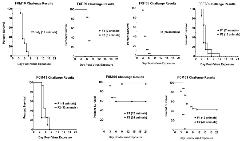
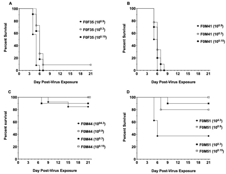
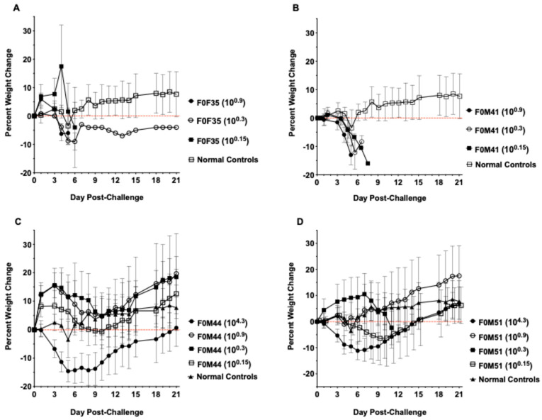
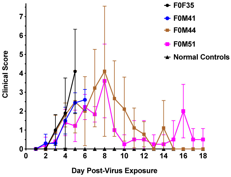
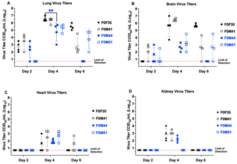
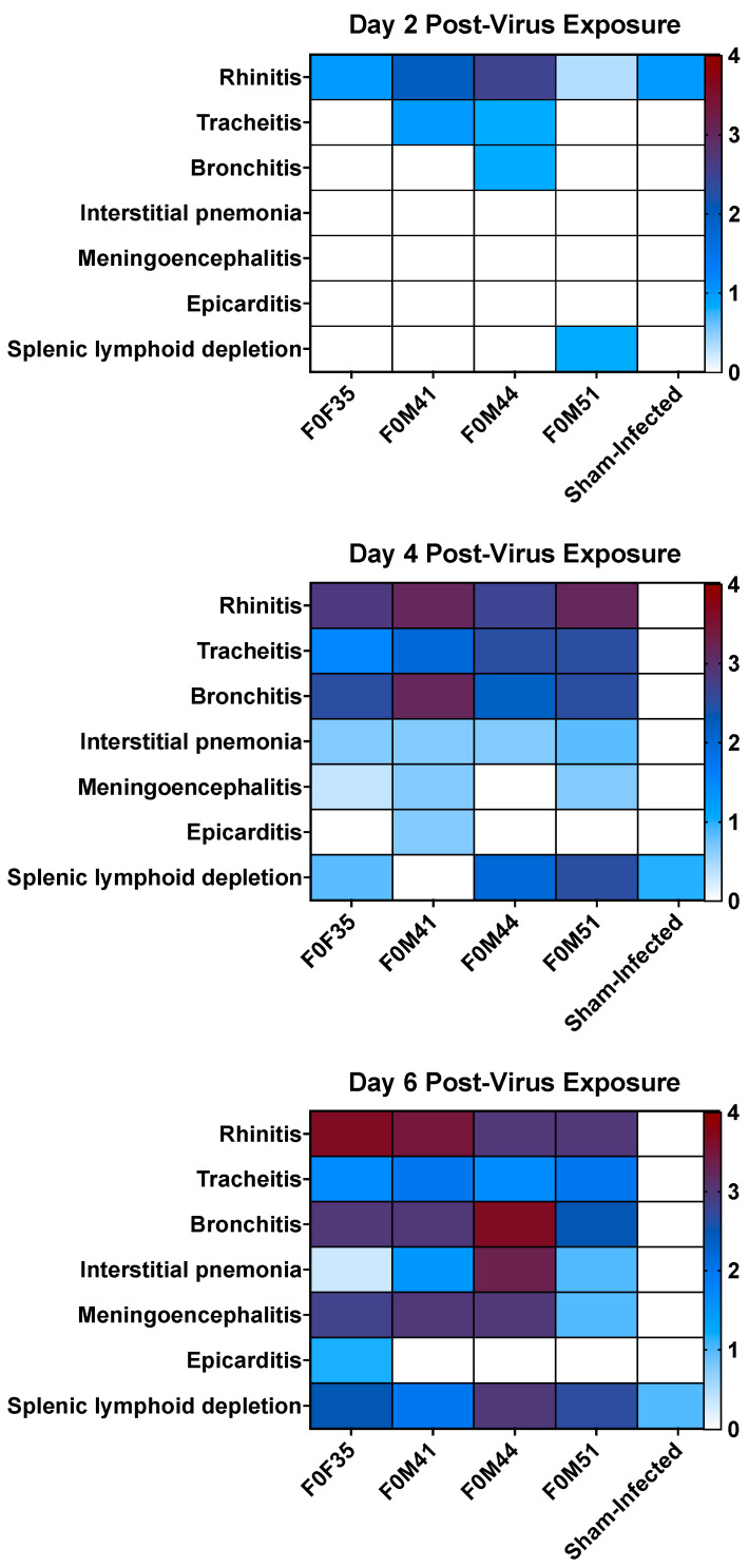
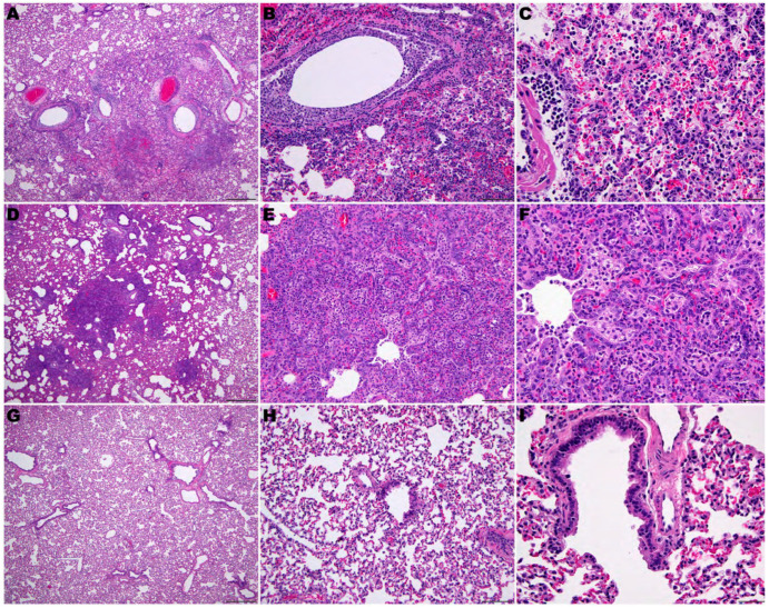
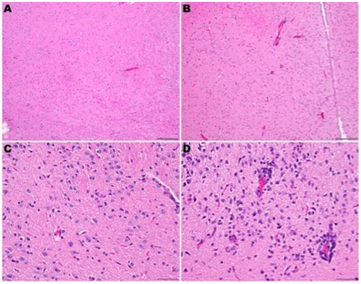
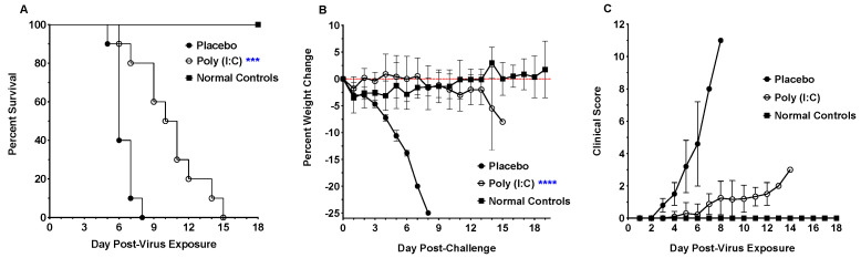
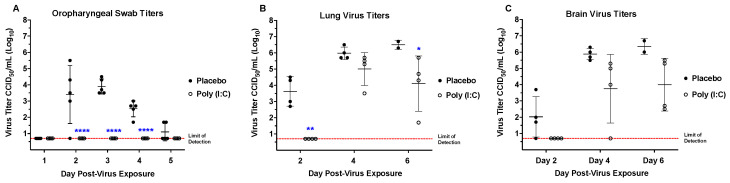
Animal models that are susceptible to SARS-CoV-2 infection and develop clinical signs like human COVID-19 are desired to understand viral pathogenesis and develop effective medical countermeasures. The golden Syrian hamster is important for the study of SARS-CoV-2 since hamsters are naturally susceptible to SARS-CoV-2. However, infected hamsters show only limited clinical disease and resolve infection quickly. In this study, we describe development of human angiotensin-converting enzyme 2 (hACE2) transgenic hamsters as a model for COVID-19. During development of the model for SARS-CoV-2, we observed that different hACE2 transgenic hamster founder lines varied in their susceptibility to SARS-CoV-2 lethal infection. The highly susceptible hACE2 founder lines F0F35 and F0M41 rapidly progress to severe infection and death within 6 days post-infection (p.i.). Clinical signs included lethargy, weight loss, dyspnea, and mortality. Lethality was observed in a viral dose-dependent manner with a lethal dose as low as 1 × 100.15 CCID50. In addition, virus shedding from highly susceptible lines was detected in oropharyngeal swabs on days 2-5 p.i., and virus titers were observed at 105.5-6.5 CCID50 in lung and brain tissue by day 4 p.i.. Histopathology revealed that infected hACE2-hamsters developed rhinitis, tracheitis, bronchointerstitial pneumonia, and encephalitis. Mortality in highly susceptible hACE2-hamsters can be attributed to neurologic disease with contributions from the accompanying respiratory disease. In contrast, virus challenge of animals from less susceptible founder lines, F0M44 and F0M51, resulted in only 0-20% mortality. To demonstrate utility of this SARS-CoV-2 infection model, we determined the protective effect of the TLR3 agonist polyinosinic-polycytidylic acid (Poly (I:C)). Prophylactic treatment with Poly (I:C) significantly improved survival in highly susceptible hACE2-hamsters. In summary, our studies demonstrate that hACE2 transgenic hamsters differ in their susceptibility to SARS-CoV-2 infection, based on the transgenic hamster founder line, and that prophylactic treatment with Poly (I:C) was protective in this COVID-19 model of highly susceptible hACE2-hamsters.

Keywords: SARS-CoV-2; animal model; transgenic hamster; viral pathogenesis.


Conflict of interest statement

The authors declare no conflicts of interest. The funders had no role in the design of the study; in the collection, analyses, or interpretation of data; in the writing of the manuscript; or in the decision to publish the results.

Figures

Figure 1
Figure 1
Diagram of epithelium-specific expression cassette for the hACE2 gene, pK18-hACE2.
Figure 2
Figure 2
Survival curves for F1 and F2 generations of hACE2-hamster founder lines infected with SARS-CoV-2. In five of the founder lines, F0M16, F0M29, F0M35, F0M39, and F0M41, the susceptibility to a lethal SARS-CoV-2 infectious dose of 104.3 CCID50 appears similar between the F1 and F2 generations. In two founder lines, F0M44 and F0M51, the susceptibility to the virus decreases between the F1 and F2 generations. The varying susceptibilities could be a result of founders exhibiting a mosaic expression of the hACE2 transgene in different founder lines.
Figure 3
Figure 3
Survival curves for hACE2-hamster founder lines differing in susceptibility to SARS-CoV-2. Groups of 8-week-old hACE2-hamsters (n = 10/virus dose) infected with SARS-CoV-2. (A) Viral challenge in F0F35 highly susc line resulted in 90–100% mortality (B) and 100% mortality in F0M41highly susc line. (C) Challenge in the F0M44less susc line resulted in 0–10% mortality. (D) Infection in the F0M51less susc line resulted in 0–20% mortality when challenged with a series of low infectious doses and 60% mortality with a high dose.
Figure 4
Figure 4
Percent weight loss in hACE2-hamster founder lines differing in susceptibility to SARS-CoV-2. Groups of 8-week-old hACE2-hamsters (n = 10/virus dose) infected with SARS-CoV-2. (A,B) Hamsters from the F035highly susc and F041highly susc lines lost significant weight prior to mortality when challenged with a low dose (100.3 CCID50). (C,D) In the F0M44 highly susc and F0M51 highly susc lines, hamsters experienced significant weight loss only when challenged with the high dose (104.3 CCID50).
Figure 5
Figure 5
Clinical scores in hACE2-hamster founder lines differing in susceptibility to SARS-CoV-2. Groups of 8-week-old hACE2-hamsters (n = 10) infected with SARS-CoV-2. Hamsters in the F0M35highly susc and F0M41highly susc groups were infected with an infectious dose of 100.3 CCID50, whereas the animals in the F0M44less susc and F0M51less susc were infected with an infectious dose of 104.3 CCID50. The clinical scores for all 4 founder lines began to increase at day 2 p.i. and continued to rise until either mortality in F0M35highly susc and F0M41highly susc or mean peak scores of 4.1 and 3.6 at day 8 p.i. in the F0M44less susc and F0M51less susc lines, respectively. Following day 8, the F0M44less susc and F0M51less susc hamsters began to recover from infection at which point clinical scores decreased.
Figure 6
Figure 6
Virus tissue titers in hACE2-hamster founder lines differing in susceptibility to SARS-CoV-2. Groups of 8-week-old hACE2-hamsters infected with SARS-CoV-2 were sacrificed on days 2, 4, and 6 post-infection (n = 4/day). Titers for F0F35highly susc and F0M41highly susc, shown in black, are after a 100.3 CCID50 virus challenge dose. Titers for F0F35highly susc and F0M41highly susc animals, shown in black, are after a 100.3 CCID50 virus challenge dose. Titers for F0M44less susc and F0M51less susc animals, shown in blue, are after a 104.3 CCID50 challenge dose. (A) SARS-CoV-2 is first detectable at day 2 p.i. and increases to a peak in lung tissue at day 4 p.i., after which it decreases by day 6 p.i.. (B) Virus titer reaches a peak in brain tissue at day 4 p.i. and, like lung tissue, begins to decrease by day 6 p.i.. (C) Infectious virus is detectable in the cardiac tissue at day 4 p.i. for all 4 founder lines and at day 6 p.i. in 1 of F0M41highly susc animal. (D) Virus was only detected in renal tissue on day 4 p.i. in 3 of the 4 founder lines (F0F35, F0M41, and F0F44). The only significant difference observed between founder lines post-infection was in lungs on day 4 p.i. (** p < 0.01). In addition, hACE2 mRNA expression is high in in all tissues in which virus replication was observed (See Figure S1).
Figure 7
Figure 7
Summary of histological lesions in ACE2-hamster founder lines differing in susceptibility to SARS-CoV-2. Groups of 8-week-old hACE2-hamsters (n = 3/day) infected with 100.3 CCID50 SARS-CoV-2. Hamsters from the F0M44less susc line had the most respiratory lesions at day 2 p.i. and developed a moderate to severe infection of both upper and lower respiratory tracts. In addition, animals from the F0M35highly susc, F0M41highly susc, and F0M44less susc lines developed moderate meningoencephalitis. However, over the course of infection, the F0M51less susc line developed the lowest lesion scores.
Figure 8
Figure 8
Pulmonary pathology of SARS-CoV-2-infected F035Highly Susc hACE2-hamsters. (AC) SARS-CoV-2 (100.3 CCID50)-infected hamster at day 6 p.i.. (DF) SARS-CoV-2-infected hamster at day 8 p.i.. (GI) Sham-infected hamster. (A) Inflammatory reaction and necrosis centered on bronchi and bronchiole extending into adjacent alveoli. (B) Bronchiolar epithelial cell necrosis with neutrophilic and lymphocytic inflammation. Alveolar septae are thickened by inflammatory cells. (C) Edema fluid, fibrin, neutrophils, and macrophages fill alveoli and expend alveolar septa. (D) Early resolution of bronchointerstitial pneumonia. Alveolar septae are prominent around bronchioles. (E,F) Alveolar septa are prominent due to marked pneumocytes type Il hyperplasia. Alveolar spaces are filled with edema fluid, fibrin, neutrophils, and macrophages. (GI) Lung of a sham-infected hamster. (H,E) staining. (A,D,E): 40×. Bar = 500 μm. (B,E,H): 200×. Bar = 100 μm. (C,F,I): 400×.
Figure 9
Figure 9
Neuropathology of SARS-CoV-2-infected F035Highly Susc hACE2-hamsters. (A,C) Sham-infected hamster at day 6 p.i.. (B,D) SARS-CoV-2 (100.3 CCID50)-infected hamster. Figure (B): Mild lymphocytic perivascular cuffing in the thalamus. Figure (D): Gliosis and lymphocytic perivascular cuffing in the thalamus. H&E staining. (A,C): 100×. Bar = 200 μm. (C,D): 400×.
Figure 10
Figure 10
Effect of prophylactic treatment with Poly (I:C) on SARS-CoV-2 morbidity and mortality in F041Highly Susc hACE2-hamsters. Groups of 8-week-old hACE2-hamsters (n = 10) infected with 100.3 CCID50 SARS-CoV-2. (A) Poly (I:C) treatment significantly increased the MDD for treated hamsters. (B) Treatment with Poly(I:C) prevented weight loss over the course of the infection. (C) Clinical scores of treated hamsters decreased compared to placebo controls. Daily clinical signs and scores following infection consisted of rough coat (1), nasal or ocular discharge (1), hunched posture (1), abnormal gait (2), lethargy (2), and mild to moderate dyspnea (1 to 3), with each animal scored daily. (*** p < 0.01, **** p < 0.0001).
Figure 11
Figure 11
Prophylactic treatment with Poly(I:C) prevents viral shedding and reduces lung virus titers in F041Highly Susc hACE2-hamsters. Groups of 8-week-old hACE2-hamsters (n = 5/day) infected with 100.3 CCID50 SARS-CoV-2. (A) Infectious virus was not observed in oropharyngeal swabs from treated animals, in contrast to placebo-treated hamsters. (B) Virus titer was significantly reduced in the lung tissue from treated hamsters on days 2 and 6 p.i.. (C) Poly (I:C) treatment reduced virus titers in the brain, but the reduction was not significant. (* p < 0.05, ** p < 0.1, **** p < 0.0001).

Similar articles

References

    1. Chen N., Zhou M., Dong X., Qu J., Gong F., Han Y., Qiu Y., Wang J., Liu Y., Wei Y., et al. Epidemiological and Clinical Characteristics of 99 Cases of 2019 Novel Coronavirus Pneumonia in Wuhan, China: A Descriptive Study. Lancet. 2020;395:507–513. doi: 10.1016/S0140-6736(20)30211-7. - DOI - PMC - PubMed
    1. Ollarves-Carrero M.F., Rodriguez-Morales A.G., Bonilla-Aldana D.K., Rodriguez-Morales A.J. Anosmia in a Healthcare Worker with COVID-19 in Madrid, Spain. Travel Med. Infect. Dis. 2020;35:101666. doi: 10.1016/j.tmaid.2020.101666. - DOI - PMC - PubMed
    1. Huang C., Wang Y., Li X., Ren L., Zhao J., Hu Y., Zhang L., Fan G., Xu J., Gu X., et al. Clinical Features of Patients Infected with 2019 Novel Coronavirus in Wuhan, China. Lancet. 2020;395:497–506. doi: 10.1016/S0140-6736(20)30183-5. - DOI - PMC - PubMed
    1. Koutsakos M., Kedzierska K. A Race to Determine What Drives COVID-19 Severity. Nature. 2020;583:366–368. doi: 10.1038/d41586-020-01915-3. - DOI - PubMed
    1. Ziemele D., Ķauķe G., Skrējāne K., Jaunozoliņa L., Karelis G. A Fatal Case of COVID-19-Associated Acute Necrotizing Encephalopathy. Eur. J. Neurol. 2021;28:3870–3872. doi: 10.1111/ene.14966. - DOI - PMC - PubMed

Substances

LinkOut - more resources