. 2024 Sep 27;17(10):1286.
doi: 10.3390/ph17101286.

Discovery of Novel Inhibitors against ALS-Related SOD1(A4V) Aggregation through the Screening of a Chemical Library Using Differential Scanning Fluorimetry (DSF)

Affiliations

Discovery of Novel Inhibitors against ALS-Related SOD1(A4V) Aggregation through the Screening of a Chemical Library Using Differential Scanning Fluorimetry (DSF)

Maria Giannakou et al. Pharmaceuticals (Basel). .

Abstract

Background: Cu/Zn Superoxide Dismutase 1 (SOD1) is a 32 kDa cytosolic dimeric metalloenzyme that neutralizes superoxide anions into oxygen and hydrogen peroxide. Mutations in SOD1 are associated with ALS, a disease causing motor neuron atrophy and subsequent mortality. These mutations exert their harmful effects through a gain of function mechanism, rather than a loss of function. Despite extensive research, the mechanism causing selective motor neuron death still remains unclear. A defining feature of ALS pathogenesis is protein misfolding and aggregation, evidenced by ubiquitinated protein inclusions containing SOD1 in affected motor neurons. This work aims to identify compounds countering SOD1(A4V) misfolding and aggregation, which could potentially aid in ALS treatment.

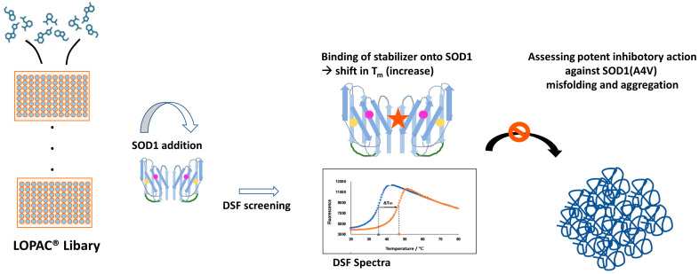
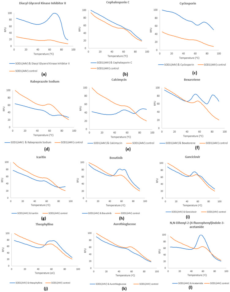
Methods: The approach employed was in vitro screening of a library comprising 1280 pharmacologically active compounds (LOPAC®) in the context of drug repurposing. Using differential scanning fluorimetry (DSF), these compounds were tested for their impact on SOD1(A4V) thermal stability.

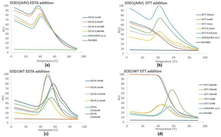
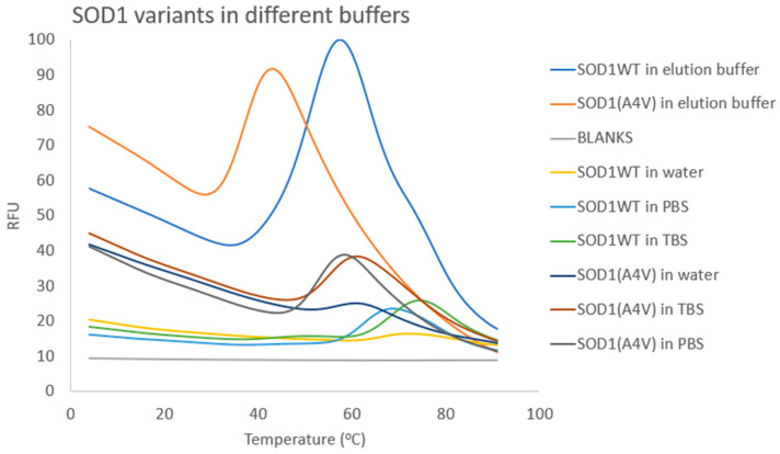
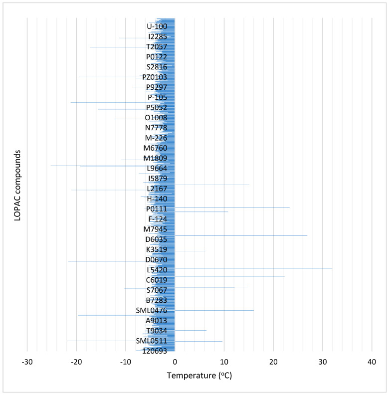
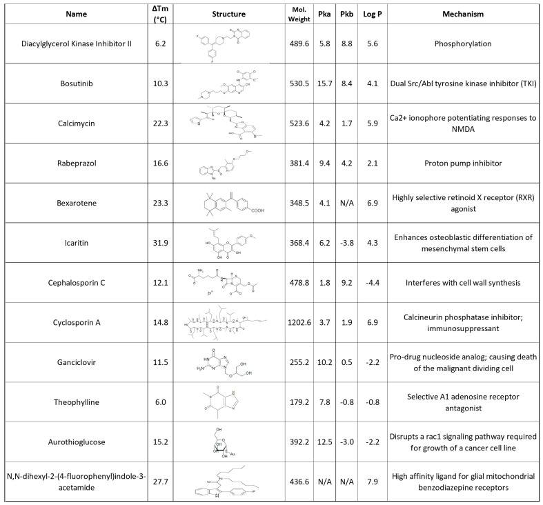
Results and conclusions: Dimer stability was the parameter chosen as the criterion for screening, since the dissociation of the native SOD1 dimer is the step prior to its in vitro aggregation. The screening revealed one compound raising protein-ligand Tm by 6 °C, eleven inducing a higher second Tm, suggesting a stabilization effect, and fourteen reducing Tm from 10 up to 26 °C, suggesting possible interactions or non-specific binding.

Keywords: ALS; DSF technique; SOD1; chemical library scanning; drug discovery; drug repurposing; protein-misfolding diseases.


Conflict of interest statement

The authors declare no conflicts of interest.

Figures

Figure 1
Figure 1
Native-PAGE results for the selected fractions of both SOD1WT (lanes 10–15) and SOD1(A4V) (10–16), aiming to provide an overview of the size of the structures that correspond to the native conformational status of the protein, the dimer (lanes 13 and 14 for both variants). EST corresponds to ESTDZ2, and was chosen to be close in size to the proteins of interest, in order to be used as a standard compound for the identification of the sizes of the proteins analyzed. ESDDZ2 is a globular protein with a molecular weight close to 28 kDa [83], while that of Flag-SOD1-6His is 35.4 kDa.
Figure 2
Figure 2
(Top) Gel filtration chromatogram of SOD1WT eluted after the first purification step in the NiNTA. Protein was monitored via UV detection (A280). Column used: Sephacryl™ S-200 (GE Healthcare, Chicago, IL, USA) with mobile phase TBS buffer pH7.4. (Bottom) SDS-PAGE of the selected fractions of purified SOD1WT after gel filtration.
Figure 3
Figure 3
(Top) Gel filtration chromatogram of SOD1(A4V) eluted after the first purification step in the NiNTA. Protein was monitored via UV detection (A280). Column used: Sephacryl™ S-200 with mobile phase TBS buffer pH7.4. (Bottom) SDS-PAGE of the selected fractions of purified SOD1WT after gel filtration.
Figure 3
Figure 3
(Top) Gel filtration chromatogram of SOD1(A4V) eluted after the first purification step in the NiNTA. Protein was monitored via UV detection (A280). Column used: Sephacryl™ S-200 with mobile phase TBS buffer pH7.4. (Bottom) SDS-PAGE of the selected fractions of purified SOD1WT after gel filtration.
Figure 4
Figure 4
Zymogram performed using the method of riboflavin and B2/NBT. Equimolar quantities of the WT and A4V SOD1 variants were used, in order to estimate the effect of the mutation A4V on the protein. Boiling and the addition of the chelator EDTA were tested in order to estimate the tolerance/stability of each variant. The boiling treatment corresponded to 5 min at 95 °C, while the EDTA treatment corresponded to 15 min incubation at the indicated EDTA concentrations before loading onto the gel occurred.
Figure 5
Figure 5
Addition of EDTA and DTT to SOD1(A4V) (a,b) and SOD1WT (c,d). SOD1 variants were diluted in the following buffer (elution buffer): 50 mM NaH2PO4, 300 mM NaCl, and 250 mM imidazole; pH 8.0. RFU corresponds to the normalized fluorescence from triplicate samples.
Figure 6
Figure 6
Examination of different buffers in regard to SOD1 stability. RFU corresponds to the normalized fluorescence for each compound from triplicate samples.
Figure 7
Figure 7
Workflow of LOPAC® screening. The compounds are arranged (after proper dilution) in a 96-well plate format and the freshly purified SOD1(A4V) is added to the mixture. Following a short incubation period, DSF spectra are extracted. The compounds presenting a positive shift in the Tm of the mixture (denoted graphically with the red star onto the protein) will further be evaluated as potent aggregation inhibitors of SOD1 in kinetics experiments. The orange and pink dots represent Cu and Zn and the green round lines the disulfide bonds.
Figure 8
Figure 8
The difference in Tm values of SOD1(A4V) caused by the compounds of the LOPAC® library. ΔTm corresponds to the function: Tm(SOD1(A4V)) − Tm(SOD1(A4V) upon a compound’s presence).
Figure 9
Figure 9
LOPAC® compounds assessed as being able to cause a significant positive ΔTm on SOD1(A4V) and as being worthy of further examination. In this table, some physicochemical PK properties and their mechanism of actions are also presented. (N/A represents not applicable measurement for the specific property).
Figure 10
Figure 10
DSF spectra of the selected compounds. RFU corresponds to the normalized fluorescence for each compound from triplicate samples. The molecular ratio is SOD1(A4V):compound, 1:20 and these are (a) diakyl glycerol kinase inhibitor II, (b) cephalosporin C, (c) cyclosporin, (d) rabeprazole sodium, (e) calcimycin, (f) bexarotene, (g) icaritin, (h) bosutinib, (i) ganciclovir, (j) theophylline, (k) aurothioglucose, (l) N,N-dihexyl-2-(4-fluorophenyl)indole-3-acetamide.
Figure 11
Figure 11
Docking pockets on the SOD1(A4V) surface are illustrated with yellow color. Green and purple colors denote the two monomers that create the dimer. Specifically, (a) Pocket 1 is located near Cys 11 residue, (b) Pocket 2 at the surface-exposed Trp32 site, (d) Pocket 3 is present close to the region where the intra-disulfide bond between the residues Cys57 and Cys146 is formed, and (c) Pocket 4 is positioned next to the electrostatic loop.

Similar articles

References

    1. Deng H.-X., Hentati A., Tainer J.A., Iqbal Z., Cayabyab A., Hung W.Y., Getzoff E.D., Hu P., Herzfeldt B., Roos R.P. Amyotrophic lateral sclerosis and structural defects in Cu,Zn superoxide dismutase. Science. 1993;261:1047–1051. doi: 10.1126/science.8351519. - DOI - PubMed
    1. Hardiman O., Al-Chalabi A., Chio A., Corr E.M., Logroscino G., Robberecht W., Shaw P.J., Simmons Z., van den Berg L.H. Amyotrophic lateral sclerosis. Nat. Rev. Dis. Primers. 2017;3:17071. doi: 10.1038/nrdp.2017.71. - DOI - PubMed
    1. Mehta P.R., Jones A.R., Opie-Martin S., Shatunov A., Iacoangeli A., Al Khleifat A., Smith B.N., Topp S., Morrison K.E., Shaw P.J., et al. Younger age of onset in familial amyotrophic lateral sclerosis is a result of pathogenic gene variants, rather than ascertainment bias. J. Neurol. Neurosurg. Psychiatry. 2019;90:268–271. doi: 10.1136/jnnp-2018-319089. - DOI - PMC - PubMed
    1. Wiedau-Pazos M., Goto J.J., Rabizadeh S., Gralla E.B., Roe J.A., Lee M.K., Valentine J.S., Bredesen D.E. Altered Reactivity of Superoxide Dismutase in Familial Amyotrophic Lateral Sclerosis. Science. 1996;271:515–518. doi: 10.1126/science.271.5248.515. - DOI - PubMed
    1. Renton A.E., Chiò A., Traynor B.J. State of play in amyotrophic lateral sclerosis genetics. Nat. Neurosci. 2014;17:17–23. doi: 10.1038/nn.3584. - DOI - PMC - PubMed

LinkOut - more resources