. 2024 Oct 18;25(20):11225.
doi: 10.3390/ijms252011225.

Adipose-Derived Stem Cells as Carrier of Pro-Apoptotic Oncolytic Myxoma Virus: To Cross the Blood-Brain Barrier and Treat Murine Glioma

Affiliations

Adipose-Derived Stem Cells as Carrier of Pro-Apoptotic Oncolytic Myxoma Virus: To Cross the Blood-Brain Barrier and Treat Murine Glioma

Joanna Jazowiecka-Rakus et al. Int J Mol Sci. .

Abstract

Treatment of glioblastoma is ineffective. Myx-M011L-KO/EGFP, a myxoma virus actively inducing apoptosis in BTICs linked to recurrence, offers innovative treatment. We loaded this construct into adipose-derived stem cells (ADSCs) to mitigate antiviral host responses and enable systemic delivery. The apoptotic and cytotoxic effects of the construct were studied using murine and human glioblastoma cell lines. Before implementing systemic delivery, we delivered the construct locally using ADSC to verify elimination of orthotopic murine glioma lesions. vMyx-M011L-KO/EGFP was cytotoxic to a murine cell line, preventing effective virus multiplication. In three human glioma cell lines, viral replication did occur, coupled with cell killing. The knock-out construct induced apoptotic cell death in these cultures. ADSCs infected ex vivo were shown to be sufficiently migratory to assure transfer of the therapeutic cargo to murine glioma lesions. Virus-loaded ADSCs applied to the artificial blood-brain barrier (BBB) yielded viral infection of glioma cells grown distally in the wells. Two rounds of local administration of this therapeutic platform starting 6 days post tumor implantation slowed down growth of orthotopic lesions and improved survival (total recovery < 20%). ADSCs infected ex vivo with vMyx-M011L-KO/EGFP show promise as a therapeutic tool in systemic elimination of glioma lesions.

Keywords: adipose tissue-derived stem cells (ADSCs); blood–brain barrier; glioblastoma; myxoma virus; oncolytic virotherapy.


Conflict of interest statement

The authors declare no conflicts of interest.

Figures

Figure 1
Figure 1
Permissiveness and cell viability. Cell lines: RK-13 and ADSCs as well as GL261, T98G, LN18, and U-251MG (gliomas) infected with vMyx-M011L-KO/EGFP; (A) Representative images of infected cells (MOI = 5) after 24 h p.i. stained and visualized by fluorescence microscopy (Zeiss LSM 710 confocal workstation; magn. 20×; scale bar = 50 µm); blue: DAPI dye (nuclei); green: EGFP fluorescence; (B) Flow cytometric quantitation (12–48 h p.i.) of infected EGFP-positive cells in tested cell lines (MOI = 5); Replication of the virus in tested cell lines by determining (C) the single- (MOI = 5) and (D) multi-step virus growth curve (MOI = 0.02). Cell viability assay (MTS) for bEnd.3, RK-13, ADSCs and glioblastoma GL261, T98G, LN18 and U-251MG lines and four different MOI values: (E) 24 h, (F) 48 h and (G) 72 h time points p.i. and eight independently infected wells were examined. Error bars represent SD of the measurements.
Figure 2
Figure 2
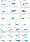
Analysis of Apoptosis by Flow Cytometry. Cells were seeded at a density of 1 × 10⁵ cells/well using 6-well plates and vMyx-M011L-KO/EGFP (MOI = 5) was added to the cultured cells. After 12, 24 h and 48 h (RK-13, ADSCs, and GL261-luc) and after 12, 24, 48 and 72 h p.i. (LN18, T98G, U-251 MG and bEnd.3) cells were collected and washed twice with PBS and staining buffer. Cells were then stained with Annexin V-Pacific Blue™ and 7-AAD and analyzed for apoptosis/necrosis using flow cytometry (BD FACSLyric™). Annexin V was detected using the Pacific Blue channel and 7-AAD using the PerCP-Cy5.5 channel and a region for live cells was defined. Non-infected cells were used as a control. Q1—early apoptotic cells; Q2—viable cells; Q3—late apoptotic cells; and Q4—necrotic cells.
Figure 3
Figure 3
Expression of apoptotic proteins by Western blot analysis of lysates obtained from cell cultures infected (MOI = 5) with vMyx-EGFP/tdTr or vMyx-M011L-KO/EGFP constructs. Lysates were prepared from whole cell extracts after 12, 24, 48 and 72 h p.i. (RK-13, ADSCs, T98G, U-251 MG and LN18) and after 3, 6, 12, 24, and 48 h p.i. (GL261luc; control samples run on separate gel, same experiment). Full-length (116 kDa) and cleaved forms (89 kDa) of poly ADP-ribose polymerase (PARP), as well as endogenous levels of the full length caspase-3 (35 kDa) and the large fragment of caspase-3 resulting from cleavage (19/17 kDa) were assessed by immunoblotting using β-actin as a loading control. See also Figure S1.
Figure 4
Figure 4
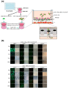
In vitro blood–brain barrier (BBB) migration studies. (A) Schematic representation of the barrier formed by plating bEnd.3 cells (murine endothelial cells isolated from endothelioma) on inserts of the 24-well plate (Matrigel®-covered 8 µm pore inserts). Studies of virus migration through the BBB were started when transendothelial electrical resistance (TEER) of the BBB (measured with an ohmmeter) reached ca. 20 Ω × cm2. The virus migration assay was performed by administering three equivalent doses of ADSCs infected with vMyx-M011L-KO/EGFP (MOI = 5) or unshielded virus every 24 h on pre-prepared inserts. (B) The migration was assessed within a 24–120 h period after the addition of the virus construct or virus-loaded ADSCs (microscopic observation in visible light and using the green fluorescence channel (EGFP), ZEISS AxioVert microscope, 20× magnification, scale bar = 100 µm).
Figure 5
Figure 5
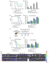
The therapeutic effect on experimentally-induced murine glioma lesions following intracranial treatment with shielded or unshielded vMyx-M011L-KO/EGFP constructs. (A) Inhibited lesion formation and prolonged survival: mice with orthotopically administered GL261-luc glioma cells were subjected to simultaneous (consecutive) injection of either unshielded or ADSC-shielded vMyx-M011L-KO/EGFP knockout construct. (B) ROI-based bioluminescence (BLI) analysis of total photon flux in mouse heads. IVIS-detected signal at 4 time points following GL261-luc implantation (mean +/− SD from 6 mice/group). (C) One-dose therapy: mice with similar intracranial BLI randomly divided (6th day post inoculation) into groups were injected intratumorally (day 7) with unshielded vMyx-M011L-KO/EGFP construct or ADSC-shielded construct. (D) One-dose therapy: ROI-based BLI analysis of total photon flux in mouse heads: IVIS-detected signal at four time points following GL261-luc implantation (mean +/− SD from 6 mice/group). (E) Timetable of two-dose therapy. (F) Two-dose therapy: mice with similar intracranial BLI randomly divided (6th day post inoculation) into groups were injected intratumorally (days 7 and 14) with unshielded vMyx-M011L-KO/EGFP construct or ADSC-shielded construct. (G) Two-dose therapy: ROI-based BLI analysis of total photon flux in mouse heads: IVIS-detected signal at three time points following GL261-luc implantation (mean +/− SD from 6 or 7 mice/group). (H) IVIS-detected signal following two-dose therapy: ROI-based analysis of total photon flux in mouse heads. BLI expressed as radiance (photons/sec/cm2/sr). Single radiance scale shown to cover the whole span of bioluminescence. The data show mean ± SD of two independent experiments.

Similar articles

References

    1. Ilic I., Ilic M. International patterns and trends in the brain cancer incidence and mortality: An observational study based on the global burden of disease. Heliyon. 2023;9:e18222. doi: 10.1016/j.heliyon.2023.e18222. - DOI - PMC - PubMed
    1. Salvato I., Marchini A. Immunotherapeutic Strategies for the Treatment of Glioblastoma: Current Challenges and Future Perspectives. Cancers. 2024;16:1276. doi: 10.3390/cancers16071276. - DOI - PMC - PubMed
    1. Sterner R.C., Sterner R.M. CAR-T cell therapy: Current limitations and potential strategies. Blood Cancer J. 2021;11:69. doi: 10.1038/s41408-021-00459-7. - DOI - PMC - PubMed
    1. Achard C., Surendran A., Wedge M.E., Ungerechts G., Bell J., Ilkow C.S. Lighting a Fire in the Tumor Microenvironment Using Oncolytic Immunotherapy. EBioMedicine. 2018;31:17–24. doi: 10.1016/j.ebiom.2018.04.020. - DOI - PMC - PubMed
    1. Pisklakova A., McKenzie B., Zemp F., Lun X., Kenchappa R.S., Etame A.B., Rahman M.M., Reilly K., Pilon-Thomas S., McFadden G., et al. M011L-deficient oncolytic myxoma virus induces apoptosis in brain tumor-initiating cells and enhances survival in a novel immunocompetent mouse model of glioblastoma. Neuro-Oncol. 2016;8:1088–1098. doi: 10.1093/neuonc/now006. - DOI - PMC - PubMed

LinkOut - more resources