. 2024 Oct 17;25(20):11152.
doi: 10.3390/ijms252011152.

Alpha- to Beta-Cell Transdifferentiation in Neonatal Compared with Adult Mouse Pancreas in Response to a Modest Reduction in Beta-Cells Using Streptozotocin

Affiliations

Alpha- to Beta-Cell Transdifferentiation in Neonatal Compared with Adult Mouse Pancreas in Response to a Modest Reduction in Beta-Cells Using Streptozotocin

Jiwon Hahm et al. Int J Mol Sci. .

Abstract

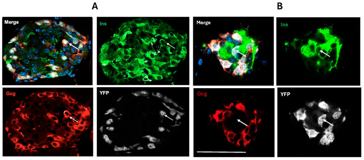
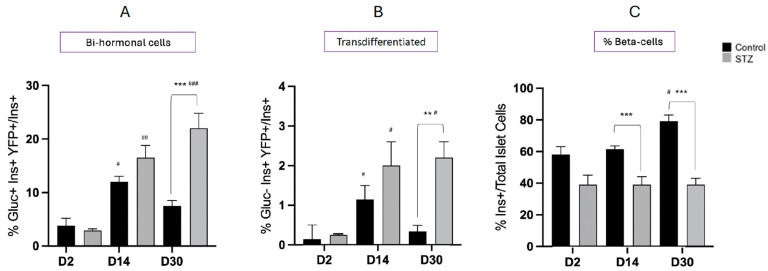
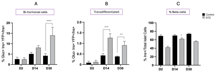
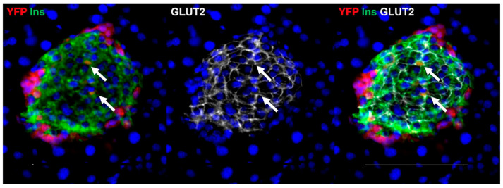
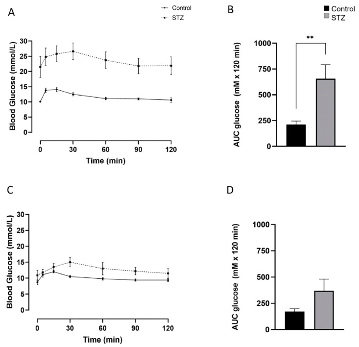
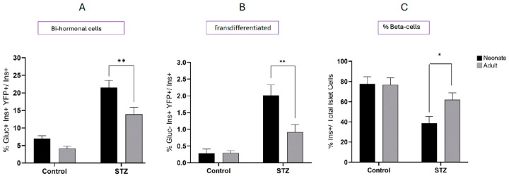
Following the near-total depletion of pancreatic beta-cells with streptozotocin (STZ), a partial recovery of beta-cell mass (BCM) can occur, in part due to the alpha- to beta-cell transdifferentiation with an intermediary insulin/glucagon bi-hormonal cell phenotype. However, human type 2 diabetes typically involves only a partial reduction in BCM and it is not known if recovery after therapeutic intervention involves islet cell transdifferentiation, or how this varies with age. Here, we used transgenic mouse models to examine if islet cell transdifferentiation contributes to BCM recovery following only a partial depletion of BCM. Cell lineage tracing was employed using Glucagon-Cre/yellow fluorescent protein (YFP) transgenic mice treated with STZ (25 mg/kg-neonates; 70 mg/kg-adults) or vehicle alone on 3 consecutive days. Mice were euthanized 2-30 days later with a prior glucose tolerance test on day 30, and immunofluorescence histology performed on the pancreata. Beta-cell abundance was reduced by 30-40% two days post STZ in both neonates and adults, and subsequently partially recovered in adult but not neonatal mice. Glucose tolerance recovered in adult females, but not in males or neonates. Bi-hormonal cell abundance increased 2-3-fold in STZ-treated mice vs. controls in both neonates and adults, as did transdifferentiated cells expressing insulin and the YFP lineage tag, but not glucagon. Transdifferentiated cell presence was an order of magnitude lower than that of bi-hormonal cells. We conclude that alpha- to beta-cell transdifferentiation occurs in mice following only a moderate depletion in BCM, and that this was accompanied by a partial recovery of BCM in adults.

Keywords: alpha-cell; beta-cell; bi-hormonal; diabetes; mouse; pancreas; streptozotocin; transdifferentiation.


Conflict of interest statement

The authors declare no conflicts of interest.

Figures

Figure 1
Figure 1
Immunohistochemical visualization of bi-hormonal cells (arrows) co-staining for insulin (Ins, green), glucagon (Gcg, red), and YFP (white) (A); and transdifferentiated cells (arrows) co-staining for insulin and YFP but not glucagon (B) within adult mouse pancreas. Cell nuclei are visualized with DAPI in the merged image. The size bar represents 100 μm in (A) and 50 μm in (B).
Figure 2
Figure 2
Immunohistochemical visualization of alpha- to beta-cell transdifferentiation in small islets (A,B) and an endocrine cell cluster (C) within adult mouse pancreas. Insulin (Ins) presence is shown in red and YFP presence in green. Cell nuclei are visualized with DAPI. Arrows indicate examples of alpha- to beta-cell transdifferentiation. The size bar represents 50 μm.
Figure 3
Figure 3
Immunohistochemical visualization of glucagon (Gcg, red) and insulin (Ins, green) in neonatal mouse islets two days following treatment with vehicle alone (A) or STZ (B). Cell nuclei are visualized with DAPI. A 31% mean reduction in insulin-containing beta-cells occurred following STZ treatment with an increased number of glucagon-containing alpha-cells extending into the core of the islet in addition to the rim. The size bar represents 100 μm.
Figure 4
Figure 4
Changes in the abundance of glucagon/insulin bi-hormonal cells (A), alpha- to beta-transdifferentiated cells (B), and the percent insulin containing beta-cells/islet (C) in neonatal mouse pancreata 2 days (D2), 14 days (D14), or 30 days (D30) following treatment with STZ or vehicle alone (Control). Bi-hormonal cells were recognized as co-staining for glucagon (Gluc+), insulin (Ins+), and YFP (YFP+) and are expressed relative to total Ins+ cells. Transdifferentiated cells were recognized as co-staining for insulin and YFP, but not glucagon. Values show mean ± SEM. # p < 0.05, ## p < 0.01, ### p < 0.001 vs. D2; ** p < 0.01, *** p < 0.001 vs. control.
Figure 5
Figure 5
(A) Glucose excursions during intraperitoneal glucose tolerance tests in neonatal mice at D30 following treatment with STZ or vehicle alone (control); and (B) the glucose area under the curve (AUC). Values show mean ± SEM. ** p < 0.01 vs. Control.
Figure 6
Figure 6
Changes in the abundance of glucagon/insulin bi-hormonal cells (A), alpha- to beta-transdifferentiated cells (B), and the percent insulin containing beta-cells/islet (C) in adult mouse pancreata 2 days (D2), 14 days (D14), or 30 days (D30) following treatment with STZ or vehicle alone (control). Bi-hormonal cells were recognized as co-staining for glucagon (Gluc+), insulin (Ins+), and YFP (YFP+) and are expressed relative to total Ins+ cells. Transdifferentiated cells were recognized as co-staining for insulin and YFP, but not glucagon. Values show mean ± SEM. # p < 0.05 vs. D2; * p < 0.05, ** p < 0.01, *** p < 0.001 vs. control.
Figure 7
Figure 7
Immunohistochemical visualization of insulin (Ins, green), YFP (red), and Glut2 (white) in an adult mouse islet. Cell nuclei are visualized with DAPI. Alpha- to beta-transdifferentiated cells shown by co-localization of Ins and YFP also express Glut2 (arrows). The size bar represents 100 μm.
Figure 8
Figure 8
Glucose excursions during intraperitoneal glucose tolerance tests and area under the curve (AUC) in adult male (A,B) and female (C,D) mice at D30 following treatment with STZ or vehicle alone (control). Values show mean ± SEM. ** p < 0.01 vs. control.
Figure 9
Figure 9
Comparison of glucagon/insulin bi-hormonal (A), transdifferentiated cell presence (B), and the percent insulin containing beta-cells/islet (C) between neonatal and adult mouse pancreata 30 days following treatment with STZ or vehicle alone (control). Values show mean ± SEM. * p < 0.05, ** p < 0.01 vs. neonates.

Similar articles

References

    1. Wang Q., Huang Y.X., Liu L., Zhao X.H., Sun Y., Mao X., Li S.W. Pancreatic islet transplantation: Current advances and challenges. Front. Immunol. 2024;15:1391504. doi: 10.3389/fimmu.2024.1391504. - DOI - PMC - PubMed
    1. Dor Y., Brown J., Martinez O.I., Melton D.A. Adult pancreatic beta-cells are formed by self-duplication rather than stem-cell differentiation. Nature. 2004;429:41–46. doi: 10.1038/nature02520. - DOI - PubMed
    1. Meier J.J., Butler A.E., Saisho Y., Monchamp T., Galasso R., Bhushan A., Rizza R.A., Butler P.C. Beta-cell replication is the primary mechanism subserving the postnatal expansion of beta-cell mass in humans. Diabetes. 2008;57:1584–1594. doi: 10.2337/db07-1369. - DOI - PMC - PubMed
    1. Montanya E., Nacher V., Biarnés M., Soler J. Linear correlation between beta-cell mass and body weight throughout the lifespan in Lewis rats: Role of beta-cell hyperplasia and hypertrophy. Diabetes. 2000;49:1341–1346. doi: 10.2337/diabetes.49.8.1341. - DOI - PubMed
    1. Rankin M.M., Kushner J.A. Adaptive-cell proliferation is severely restricted with advanced age. Diabetes. 2009;58:1365–1372. doi: 10.2337/db08-1198. - DOI - PMC - PubMed

MeSH terms

LinkOut - more resources