Review
. 2024 Oct 12;25(20):10971.
doi: 10.3390/ijms252010971.

Membrane Ruffles: Composition, Function, Formation and Visualization

Affiliations
Review

Membrane Ruffles: Composition, Function, Formation and Visualization

Guiqin Yan et al. Int J Mol Sci. .

Abstract

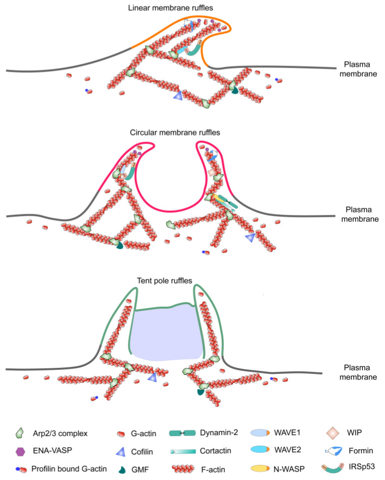
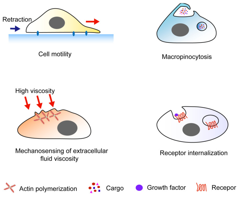
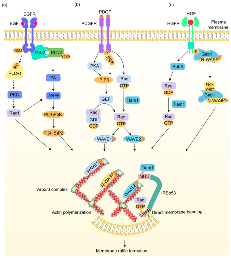
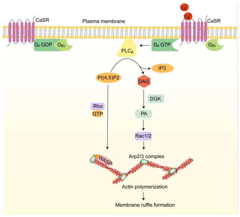
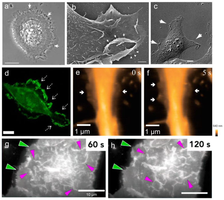
Membrane ruffles are cell actin-based membrane protrusions that have distinct structural characteristics. Linear ruffles with columnar spike-like and veil-like structures assemble at the leading edge of cell membranes. Circular dorsal ruffles (CDRs) have no supporting columnar structures but their veil-like structures, connecting from end to end, present an enclosed ring-shaped circular outline. Membrane ruffles are involved in multiple cell functions such as cell motility, macropinocytosis, receptor internalization, fluid viscosity sensing in a two-dimensional culture environment, and protecting cells from death in response to physiologically compressive loads. Herein, we review the state-of-the-art knowledge on membrane ruffle structure and function, the growth factor-induced membrane ruffling process, and the growth factor-independent ruffling mode triggered by calcium and other stimulating factors, together with the respective underlying mechanisms. We also summarize the inhibitors used in ruffle formation studies and their specificity. In the last part, an overview is given of the various techniques in which the membrane ruffles have been visualized up to now.

Keywords: calcium; cell motility; growth factor; macropinocytosis; membrane ruffles; optic imaging; viscosity sensing.


Conflict of interest statement

The authors declare no conflicts of interest.

Figures

Figure 1
Figure 1
Structure and composition of membrane ruffles. The linear ruffles assemble at the leading edge of a cell membrane, with columnar spike-like and veil-like structures. Circular dorsal ruffles have no supporting columnar structures but have veil-like structures, connecting from end to end, that present an enclosed ring-shaped circular outline. Tent pole ruffles are supported by two distinct filamentous extensions that stand erect on the cell surface at the outer edge of the fold, acting like “tent poles” and holding up the large F-actin sheet which serves as the veil of the fold between them. Some clarified actin-binding proteins associated with membrane ruffles are illustrated in the figure.
Figure 2
Figure 2
Biological functions of membrane ruffles. Linear membrane ruffles are involved in all these processes. Circular dorsal ruffles (CDR) are primarily involved in cell migration, macropinocytosis, and receptor internalization. Tent pole ruffles mainly function in macropinocytosis and receptor internalization.
Figure 3
Figure 3
The initiation mechanism of membrane ruffles induced by growth factors. (a) Upon epidermal growth factor(EGF) stimulation, the receptor autophosphorylates at tyrosine 992, which facilitates phospholipase Cγ1 (PLCγ1) recruitment via its SH2 domain. PLCγ1 activation subsequently activates Rac1, which may be mediated by protein kinase C (PKC). Rac1 induces IRSp53 to bind to the Wiskott-Aldrich syndrome protein (WASP) family verprolin-homologous protein-2 (WAVE2), which activates the Arp2/3 complex, then stimulates actin polymerization and membrane ruffle formation. In addition, epidermal growth factor receptors (EGFR) activation leads to Grb2 binding, which associates with phospholipase D2 (PLD2) at the Y169 site, enhancing ruffle formation through increased phosphatidic acid (PA) production. The presence of PA also activates ADP-ribosylation factor (ARF) 6, an activator of phosphatidylinositol 4-phosphate 5-kinase (PI(4)P5K) that produces phosphatidylinositol 4,5-bisphosphate (PI(4,5)P2) and activates the Arp2/3 complex. Rac1 can also activate ARF6. (b) After platelet-derived growth factor (PDGF) stimulation, protein kinase A (PKA) activation facilitates phosphatidylinositol 3,4,5-trisphosphate (PIP3) accumulation. On the other hand, platelet-derived growth factor receptor (PDGFR) activation triggers phosphoinositide 3-kinase (PI3K), promoting PIP3 synthesis. PIP3 then recruits guanine nucleotide exchange factors (GEFs) to the membrane, leading to Rac release from the RhoGDI and subsequent activation. Activated Rac then binds to WAVE1. PDGF stimulation also activates Ras-GTP, which binds to Tiam1 and signals to Rac-GTP. Tiam1 interacts with IRSp53, enhancing the binding of Rac-IRSp53-WAVE2. WAVE1/2 activates the Arp2/3 complex. (c) Hepatocyte growth factor (HGF) activation recruits a Gab1-N-WASP complex to the membrane, with Gab1 phosphorylation at Y407 creating a docking site for Nck proteins. Nck then binds to it through the SH3 domain for N-WASP activation, which then recruits the Arp2/3 complex. Additionally, Rab5, as a downstream effector, activates Rac via Tiam1, which then induces circular dorsal ruffles (CDR) formation via Rac-IRSp53-WAVE2. Dashed arrows indicate mechanisms yet to be fully elucidated.
Figure 4
Figure 4
The growth factor-independent membrane ruffling triggered by extracellular calcium. In response to extracellular calcium, calcium-sensing receptor (CaSR) activates the Gα protein, leading to the dissociation of the Gβγ subunit. Active Gα-GTP then activates phospholipase C (PLC), resulting in the breakdown of PI(4,5)P2 into diacylglycerol (DAG) and inositol 1,4,5-trisphosphate (IP3). PI(4,5)P2 collaborates with active Rho GTPases to release WASP, initiating Arp2/3-mediated actin assembly. DAG is phosphorylated to form PA, which activates Rac1/2, promoting the nucleation of branched actin networks and the formation of membrane ruffles.
Figure 5
Figure 5
The micrographs of membrane ruffles visualized by various techniques. Membrane ruffles (white arrows) formed in keratinocytes captured by phase-contrast microscopy (a) and scanning electron microscopy (SEM) (b) [44]. Cos1 cells stimulated with epidermal growth factor (EGF) after 5 min imaged by differential interference contrast (DIC) microscopy (c) [144] Human monocyte-derived macrophage imaged by confocal microscopy (d) [145]. Arrows indicate the membrane ruffle area. Time-lapse images of live hippocampal neurons visualized by high-speed atomic force microscopy (HS-AFM) (e,f) [146]. The arrows points to the thin, sheet-like ruffling. Dorsal membrane ruffles (magenta arrows) formed in human retinal pigmented epithelial (RPE1) cells imaged by lattice light-sheet microscopy (LLSM) (g,h) [83]. The green arrows indicate the increased protrusion. Scale bars: 10 μm in (a,c,d,g,h); 1 μm in (b,e,f).

Similar articles

References

    1. Ridley A.J. Membrane ruffling and signal transduction. BioEssays News Rev. Mol. Cell. Dev. Biol. 1994;16:321–327. doi: 10.1002/bies.950160506. - DOI - PubMed
    1. Pittman M., Iu E., Li K., Wang M., Chen J., Taneja N., Jo M.H., Park S., Jung W.H., Liang L., et al. Membrane Ruffling is a Mechanosensor of Extracellular Fluid Viscosity. Nat. Phys. 2022;18:1112–1121. doi: 10.1038/s41567-022-01676-y. - DOI - PMC - PubMed
    1. Patel P.C., Harrison R.E. Membrane ruffles capture C3bi-opsonized particles in activated macrophages. Mol. Biol. Cell. 2008;19:4628–4639. doi: 10.1091/mbc.e08-02-0223. - DOI - PMC - PubMed
    1. Hoon J.L., Wong W.K., Koh C.G. Functions and regulation of circular dorsal ruffles. Mol. Cell. Biol. 2012;32:4246–4257. doi: 10.1128/MCB.00551-12. - DOI - PMC - PubMed
    1. Hasegawa T. Production of a unique antibody specific for membrane ruffles and its use to characterize the behavior of two distinct types of ruffles. J. Cell Biol. 1993;120:1439–1448. doi: 10.1083/jcb.120.6.1439. - DOI - PMC - PubMed

LinkOut - more resources