. 2024 Oct 25;11(10):ENEURO.0307-24.2024.
doi: 10.1523/ENEURO.0307-24.2024. Print 2024 Oct.

Depicting Primate-Like Granular Dorsolateral Prefrontal Cortex in the Chinese Tree Shrew

Affiliations

Depicting Primate-Like Granular Dorsolateral Prefrontal Cortex in the Chinese Tree Shrew

Xiu-Peng Nie et al. eNeuro. .

Abstract

It remains unknown whether the Chinese tree shrew, regarded as the closest sister of primate, has evolved a dorsolateral prefrontal cortex (dlPFC) comparable with primates that is characterized by a fourth layer (L4) enriched with granular cells and reciprocal connections with the mediodorsal nucleus (MD). Here, we reported that following AAV-hSyn-EGFP expression in the MD neurons, the fluorescence micro-optical sectioning tomography revealed their projection trajectories and targeted brain areas, such as the hippocampus, the corpus striatum, and the dlPFC. Cre-dependent transsynaptic viral tracing identified the MD projection terminals that targeted the L4 of the dlPFC, in which the presence of granular cells was confirmed via cytoarchitectural studies by using the Nissl, Golgi, and vGlut2 stainings. Additionally, the L5/6 of the dlPFC projected back to the MD. These results suggest that the tree shrew has evolved a primate-like dlPFC which can serve as an alternative for studying cognition-related functions of the dlPFC.

Keywords: dorsolateral prefrontal cortex (dlPFC); fourth layer (L4); granular cell; medial prefrontal cortex (mPFC); mediodorsal nucleus (MD); orbital frontal cortex (OFC); primate; tree shrew.


Conflict of interest statement

The authors declare no competing financial interests.

Figures
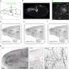
Figure 1.
Figure 1.
The fMOST delineates the MD→PFC in the Chinese tree shrew. a, Injection of AAV-hSyn-EGFP into the mediodorsal nucleus (MD). b, c The fMOST clearly visualized the EGFP-labeled neurons within the MD and the MD projection trajectories and targeting brain regions. d–f, The MD projection to the caudate nucleus (Cd), the putamen (Pu), and densely to the dorsal, medial, and orbital subregions of the frontal cortex (FC). g, The MD projection terminals located in the dorsal frontal cortex (dPFC), forming presynaptic boutons in the cortical layers of the dPFC. fMOST, fluorescence micro-optical sectioning tomography; PFC, prefrontal cortex; Ent, entorhinal cortex. AD, anterodorsal nucleus; PC, paracentral nucleus; sm, stria medullaris; Sub, submedius nucleus; fr, fasciculus retroflexus; PF, parafascicular nucleus; LHB, lateral habenula. mPFC, medial PFC; OFC, orbital FC; cc, corpus callosum; Pc, central nucleus of the pulvinar; Pd, dorsal nucleus of the pulvinar; Pv, ventral nucleus of the pulvinar.
Figure 2.
Figure 2.
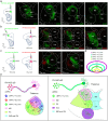
Cre-dependent tracing indicates the MD-projection to the L4 of the dPFC in tree shrews. a, AAV-Ef1α-DIO-EYFP and AAV1- Ef1α-Cre were injected into the MD, but AAV- Ef1α-DIO-mCherry alone was injected into the dPFC. b, The EYFP-labeled neurons were observed within the MD of the viral injecting side and the EYFP-labeled terminals were found in the contralateral MD. c, The mCherry-labeled neurons in the dPFC were resulted from anterograde transferring of the Cre from the MD that met the expression of AAV-Ef1α-DIO-mCherry. d, e, These mCherry-labeled neurons were mainly located in the L4 of the dPFC where the MD-projected terminals were densely distributed. MD, mediodorsal nucleus; IMD, intermediodorsal nucleus; PC, paracentral nucleus; dPFC, dorsal prefrontal cortex.
Figure 3.
Figure 3.
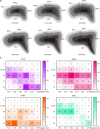
Definition of the dorsolateral prefrontal cortex (dlPFC) in the tree shrew. a, The colors indicated layer 2/3 (blue), layer 4 (red), layer 5 (green), and layer 6 (yellow) in the PFC. Each dot represents a neuron that received Cre from the MD to drive mCherry expression. Each panel indicated the anterior–posterior coordinates (AP) from the bregma: +7.63, +7.23, +6.83, +6.43, +6.03, and +5.76 mm. b, The distribution of neuronal numbers in the layers that received the MD projections in the lateral PFC (lPFC), dorsal PFC (dPFC), medial PFC (mPFC), and orbital FC (OFC) at the AP coordinates. Notably, the pattern of the lPFC and dPFC was highly similar and pooled together as the dlPFC in the tree shrew.
Figure 4.
Figure 4.
The granular dlPFC in the tree shrew. a, The L4 (IV) of the dlPFC distributed with the vesicular glutamate transporter 2 (vGlut2)-positive terminals ascending from the diencephalon including the MD neurons. Many of the L4 neurons were parvalbumin (PV) positive. b, The frontal motor cortex (MC) lacked the L4 and did not have the ascending vGlut2-positive terminals. The PV-positive cells distributed in the L2/3 and the L5. c, The visual cortex (VC) L4 densely distributed with the ascending vGlut2-positive terminals. Many of the L4 neurons were PV positive. d, e, Comparison between the dlPFC and the frontal MC. The calbindin (CB)-positive cells were mainly distributed in the L2/3 of both the regions, but the CB-positive terminals were densely distributed in the L6 of the MC only. The T-box brain 1 (Tbr1)-positive cells were similarly distributed in the L5/6 of both the regions but denser in the frontal MC. f, The Nissl staining of the dlPFC. The green circle indicated the granular cells (smaller in size and irregular in shape). g, The Golgi staining of the dlPFC. The red arrows indicated the pyramidal cells and the blue ones suggested the granular cells. All sections were coronal with a calibration bar (white horizontal), 100 µm.
Figure 5.
Figure 5.
Organization of the dlPFC neurons projecting to the thalamic nuclei. a–c, From the sagittal view of the tree shrew brain, RV-ΔG-N2C-EGFP was injected into the MD. The EGFP-labeled neurons in the MD were the receivers, from which retrograde tracing visualized the starters that were the EGFP-labeled neurons densely located in the L5/6 of the dlPFC and the mPFC, and fewer in the OFC. d–f, RV-ΔG-N2C-EGFP was injected into the paracentral nucleus (PC). The EGFP-labeled neurons in the PC were the receivers, from which retrograde tracing visualized the starters that were the EGFP-labeled neurons located in the L5/6 of the dlPFC and the mPFC, and few in the OFC. g–i, RV-ΔG-N2C-EGFP was injected into the intermediodorsal nucleus (IMD). The EGFP-labeled neurons in the IMD were the receivers, from which retrograde tracing visualized the starters that were the EGFP-labeled neurons sparsely located in the L5/6 of the dlPFC and the mPFC, and very few in the OFC. CM, central medial nucleus; AO, anterior olfactory nucleus.
Figure 6.
Figure 6.
Organization of the thalamic neurons projecting to the dlPFC and the mPFC. a, The non-transsynaptic retrograde tracing virus RV-N2C-ΔG-EGFP was injected into the mPFC, and the EGFP-labeled neurons were found in the MD, IMD, and CM of the thalamus at the AP coordinates from −2.21 to −3.48 mm, suggesting that these EGFP-labeled neurons in the thalamus projected to the mPFC. b, The RV-N2C-ΔG-EGFP and RV-N2C-ΔG-DsRed were injected into the dlPFC and the mPFC, respectively. The EGFP-labeled neurons were found in the MD, PC, and Sub, but sparsely also in the CM. The DsRed-labeled neurons were found in the MD, PC, CM, and IMD. These labeled neurons were projected to the dlPFC (EGFP) and the mPFC (DsRed), respectively. c, The RV-N2C-ΔG-EGFP and RV-N2C-ΔG-DsRed were injected into the mPFC at the AP coordinates +6.5 or +5.2 mm. EGFP-labeled neurons were found in the MD and PC. The DsRed-labeled neurons were found only in the PC. d, Summary of the thalamic projection areas to the dlPFC or the mPFC. The MD and PC are the common areas projected to both the dlPFC and the mPFC. e, The topographic distribution of the thalamic projecting neurons to the different AP coordinates of the dlPFC and the mPFC. Scale bar, 200 μm. MD, mediodorsal nucleus; PC, paracentral nucleus; Sub, submedius nucleus; IMD, intermediodorsal nucleus; CM, central medial nucleus.
Figure 7.
Figure 7.
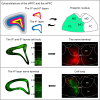
Summary of the granular dlPFC→MD in the Chinese tree shrew. This study found that the Chinese tree shrew has evolved the granular dlPFC with reciprocal connections with the MD. MD, mediodorsal nucleus; PC, paracentral nucleus; IMD, intermediodorsal nucleus; CM, central medial nucleus.

Similar articles

References

    1. Arnsten AF (2015) Stress weakens prefrontal networks: molecular insults to higher cognition. Nat Neurosci 18:1376–1385. 10.1038/nn.4087 - DOI - PMC - PubMed
    1. Berger RJ, Walker JM (1972) A polygraphic study of sleep in the tree shrew (Tupaia glis). Brain Behav Evol 5:54–69. 10.1159/000123737 - DOI - PubMed
    1. Brodmann K (1909) Vergleichende Lokalisationslehre der Grosshirnrinde in ihren Prinzipien dargestellt auf Grund des Zellenbaues. Lepzig: Barth.
    1. Buettner-Janusch J (1963) Evolutionary and genetic biology of primates. London and New York: Academic Press.
    1. Cai JX, Xu L, Hu XT, Ma YY, Su W, Xiao KY (1993) The relation between evolution of spatial working memory function and of morphology of the dorsolateral prefrontal cortex among the rhesus monkey, slow loris and tree shrew. Zool Res 14:158–165.

LinkOut - more resources