. 2024 Oct 24;15(1):9194.
doi: 10.1038/s41467-024-53512-x.

Structural insights into the inhibition mechanism of fungal GWT1 by manogepix

Affiliations

Structural insights into the inhibition mechanism of fungal GWT1 by manogepix

Xinli Dai et al. Nat Commun. .

Abstract

Glycosylphosphatidylinositol (GPI) acyltransferase is crucial for the synthesis of GPI-anchored proteins. Targeting the fungal glycosylphosphatidylinositol acyltransferase GWT1 by manogepix is a promising antifungal strategy. However, the inhibitory mechanism of manogepix remains unclear. Here, we present cryo-EM structures of yeast GWT1 bound to the substrate (palmitoyl-CoA) and inhibitor (manogepix) at 3.3 Å and 3.5 Å, respectively. GWT1 adopts a unique fold with 13 transmembrane (TM) helixes. The palmitoyl-CoA inserts into the chamber among TM4, 5, 6, 7, and 12. The crucial residues (D145 and K155) located on the loop between TM4 and TM5 potentially bind to the GPI precursor, contributing to substrate recognition and catalysis, respectively. The antifungal drug, manogepix, occupies the hydrophobic cavity of the palmitoyl-CoA binding site, suggesting a competitive inhibitory mechanism. Structural analysis of resistance mutations elucidates the drug specificity and selectivity. These findings pave the way for the development of potent and selective antifungal drugs targeting GWT1.


Conflict of interest statement

The authors declare no competing interests.

Figures

Fig. 1
Fig. 1. The characterization of GWT1.
a The cartoon representations of the substrates and products of the GWT1 enzyme are displayed. The chemical structure of manogepix, an inhibitor of GWT1 protein, is demonstrated. The hydroxyl group that accepts the palmitoyl group (yellow) is highlighted in the red circle. b Topology of fGWT1 presents the specific sites of GFP insertion. Through screening, A fusion of GFP at the Y422 position construct exhibited the most homogeneous protein properties. c FACS (fluorescence-activated cell sorting) of HEK293T△PIGW cells expressing fGWT1 (light green) using CD59 as the marker with gating strategies. HEK293TWT cells are highlighted in red, while individual HEK293T△PIGW cells are marked in blue. Those HEK293T△PIGW cells responding to fGWT1 are labeled in light green. The background served as a blank control, referring to HEK293T△PIGW cells not stained with CD59 antibodies. d Fluorescent confocal microscopy analyses were conducted to assess fGWT1 activity, with the construct and cell line indicated alongside the images. The first, second, and third columns illustrate confocal fluorescence microscopy images of CD59, DAPI, and merged views, respectively. A scale bar of 10 μm is included. The confocal microscopy experiment was repeated at least three times.
Fig. 2
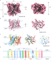
Fig. 2. Cryo-EM structure of GWT1 bound palmitoyl-CoA.
a The density map of the monomeric GWT1 bound to palmitoyl-CoA, front views from within the plane of the membrane (left) and the intracellular side of the membrane (right). The approximate position of the ER membrane is indicated with light blue shading. The elongated density is shown in green. b The structure of the monomeric GWT1 in complex with palmitoyl-CoA monomer is illustrated in cartoon representations, offering perspectives from within the plane of the membrane (left) and the intracellular side of the membrane (right). The approximate position of the ER membrane is highlighted with light blue shading. c. The structural models of GWT1 bound to palmitoyl-CoA monomer are shown in the front view and top view. Helices are shown as cylinders and depicted in rainbow colors. Palmitoyl-CoA is encircled by a transmembrane spiral marked with a blue circle, and a red star indicates palmitoyl-CoA. d, The topology of palmitoyl-CoA-bound GWT1 is represented using colors like in panel (c).
Fig. 3
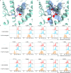
Fig. 3. Overall structure of GWT1 and palmitoyl-CoA-binding site.
a Three significant openings leading to the reaction chamber of palmitoyl-CoA include a cytosolic tunnel (highlighted in green), an ER-luminal tunnel (highlighted in blue), and the overall structure of palmitoyl-CoA (highlighted in orange). The electrostatic surface is illustrated. b The structural models of GWT1 bound to palmitoyl-CoA. Helices are represented as cylinders, and the palmitoyl-CoA molecule is depicted as a green ball and stick. The approximate position of the ER membrane is highlighted with light blue shading. c The cut-away view illustrates the binding pocket of the substrate palmitoyl-CoA within the reaction chamber. The regions corresponding to the “head”, “neck”, and “tail” of palmitoyl-CoA are highlighted with orange, purple, and blue frames, respectively. The palmitoyl-CoA molecule is shown as a ball and stick (depicted in green). df Amino acids located at the sites surrounding the head (highlighted in orange frame), neck (highlighted in light purple frame), and tail (highlighted in blue frame) of palmitoyl-CoA are shown as sticks with carbon atoms colored in green. gi Sequence logos were generated using the WebLogo server (https://weblogo.berkeley.edu/logo.cgi) for a region encompassing conserved binding pocket of palmitoyl-CoA. Amino acids are color-coded based on their chemical properties: polar amino acids (G, S, T, Y, C, Q, N) are represented in green; basic amino acids (K, R, H) are depicted in blue; acidic amino acids (D, E) are shown in red; and hydrophobic amino acids (A, V, L, I, P, W, F, M) are illustrated in black.
Fig. 4
Fig. 4. The proposed luminal access cavity for GlcN-PI.
a, d The cryo-EM map of GWT1 is displayed from both side (a) and top (d) perspectives, with the luminal access cavity highlighted by a blue dashed oval. b, e An additional density within the highly conserved luminal access cavity. c Molecular lipophilicity potential (MLP) of the luminal access cavity. The surfaces are colored by lipophilicity potential calculated by Chimera X. The putative entry route for GlcN-PI is indicated by a dashed blue path. f Comparison of catalytic sites of GWT1 and HGSNAT. The predicted catalytic sites of HGSNAT are N286 and H297, while the putative catalytic sites of GWT1 are D145 and K155. g Expressing fGWT1 in HEK293T△PIGW cells restores CD59 staining in the FACS assay. HEK293T△PIGW cells, where GPI-AP biosynthesis is blocked, serve as the staining control (gray). Loss-of-function mutants are highlighted in brilliant blue. Coordination residue mutants of the luminal access cavity are highlighted in purple.
Fig. 5
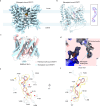
Fig. 5. Cryo-EM structure of GWT1 in complex with manogepix.
ab Density map (a) and structure (b) of GWT1 in complex with manogepix. The density map and structure of GWT1 are colored blue, while the density map of manogepix is shown in yellow and its structure is in lilac. c Superposition of the palmitoyl-CoA-bound GWT1 complex and the manogepix-bound GWT1 complex. The palmitoyl-CoA-bound GWT1 complex and the manogepix-bound GWT1 complex are shown as cylinder with colors of pink and blue. d Sliced view of the molecular surface of fGWT1, highlighting the bound manogepix (blue) and palmitoyl-CoA (pink). ef Close-up views of the manogepix (depicted in lilac), showing cryo-EM density (highlighted in yellow) and contacting GWT1 residues side chain (rendered in white).
Fig. 6
Fig. 6. Drug resistance mutation in GWT1.
a Previously reported drug resistance sites for GWT1 are shown as spheres and colored in gray-blue. b All drug resistance sites, including those previously reported and newly identified, are shown. c Evaluating the sensitivity of manogepix by staining CD59 in the FACS assay, expressing fGWT1 in HEK293T△PIGW cells. Cells treated with manogepix (orange) or without manogepix (blue) were analyzed by flow cytometry using gating strategies, with negative controls marked in red. All experiments were conducted independently three times (n = 3).
Fig. 7
Fig. 7. Putative catalytic model and inhibitory mechanism of GWT1.
a Hypothetical model for GWT1-catalysed GlcN-(acyl)PI formation. Highlighting the substrate-donor (Palmitoyl-CoA) and acceptor (GlcN-PI) binding pocket. The binding sites for palmitoyl-CoA are colored in blue, while the putative GlcN-PI binding sites are colored in orange. b The putative post-catalytic state contains two products including GlcN-(acyl)PI and free CoA. c The diagram of the inhibition mechanism model. The inhibitor competitively occupies the binding cavity of the lipid tail of palmitoyl-CoA, blocking the palmitoyl-CoA entry.

Similar articles

References

    1. Lockhart, S. R., Chowdhary, A. & Gold, J. A. W. The rapid emergence of antifungal-resistant human-pathogenic fungi. Nat. Rev. Microbiol.21, 818–832 (2023). - PMC - PubMed
    1. Perfect, J. R. The antifungal pipeline: a reality check. Nat. Rev. Drug Discov.16, 603–616 (2017). - PMC - PubMed
    1. Fisher, M. C., Hawkins, N. J., Sanglard, D. & Gurr, S. J. Worldwide emergence of resistance to antifungal drugs challenges human health and food security. Science360, 739–742 (2018). - PubMed
    1. Parums, D. V. Editorial: The World Health Organization (WHO) fungal priority pathogens list in response to emerging fungal pathogens during the COVID-19 pandemic. Med. Sci. Monit.28, e939088 (2022). - PMC - PubMed
    1. Vogt, M. S., Schmitz, G. F., Varon Silva, D., Mosch, H. U. & Essen, L. O. Structural base for the transfer of GPI-anchored glycoproteins into fungal cell walls. Proc. Natl Acad. Sci. Usa.117, 22061–22067 (2020). - PMC - PubMed

MeSH terms

LinkOut - more resources