. 2024 Nov 27;52(21):13019-13035.
doi: 10.1093/nar/gkae918.

PCNA-binding activity separates RNF168 functions in DNA replication and DNA double-stranded break signaling

Affiliations

PCNA-binding activity separates RNF168 functions in DNA replication and DNA double-stranded break signaling

Yang Yang et al. Nucleic Acids Res. .

Abstract

RNF168 orchestrates a ubiquitin-dependent DNA damage response to regulate the recruitment of repair factors, such as 53BP1 to DNA double-strand breaks (DSBs). In addition to its canonical functions in DSB signaling, RNF168 may facilitate DNA replication fork progression. However, the precise role of RNF168 in DNA replication remains unclear. Here, we demonstrate that RNF168 is recruited to DNA replication factories in a manner that is independent of the canonical DSB response pathway regulated by Ataxia-Telangiectasia Mutated (ATM) and RNF8. We identify a degenerate Proliferating Cell Nuclear Antigen (PCNA)-interacting peptide (DPIP) motif in the C-terminus of RNF168, which together with its Motif Interacting with Ubiquitin (MIU) domain mediates binding to mono-ubiquitylated PCNA at replication factories. An RNF168 mutant harboring inactivating substitutions in its DPIP box and MIU1 domain (termed RNF168 ΔDPIP/ΔMIU1) is not recruited to sites of DNA synthesis and fails to support ongoing DNA replication. Notably, the PCNA interaction-deficient RNF168 ΔDPIP/ΔMIU1 mutant fully rescues the ability of RNF168-/- cells to form 53BP1 foci in response to DNA DSBs. Therefore, RNF168 functions in DNA replication and DSB signaling are fully separable. Our results define a new mechanism by which RNF168 promotes DNA replication independently of its canonical functions in DSB signaling.


Figures

Graphical Abstract
Graphical Abstract
Figure 1.
Figure 1.
RNF168 is recruited to DNA replication and DSB via separable mechanisms. (A) U2OS cells were transiently co-transfected with expression constructs encoding HA-PCNA and FLAG-RNF168 WT or FLAG-RNF168 ΔMIU2. Forty-eight hours post-transfection, cells were treated conditionally with 2 mM HU or with 10 Gy IR. Cells were fixed 2 h following HU or 30 min following IR treatments, then stained with anti-HA and anti-FLAG antibodies prior to analysis by immunofluorescence confocal microscopy. Panel (A) shows images of representative HA-PCNA and FLAG-RNF168 co-expressing cells from all experimental conditions. (B) Bar chart showing enumeration of cells containing HA-PCNA-co-localizing FLAG-RNF168 foci. Each data point represents the mean of results from three separate experiments and the error-bars represent the standard deviation. (C) U2OS cells were transiently co-transfected with constructs encoding FLAG-RNF168 WT or FLAG-RNF8 WT. Forty-eight hours post-transfection, cells were treated conditionally with 2 mM HU. Cells were fixed 2 h following HU or treatments, then stained with anti-HA and anti-PCNA antibodies prior to analysis by immunofluorescence confocal microscopy. The individual images are of representative cells. Cells transfected with FLAG-RNF168 plasmids co-expressed both PCNA and RNF168 proteins. Unexpectedly, however, in cultures co-transfected with FLAG-RNF8 plasmid, immunoreactivity with anti-PCNA and anti-FLAG antibodies was mutually exclusive (suggesting that FLAG-RNF8 is not expressed at detectable levels in PCNA-positive S-phase cells). (D) U2OS cells were transiently transfected with constructs encoding FLAG-RNF168 WT or FLAG-RNF8 WT. Twenty-four hours post-transfection, cells were treated conditionally with 300 ng/ml NCS. Cells were fixed 1 h following NCS or treatments, then stained with anti-FLAG and anti-γH2AX antibodies prior to analysis by immunofluorescence confocal microscopy. The individual images are of representative cells showing that both RNF8 and RNF168 co-localize with γH2AX. (E) Parental RNF8+/+ U2OS cells, RNF8−/− U2OS cells or RNF8−/− cells transiently transfected with an RNF8 expression plasmid (for 24 h) were treated with 300 ng/ml NCS. One hour after NCS treatment, cells were fixed and stained with 53BP1 antibodies prior to analysis by immunofluorescence confocal microscopy. The individual images are of representative cells showing RNF8-dependency of 53BP1 focus formation. (F) RNF8+/+ cells or RNF8−/− derivatives were transiently transfected with an RNF168 expression plasmid. Twenty-four hours post-transfection, cells were pulse-labeled with 10 mM EdU for 1 h to label sites of ongoing DNA synthesis. Cells were fixed and stained with antibodies against EdU and FLAG prior to analysis by immunofluorescence confocal microscopy. The individual images are of representative cells showing RNF8-independent co-localization of RNF168 and EdU. (G) Bar chart showing enumeration of cells containing EdU-co-localizing FLAG-RNF168 foci in RNF8+/+ and RNF8−/− cells. Each data point represents the mean of results from three separate experiments and the error-bars represent the standard deviation. Unpaired Student’s t-test demonstrated no statistically significant co-localization between RNF8 and EdU incorporation (P = 0.5867).
Figure 2.
Figure 2.
RNF168 contains a DPIP domain. (A) Domain organization of RNF168 indicating key functional domains and a degenerate PIP-like sequence residing in a disordered C-terminal region. The protein disorder profile was generated using the Protein Disorder Prediction (PrDOS) tool at http://prdos.hgc.jp/cgi-bin/top.cgi. (B) AlphaFold model of the PCNA–RNF168 DPIP complex reveals a similar binding model to that of the PCNA-Pol Kappa PIP complex. Left panel shows the top predicted model of the trimeric PCNA–RNF168 DPIP complex. Right panel shows the overlay of the AlphaFold model of the trimeric PCNA–RNF168 DPIP complex with the crystal structure of the trimeric PCNA-Polκ PIP (PDB: 2zvl), with the PCNA and Polk PIP in the crystal structure colored in cyan and yellow, respectively. (C) H1299 cells were transiently co-transfected with expression constructs encoding HA-PCNA and FLAG-RNF168 WT or FLAG-RNF168 ΔDPIP. Forty-eight hours post-transfection cells were fixed, then stained with anti-HA and anti-FLAG antibodies prior to analysis by immunofluorescence confocal microscopy. The photographs are of representative cells co-expressing HA-PCNA and WT or ΔDPIP forms of FLAG-RNF168. The bar chart shows enumeration of cells containing HA-PCNA-co-localizing FLAG-RNF168 foci. Each data point represents the mean of results from three separate experiments and the error-bars represent the standard deviation. (D) RNF168−/− U2OS cells were infected with adenoviral vectors encoding FLAG-RNF168 WT or FLAG-RNF168 ΔDPIP. Twenty-four hours post-infection cells were fixed and subject to PLA to compare proximities of WT and ΔDPIP RNF168 with endogenous PCNA. The photographs are of representative DAPI-stained nuclei from each experimental condition. The immunoblots show levels of ectopically expressed WT and ΔPIP RNF168 in this experiment. The bar chart showing enumeration of cells containing eight or more PCNA / FLAG-RNF168 PLA foci. Each data point represents the mean of results from three separate experiments and the error-bars represent the standard deviation. Ordinary one-way analysis of variance (ANOVA) demonstrated statistically significant difference between RNF168 WT and PIP (P = 0.0049). (E) Replicate plates of H1299 cells were co-infected with adenovirus vectors encoding HA-PCNA and RNF168 WT or RNF168 ΔDPIP. Twenty-four hours post infection, some cultures were treated with HU (2 mM, 2 h), camptothecin (CPT, 100 nM, 2 h), ATM inhibitor KU55933 100 nM, 2 h) or the WEE1 inhibitor MK1775 (10 μM, 2 h). Chromatin extracts from the treated and untreated (control) cells were normalized for protein content and immunoprecipitated with anti-HA antibodies. Anti-HA immunoprecipitates were resolved on SDS-PAGE, transferred to nitrocellulose and analyzed by immunoblotting with the indicated antibodies. (F) Replicate plates of RNF8−/− U2OS cells were transiently infected with adenovirus vectors encoding FLAG-RNF168 WT, FLAG-RNF168 ΔDPIP or FLAG-RNF168 ΔMIU2, or with a control ‘empty’ adenoviral vector. Forty-eight hours post-infection chromatin extracts were prepared, normalized for protein content and immunoprecipitated with anti-FLAG antibodies. Anti-FLAG immunoprecipitates were resolved on SDS-PAGE, transferred to nitrocellulose and analyzed by immunoblotting with the indicated antibodies.
Figure 3.
Figure 3.
PCNA ubiquitylation facilitates RNF168-binding. (A) ITC was conducted by titrating synthetic peptide (p21, GRKRRQTSMTDFYHSKRRLIFS-amide where underlined residues denote PIP box; syringe, 150–300 μM) into a solution of PCNA (cell; 30 μM) in TBS. Control experiments using peptide injected into buffer alone showed minimal heats with no evidence of titration. Analysis of the isotherm yielded Kd = 76.3 nM and stoichiometry = 0.93 (Microcal Origin software). (B) Replicate plates of H1299 cells were infected with adenovirus vectors encoding FLAG-RNF168 WT and HA-PCNA (or an ‘empty’ adenovirus vector for control). Thirty-six hours post-infection, chromatin extracts were prepared, normalized for protein content and immunoprecipitated with anti-FLAG antibody in the presence of different concentrations of peptides corresponding to the p21 or Polη PIP boxes. Anti-FLAG immunoprecipitates were resolved on SDS-PAGE, transferred to nitrocellulose and analyzed by immunoblotting with the indicated antibodies. (C) Replicate plates of H1299 cells were co-infected with adenovirus vectors encoding HA-PCNA and FLAG-RNF168 WT or HA-PCNA and RNF168 ΔDPIP. Some cells received an ‘empty’ adenovirus vector instead of HA-PCNA (for control). Thirty-six hours post-infection, some plates were treated with 2 mM HU for 2 h. Chromatin extracts were prepared, normalized for protein content and immunoprecipitated with anti-HA antibody in the presence or absence of the p21 PIP box peptide (1 mM). Anti-HA immune complexes were resolved on SDS-PAGE, transferred to nitrocellulose and analyzed by immunoblotting with the indicated antibodies. (D) Replicate plates of H1299 cells were infected with adenovirus vectors encoding wild-type PCNA (HA-PCNA WT), ubiquitylation-resistant PCNA (HA-PCNA K164R) or a PCNA-ubiquitin fusion (HA-PCNA-Ub) in combination with FLAG-RNF168 WT adenovirus. Some cells received an ‘empty’ adenovirus vector instead of HA-PCNA viruses(for control). Thirty-six hours post-infection, chromatin extracts were prepared, normalized for protein content and immunoprecipitated with anti-HA antibody. Anti-HA immune complexes were resolved on SDS-PAGE, transferred to nitrocellulose and analyzed by immunoblotting with the indicated antibodies. (E) Replicate plates of H1299 cells were transfected with expression plasmids encoding WT or mutant forms of RNF168. The transfected cultures were then infected with adenovirus vector encoding wild-type PCNA (HA-PCNA WT) or with an ‘empty’ adenovirus vector (for control). Thirty-six hours post-infection, chromatin extracts were prepared, normalized for protein content and immunoprecipitated with anti-HA antibody. Anti-HA immune complexes were resolved on SDS-PAGE, transferred to nitrocellulose and analyzed by immunoblotting with the indicated antibodies.
Figure 4.
Figure 4.
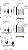
RNF168 PCNA-interacting motifs are required to sustain ongoing DNA replication fork progression and recovery from replicative stress. (A) RNF168−/− U2OS cells were stably transduced with retroviral vectors encoding RNF168 WT, RNF168 ΔDPIP and RNF168 ΔMIU2, RNF168 ΔMIU1, RNF168 ΔDPIP/ΔMIU1, RNF168 Super-PIP under transcriptional control of a doxycycline-inducible promoter, or with the ‘empty’ retroviral vector for control. Replicate plates of cells were treated with 20 ng/ml doxycycline. Twenty-four hours after doxycycline treatment, exponentially growing cells were pulse-labeled with CldU and IdU. DNA fibers prepared from the labelled cells were immuno-stained with antibodies against CldU/IdU-treated and DNA replication dynamics were determined based on relative lengths of CldU/IdU-labeled tracts. Statistical significance was calculated using a Mann–Whitney rank sum test. (B) Enumeration of ongoing and stalled DNA replication fork structures in cells complemented with wild-type and mutant forms of RNF168. Statistical significance was calculated using an unpaired Student’s T-test (two-sided, equal variance) (C and D) RNF168−/− U2OS cells complemented with different RNF168 mutants were treated sequentially with IdU (to label ongoing forks), then with HU (to induce DNA replication fork arrest). After HU washout, cells were pulse-labelled with IdU (to monitor rates of DNA replication fork recovery). DNA replication dynamics were determined as described in (A) and (B). (E) RNF168−/− U2OS cells complemented with different RNF168 mutants were transfected with control or 53BP1 siRNA, treated sequentially with CldU and IdU, then exposed to 4 mM HU for 5 h to induce DNA replication fork arrest. Replication fork stability was determined based on the ratio between the relative lengths of the CldU/IdU-labeled tracts. Statistical significance was calculated using a Mann–Whitney rank sum test.
Figure 5.
Figure 5.
RNF168–PCNA interactions are dispensable for DSB signaling to 53BP1. (A) Immunoblotting of DSB markers showing kinetics of the DDR in U2OS cells treated with NCS (100 ng/ml). (B and D) U2OS cells containing mCherry-PCNA and Venus-53BP1 reporters were electroporated with siRNF168 or with non-targeting siCon RNA for controls. Control and RNF168-depleted cells were re-plated after electroporation and 12 h later cells were infected with 1 × 108 pfu/ml of Ad-RNF168 WT or AdCon. Cultures were subject to live cell imaging every 15 min for 1200 min and were treated with NCS (100 ng/ml) at 240 min. The panels in (B) show 53BP1 foci in a representative Ad-RNF68 WT-infected cell that was in S-phase at the time of NCS treatment and was imaged repeatedly for 960 min. Panel (C) shows mean numbers of 53BP1 foci from three independent experiments in which 50 cells were individually tracked and imaged every 15 min for the indicated number of cycles both prior to and after NCS-treatment. The arrow indicates when NCS was added to the cultures. Note that In RNF168-expressing cells (‘siCon + AdCon’ and ‘siRNF168 + Ad RNF168’), numbers of 53BP1 foci reach a plateau 1 h following NCS treatment. Panel (D) is an immunoblot confirming efficient knockdown of endogenous RNF168 and effective reconstitution of RNF168-depleted cells with Adenovirus-encoded RNF168 (Ad-RNF168 WT). (E) Immunoblot confirming expression of wild-type and mutant forms of RNF168 in adenovirus-reconstituted RNF168−/− cells. (F) Confocal Immunofluorescence microscopy images showing patterns of γH2AX, 53BP1 and EdU foci in representative S-phase nuclei from NCS-treated RNF168−/− cells reconstituted with AdRNF168 WT, AdRNF168 ΔDPIP/ΔMIU1 or AdCon for control. (G) The upper panels show the fraction of cells that contained 53BP1 foci in S-phase (EdU-positive) and non-S-phase cells. The lower panels show the degree of enrichment of 53BP1 in γH2AX foci, calculated as described in the ‘Materials and methods’ section.

Similar articles

References

    1. Smeenk G., Mailand N.. Writers, readers, and erasers of histone ubiquitylation in DNA double-strand break repair. Front. Genet. 2016; 7:122. - PMC - PubMed
    1. Stewart G.S. Solving the RIDDLE of 53BP1 recruitment to sites of damage. Cell Cycle. 2009; 8:1532–1538. - PubMed
    1. Mattiroli F., Penengo L.. Histone ubiquitination: an integrative signaling platform in genome stability. Trends Genet. 2021; 37:566–581. - PubMed
    1. Stewart G.S., Panier S., Townsend K., Al-Hakim A.K., Kolas N.K., Miller E.S., Nakada S., Ylanko J., Olivarius S., Mendez M.et al. .. The RIDDLE syndrome protein mediates a ubiquitin-dependent signaling cascade at sites of DNA damage. Cell. 2009; 136:420–434. - PubMed
    1. Fradet-Turcotte A., Canny M.D., Escribano-Diaz C., Orthwein A., Leung C.C., Huang H., Landry M.C., Kitevski-LeBlanc J., Noordermeer S.M., Sicheri F.et al. .. 53BP1 is a reader of the DNA-damage-induced H2A Lys 15 ubiquitin mark. Nature. 2013; 499:50–54. - PMC - PubMed

MeSH terms

Substances