. 2024 Oct 11;3(10):pgae462.
doi: 10.1093/pnasnexus/pgae462. eCollection 2024 Oct.

Engineering a cleaved, prefusion-stabilized influenza B virus hemagglutinin by identification and locking of all six pH switches

Affiliations

Engineering a cleaved, prefusion-stabilized influenza B virus hemagglutinin by identification and locking of all six pH switches

Jarek Juraszek et al. PNAS Nexus. .

Abstract

Vaccine components based on viral fusion proteins require high stability of the native prefusion conformation for optimal potency and manufacturability. In the case of influenza B virus hemagglutinin (HA), the stem's conformation relies on efficient cleavage. In this study, we identified six pH-sensitive regions distributed across the entire ectodomain where protonated histidines assume either a repulsive or an attractive role. Substitutions in these areas enhanced the protein's expression, quality, and stability in its prefusion trimeric state. Importantly, this stabilization enabled the production of a cleavable HA0, which is further processed into HA1 and HA2 by furin during exocytic pathway passage, thereby facilitating correct folding, increased stability, and screening for additional stabilizing substitutions in the core of the metastable fusion domain. Cryo-EM analysis at neutral and low pH revealed a previously unnoticed pH switch involving the C-terminal residues of the natively cleaved HA1. This switch keeps the fusion peptide in a clamped state at neutral pH, averting premature conformational shift. Our findings shed light on new strategies for possible improvements of recombinant or genetic-based influenza B vaccines.


Figures

Fig. 1.
Fig. 1.
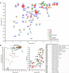
Influenza B virus HA protein structure. A) Schematic representation of HA with indicated RR1, including FP and HR, RR2, CH, transmembrane domain, and cytoplasmic domain. Structures of influenza B virus monomer in prefusion (26) and postfusion (27) conformation colored according to upper schematic description. B-loop and B-helix are indicated and histidines (H) in regions of instability. B) Structure of trimeric prefusion HA with one protomer colored according to A and location of regions of instability with zoomed-in colored panels to the right of the C) head switch (top view) and D) hinge, E) neck, and F) stem switch (side view). G) Top view of stem switch at the plane of the helical kink of helix C and D showing the repulsive cluster with E113 and the two pH-sensitive switches around H22 and H114 in an unprotonated situation (left panel) and possible impact after protonation (right panel). Colored according to A.
Fig. 2.
Fig. 2.
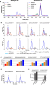
Characterization of HA variants containing stabilizing substitutions. HA trimer expression using analytical SEC and thermal stability using DSF of cell culture supernatant of Expi293F cells expressing influenza B virus HA (Fig. S2). A) A plot of trimer expression level against trimer:monomer ratio for each mutant is shown. The radius of markers is relative to the melting temperatures (Tm50) as measured by DSF. WT is indicated in brown at the cross hair. Markers are colored by different regions, including the regions of instability. Labels show residue number for HA1 (bold italic in blue) and HA2 (black). B) Plot of trimer expression level against trimer:monomer ratio for combinations of stabilizing substitutions with marker radius relative to Tm50. Markers are shown as pie charts with the colors according to the region of the stabilizing substitution as in (A) (left panel). Fully trimeric variants are boxed in the left panel and represented in the right panel plot showing trimer expression level against Tm50. Markers are colored pie charts with color shades corresponding to the region of the stabilizing mutations.
Fig. 3.
Fig. 3.
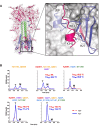
Stability of cleaved and uncleaved HAs. A) Impact of introduction of furin cleavage sites to analytical SEC profiles of culture supernatants of Expi293F cells expressing influenza B virus HA variants based on WT influenza B virus HA with monobasic or polybasic cleavage sites (left panel) or HA stabilized by substitutions in five regions of instability with different furin site designs (right panels). B) Impact of different levels of stabilization on trimer formation in uncleaved (upper panels) and cleaved (lower panels) HA variants for four different influenza B strains. Analytical SEC profiles of culture supernatant of Expi293F cells expressing selected influenza B WT (red broken line) repaired (light blue line in right upper panels), stabilization of neck and hinge switch (HA1 H311W and HA2 Q64W), stabilization of repulsive cluster, head, and stem switch (HA2 E113W, HA1 K209T, and HA2 H22F) or stabilization in five regions of instability (HA1 K209T, HA2 H22F, Q64Y, G68Y, and E113W). Upper panels contain a monobasic cleavage site which is not cleaved. Bottom panels are cleaved variants with an engineered RSV p27 domain which is cleaved out by furin at low pH. Peaks representing monomeric and trimeric species are indicated by an “M” and a “T,” respectively. C) Temperature stability as determined by DSF for WT and stabilized uncleaved HA variants. Colors as in B. D) Analytical SEC profiles of purified uncleaved (solid line) and cleaved (broken line) stabilized B/Iowa/06/2017 and B/Ohio/01/2005 HA. E) Temperature stability as determined by DSF of purified uncleaved and cleaved stabilized HAs. Stabilized variants including repair mutations are indicated by the asterisk symbol. All strains in the panels are from Victoria lineage except for B/Florida/04/2006 which is from Yamagata lineage.
Fig. 4.
Fig. 4.
The characterization of additional HA substitutions in the stem switch and repulsive cluster region. Analytical SEC and DSF of cell culture supernatant of Expi293F cells expressing variants of stabilized cleaved B/Iowa/06/2017 HA. A) All variants contained identical stabilizing substitutions (HA1 K209T, HA2 Q64Y, G68Y, and H22F) and variations at several positions in the stem switch (S40A, S110R, and H114F) and repulsive cluster (E113W/Q). Plot showing cleaved influenza B virus HA trimer expression level against temperature stability (Tm50), compared with benchmark stabilized HA indicated with dashed lines and gray sphere (HA1 K209T, HA2 E113W, Q64Y, G68Y, and H22F). The labels indicate substitutions at positions HA2 40, 110, 113, and 114 using the color coding as in Fig. 2. B) Intact mass LC–MS analysis of HA1 chain from purified Stable_HA1HA2 labeled ARWF, as described in Fig. S5. HA1 is present as equally abundant forms HA1a and HA1b with and without C-terminal arginine. C-terminal sequence is indicated in the plot. C). pH stability in different buffers during 24 h of HA variants stabilized in head, hinge, and neck (green line), stabilized in five regions of instability (blue line), as previous with three additional stabilizing substitutions in the stem (purple line) and as previous including H27F substitution in the sixth region of instability (red line). All constructs include (HA1 K209T, HA2 G68Y, and Q64Y), Additional substitutions are indicated in the left panel for each construct with color coding as in Fig. 2. HA trimer content is indicated as a percentage of the starting material as measured by analytical SEC. For pH 4.8, the raw data are shown in Fig. S9. D) HA fusogenicity as measured in a cell–cell fusion assay in HEK293 cells by co-transfection of plasmids encoding WT or stabilized HA, human TMPRSS2, and mScarlet. Fusion was triggered 24 h posttransfection by a 10-min exposure to pH 5.0 medium, followed by a 1-h recovery at pH 7.4. Shown are overlays of red and blue (Hoechst) channels. The “combination” design has all stabilizing substitutions indicated in bold.
Fig. 5.
Fig. 5.
Cryo-EM analysis of stabilized pH-sensitive switch regions. A) Head switch with K209T. B) Hinge switch with G68Y. C) Neck switch with Q64Y. The orange circle indicates that polar interactions remain intact. D) Stem switch (left panel) with aromatic cluster. Interactions have been grouped into three tightly bound clusters. Side chains for one protomer are colored as in Fig. 1. (Right panel) Details of the mutations in the single interaction cluster (delimited with red line in the left panel). K47 forming the pi-cation interaction with F2 shown as orange dashed line. E) Stem switch with polar interactions with water molecule in red. The clouds around the sidechain indicate the van der Waals surface.
Fig. 6.
Fig. 6.
The sixth pH-sensitive switch in the HA1 C-terminal helix and intersubunit beta-sheet at the base of influenza B virus HA. A) Interacting residue and clamped FP are indicated. H27 forms a pi-cation bond with K342 and an edge-to-face aromatic interaction with neighboring W14. B) Analytical SEC of HAs with increasing number of stabilizing substitutions with H27 (red broken line) or F27 (blue solid line). Trimer (T) and monomer (M) peaks are indicated. Melting temperatures (Tm50) as measured by DSF in cell culture supernatant are indicated in each respective panel in red (H27) and blue (F27). Stabilizing residues are indicated above the panel and color coded as Fig. 1.

Similar articles

References

    1. Paul Glezen W, Schmier JK, Kuehn CM, Ryan KJ, Oxford J. 2013. The burden of influenza B: a structured literature review. Am J Public Health. 103:e43–e51. - PMC - PubMed
    1. Cotter CR, Jin H, Chen Z. 2014. A single amino acid in the stalk region of the H1N1pdm influenza virus HA protein affects viral fusion, stability and infectivity. PLoS Pathog. 10:e1003831. - PMC - PubMed
    1. Hu M, et al. . 2023. Hemagglutinin destabilization in H3N2 vaccine reference viruses skews antigenicity and prevents airborne transmission in ferrets. Sci Adv. 9:eadf5182. - PMC - PubMed
    1. Sanders RW, et al. . 2002. Stabilization of the soluble, cleaved, trimeric form of the envelope glycoprotein complex of human immunodeficiency virus type 1. J Virol. 76:8875–8889. - PMC - PubMed
    1. Krarup A, et al. . 2015. A highly stable prefusion RSV F vaccine derived from structural analysis of the fusion mechanism. Nat Commun. 6:8143. - PMC - PubMed