. 2024 Oct 17;15(1):8276.
doi: 10.1038/s41467-024-52634-6.

Substrate engagement by the intramembrane metalloprotease SpoIVFB

Affiliations

Substrate engagement by the intramembrane metalloprotease SpoIVFB

Melanie A Orlando et al. Nat Commun. .

Abstract

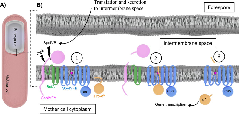
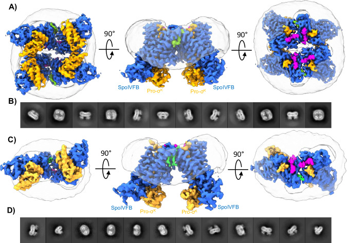
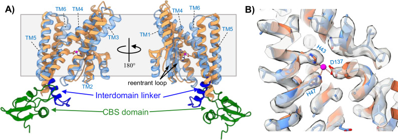
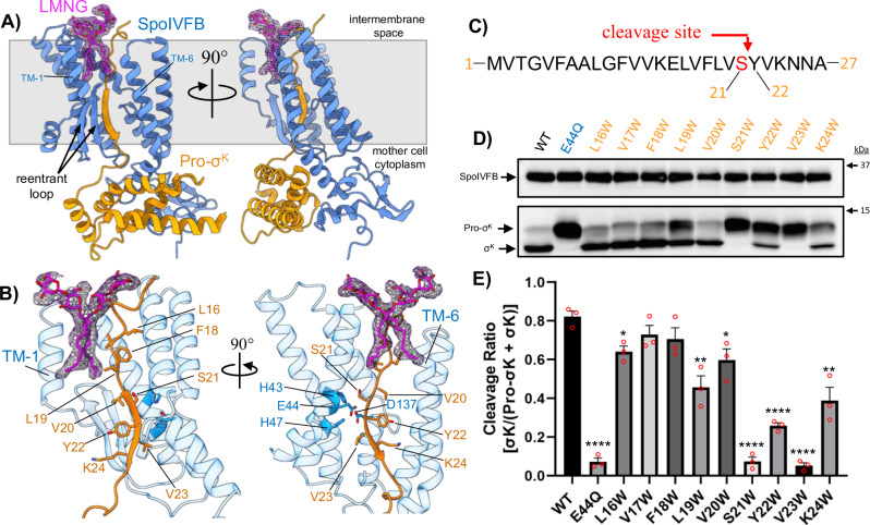
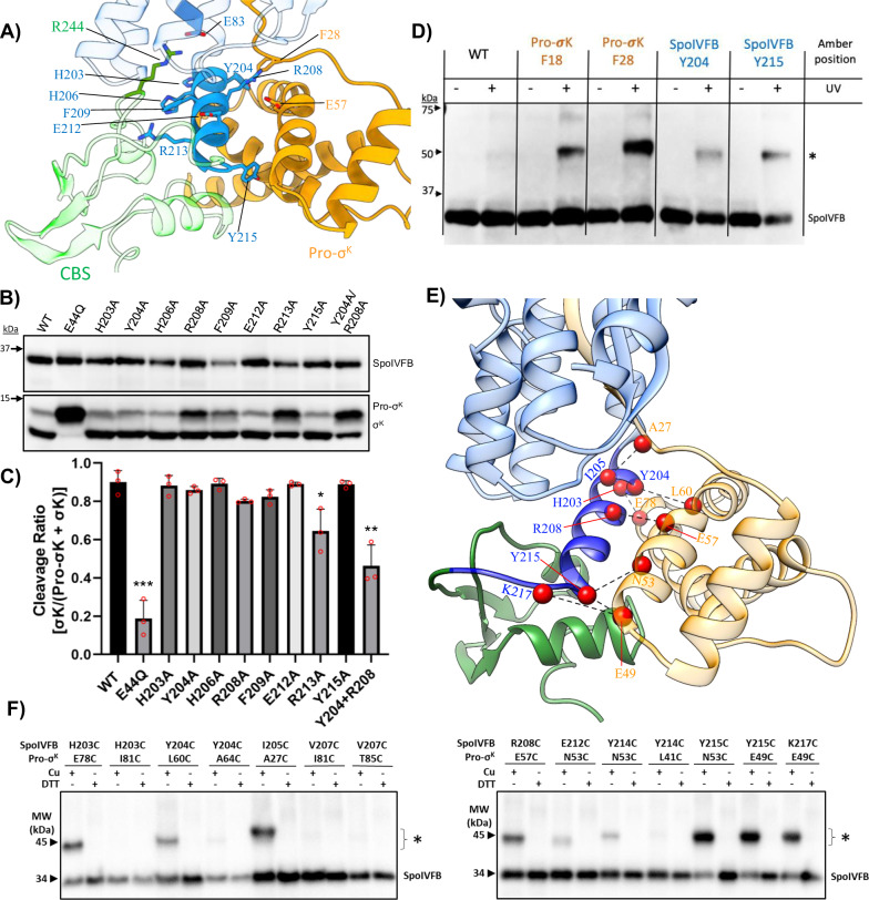
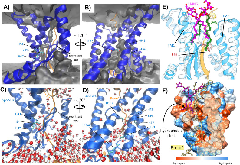
S2P intramembrane metalloproteases regulate diverse signaling pathways across all three domains of life. However, the mechanism by which S2P metalloproteases engage substrates and catalyze peptide hydrolysis within lipid membranes has remained elusive. Here we determine the cryo-EM structure of the S2P family intramembrane metalloprotease SpoIVFB from Bacillus subtilis bound to its native substrate Pro-σK. The structure and accompanying biochemical data demonstrate that SpoIVFB positions Pro-σK at the enzyme active site through a β-sheet augmentation mechanism, and reveal key interactions between Pro-σK and the interdomain linker connecting SpoIVFB transmembrane and CBS domains. The cryo-EM structure and molecular dynamics simulation reveal a plausible path for water to access the membrane-buried active site of SpoIVFB, and suggest a possible role of membrane lipids in facilitating substrate capture. These results provide key insight into how S2P intramembrane metalloproteases capture and position substrates for hydrolytic proteolysis within the hydrophobic interior of a lipid membrane.


Conflict of interest statement

The authors declare no competing interests.

Figures

Fig. 1
Fig. 1. Steps Involved in Pro-σK Cleavage by SpoIVFB.
A Sliced-view diagram of a sporulating B. subtilis cell. The mother cell is shown in red and the forespore is shown in gray. B Cartoon representation of the steps involved in relief of SpoIVFB inhibition and initiation of Pro-σK processing during B. subtilis sporulation. In the resting state (state 1) SpoIVFB is held inactive in a complex with BofA and SpoIVFA. The proteases SpoIVB and CtpB are produced in the forespore and secreted into the intermembrane space, where they proteolytically cleave SpoIVFA. Cleavage of SpoIVFA leads to relief of SpoIVFB inhibition, allowing the pro-sequence of Pro-σK to engage the active site of SpoIVFB (state 2). The proteolytic activity of SpoIVFB cleaves the pro-sequence of Pro-σK (state 3), allowing mature σK to diffuse from the vicinity of the outer forespore membrane and initiate further downstream gene transcription in the mother cell cytoplasm and progression of the sporulation process.
Fig. 2
Fig. 2. Cryo-EM Structure of SpoIVFB:Pro-σK Complexes.
A Three rotated views of the tetrameric cryo-EM structure of WT SpoIVFB:Pro-σK. SpoIVFB is colored blue, Pro-σK is colored orange, LMNG detergent is colored magenta, a lipid-like density at the interface between monomers is colored green, and the detergent micelle is shown in light transparent gray. B Selected 2D averages of the tetrameric WT SpoIVFB:Pro-σK complex showing four copies of WT SpoIVFB:Pro-σK arranged with overall 2-fold symmetry. C Three rotated views of the dimeric cryo-EM structure of E44Q SpoIVFB:Pro-σK. Coloring is the same as in (A). D Selected 2D averages of the dimeric E44Q SpoIVFB:Pro-σK complex showing two copies of E44Q SpoIVFB:Pro-σK arranged with no symmetry.
Fig. 3
Fig. 3. Structure of the SpoIVFB Intramembrane Protease.
A Comparison of the SpoIVFB structure with the previous crystal structure of mjS2P in an open conformation (PDB: 3B4R). SpoIVFB is colored with the TM helices in transparent light blue, the interdomain linker in dark blue, and the CBS domain in green. mjS2P is colored in orange. The gray box delineates the approximate boundary of the lipid membrane. B Close-up view of the SpoIVFB and mjS2P zinc-binding site. SpoIVFB is colored blue, mjS2P is colored orange, and the zinc identified in the mjS2P structure is colored magenta. The cryo-EM map of WT SpoIVFB:Pro-σK is shown as a transparent gray surface. The cryo-EM map demonstrates that no zinc ion was co-purified with WT SpoIVFB:Pro-σK. Potential zinc-coordinating residue numbers are from SpoIVFB.
Fig. 4
Fig. 4. Binding of the Pro-sequence of Pro-σK to SpoIVFB.
A Two views of a single monomer of the WT SpoIVFB:Pro-σK complex. SpoIVFB is colored blue, Pro-σK is colored orange, LMNG detergent is colored magenta with the corresponding cryo-EM map shown in magenta mesh. The gray box approximates the lipid membrane. The pro-sequence of Pro-σK engages the reentrant loop of SpoIVFB through b-sheet augmentation. The LMNG detergent molecule is positioned between TM1 and TM6, directly over the reentrant loop. B Close-up view of the Pro-σK pro-sequence engaging SpoIVFB through b-sheet augmentation. Individual residues of the pro-sequence are indicated. The three zinc-coordinating residues and catalytic E44 of SpoIVFB are highlighted in darker blue. The reentrant loop of SpoIVFB and TM1 are deleted from the image on the right to reveal the enzyme active site. C Schematic showing the amino acid sequence at the N-terminus of Pro-σK. The site of SpoIVFB-mediated cleavage is highlighted in red. D Anti-pentaHis western blot showing Pro-σK cleavage by SpoIVFB variants expressed in E. coli. The bands corresponding to SpoIVFB, Pro-σK, and processed σK are indicated to the left. Approximate locations of molecular weight markers are shown to the right. E Densitometry-based quantification of Pro-σK cleavage for the variants shown in (D). Bars represent the average cleavage ratio ([σK]/[Pro-σK + σK]) calculated from three (n = 3) independent experiments, with error bars representing standard error of the mean (SEM). Red circles indicate individual data points from the triplicate measurements. Significance from an unpaired two-sided t-test between each variant and WT is indicated with asterisks (* P ≤ 0.05, ** P ≤ 0.01, *** P ≤ 0.001, ****P ≤ 0.0001). P values for each variant are E44Q = 0.001, L16W = 0.0119, V17W = 0.1737, F18W = 0.1565, L19W = 0.0051, V20W = 0.0254, S21W = 0.0001, Y22W = 0.0001, V23W = 0.0001, K24W = 0.0041.
Fig. 5
Fig. 5. Interaction Between Pro-σK and the SpoIVFB Interdomain Linker.
A View of the interaction between the SpoIVFB interdomain linker (dark blue) and the alpha-helical cytosolic domain of Pro-σK (orange). The CBS domain of SpoIVFB is colored transparent green. B Anti-pentaHis western blot showing Pro-σK cleavage by SpoIVFB interdomain linker variants expressed in E. coli. Bands corresponding to SpoIVFB, Pro-σK, and processed σK are indicated to the right. Approximate locations of molecular weight markers are shown to the left. C Densitometry-based quantification of Pro-σK cleavage by SpoIVFB variants in the interdomain linker from (B). Bars represent the average cleavage ratio ([σK]/[Pro-σK + σK]) calculated from three (n = 3) independent experiments, with error bars representing standard error of the mean (SEM). Red circles indicate individual data points from the triplicate measurements. Significance from an unpaired two-sided t-test between each variant and WT is indicated with asterisks (* P ≤ 0.05, ** P ≤ 0.01, *** P ≤ 0.001, ****P ≤ 0.0001). P values for each variant are E44Q = 0.004, H203A = 0.7336, Y204A = 0.3242, H206A = 0.8544, R208A = 0.0520, F209A = 0.1382, E212A = 0.7979, R213A = 0.270, Y215A = 0.7856, Y204A/R208A = 0.0037. D Anti-pentaHis western blot showing in vivo photocrosslinking between SpoIVFB and Pro-σK. The position of amber stop codons in SpoIVFB or Pro-σK is indicated at the top. Crosslinked SpoIVFB species are indicated with an asterisk. Approximate molecular weight markers are shown to the left. The experiment was repeated once. E Close-up view of the interface between SpoIVFB and Pro-sK. Pairs of residues that showed strong disulfide crosslinking (F) are shown with red spheres and connecting dotted black lines. F Anti-FLAG western blots showing in vivo disulfide crosslinking between single-cysteine variants of SpoIVFB and Pro-sK. The position of cysteine residues in SpoIVFB and Pro-sK is indicated at the top, as is treatment with oxidant Cu2+(phenanthroline)3 (Cu) (1 mM) or reductant dithiothreitol (DTT) (100 mM) to promote or inhibit disulfide crosslinking, respectively. Crosslinked SpoIVFB species are indicated with an asterisk. The position of migration of protein molecular weight (MW) markers is shown. The experiment was repeated once.
Fig. 6
Fig. 6. Molecular Dynamics Analysis of the SpoIVFB:Pro-σK Complex.
A, B Rotated views of the SpoIVFB:Pro-σK complex (SpoIVFB is colored blue, Pro-σK is colored orange). A map showing the average occupancy of water calculated across one replicate 250 ns simulation is shown as a gray surface. Water fills the area in front of the membrane reentrant loop (A) and in the region of the zinc binding site (B). C, D Zoomed in views of the SpoIVFB active site from the final frame of one replicate MD simulation showing positions of waters at the end of the simulation. E Overlay of the SpoIVFB:Pro-σK cryo-EM structure (dark blue and orange) with the final frame from one replicate 250 ns MD simulation (light blue and yellow). The LMNG detergent molecule observed in the cryo-EM structure is shown in magenta ball and sticks, and a DAG lipid observed in one replicate MD simulation is shown in green ball and sticks. The acyl chains of LMNG mimic the acyl chains of the DAG lipid. F66 at the apex of the membrane reentrant loop is shown for clarity. F Surface representation of SpoIVFB with the surface colored according to hydrophobicity. The pro-sequence of Pro-σK is shown as yellow sticks, and the LMNG in the cryo-EM structure is shown in magenta sticks. The acyl chains of the detergent molecule stick into a hydrophobic pocket located behind the membrane reentrant loop.

Similar articles

References

    1. Brown, M. S., Ye, J., Rawson, R. B. & Goldstein, J. L. Regulated intramembrane proteolysis: a control mechanism conserved from bacteria to humans. Cell100, 391–398 (2000). - PubMed
    1. Sun, L., Li, X. & Shi, Y. Structural biology of intramembrane proteases: mechanistic insights from rhomboid and S2P to γ-secretase. Curr. Opin. Struct. Biol.37, 97–107 (2016). - PubMed
    1. Kristensen, S. S., Diep, D. B., Kjos, M. & Mathiesen, G. The role of site-2-proteases in bacteria: a review on physiology, virulence, and therapeutic potential. Microlife4, uqad025 (2023). - PMC - PubMed
    1. Beard, H. A., Barniol-Xicota, M., Yang, J. & Verhelst, S. H. L. Discovery of cellular roles of intramembrane proteases. ACS Chem. Biol.14, 2372–2388 (2019). - PubMed
    1. Urban, S. Mechanisms and cellular functions of intramembrane proteases. Biochimica et. biophysica acta1828, 2797–2800 (2013). - PubMed

LinkOut - more resources