

Furan-rich, biobased transfection agents as potential oligomeric candidates for intracellular plasmid DNA delivery
- PMID: 39411251
- PMCID: PMC11476585
- DOI: 10.1039/d4ra05978f
Furan-rich, biobased transfection agents as potential oligomeric candidates for intracellular plasmid DNA delivery
Abstract
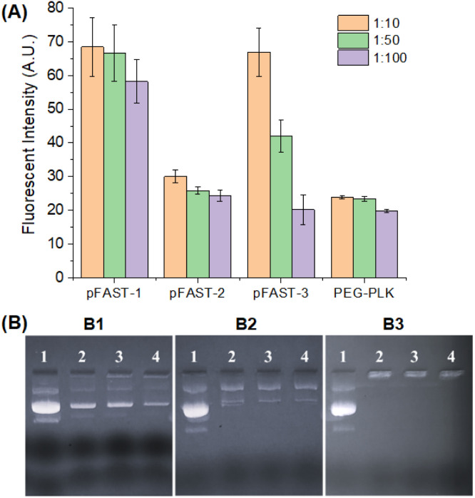
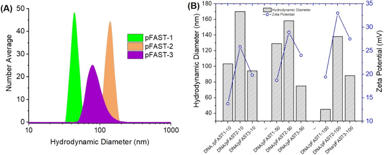
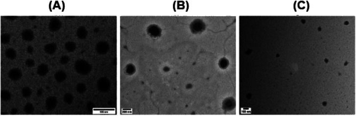
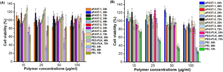
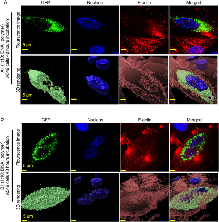
Biobased, DNA delivery vectors have been synthesized with a core motif composed of 2,5-bishydroxymethylfuran (BHMF) readily available from an important biomass feedstock 5-hydroxymethyl furfural (HMF). To generate the product, BHMF was first converted to 2,5-furan bishydroxymethyl diacrylate (2,5-FDA), which was later conjugated with different types of secondary amines. Rich in tertiary nitrogen, these oligomeric FDA-amino esters demonstrated stable electrostatic interactions with negatively charged plasmid DNA in an aqueous environment. We evaluated synthetic routes toward these plasmid DNA-binding amino esters (pFASTs), identified their nanoscale features, and attempted to establish their structure-property relationship in the context of the DNA delivery. Our preliminary studies show that the pFASTs formed stable complexes with the plasmid DNA. Dynamic light scattering indicated that the DNA polyplexes of pFASTs have hydrodynamic diameters within the size range of 100-150 nm with a surface charge (ζ-potential) ranging from -10 to +33 mV, depending on pFAST type. These oligomeric amino esters rich in furan motif were also found to successfully transfect the GFP-expressing plasmid DNA intracellularly. Collectively, this study establishes a new route to produce DNA transfection agents from sustainable resources that can be used for transferring genetic materials for humans, veterinary, and agrochemical purposes.
This journal is © The Royal Society of Chemistry.
Conflict of interest statement
Authors declare no conflict of interest.
Figures

Similar articles
-
Assessment of cholesterol-derived ionic copolymers as potential vectors for gene delivery.Biomacromolecules. 2013 Nov 11;14(11):4135-49. doi: 10.1021/bm4013088. Epub 2013 Oct 30. Biomacromolecules. 2013. PMID: 24125032
-
Diels-Alder Cycloaddition of 2,5-Bis(hydroxymethyl)furan (BHMF) and N-Phenylmaleimide Derivatives.ACS Omega. 2024 Aug 14;9(34):36380-36388. doi: 10.1021/acsomega.4c03804. eCollection 2024 Aug 27. ACS Omega. 2024. PMID: 39220524 Free PMC article.
-
Pharmaceutical and biological properties of poly(amino acid)/DNA polyplexes.J Drug Target. 1999;7(2):143-56. doi: 10.3109/10611869909085498. J Drug Target. 1999. PMID: 10617299
-
Biobased 2,5-Bis(hydroxymethyl)furan as a Versatile Building Block for Sustainable Polymeric Materials.ACS Omega. 2023 Mar 2;8(10):8991-9003. doi: 10.1021/acsomega.2c07629. eCollection 2023 Mar 14. ACS Omega. 2023. PMID: 36936293 Free PMC article. Review.
-
Formation of degradation compounds from lignocellulosic biomass in the biorefinery: sugar reaction mechanisms.Carbohydr Res. 2014 Feb 19;385:45-57. doi: 10.1016/j.carres.2013.08.029. Epub 2013 Sep 11. Carbohydr Res. 2014. PMID: 24412507 Review.
References
-
- Li Y. Tian R. Xu J. Zou Y. Wang T. Liu J. Polym. Chem. 2024;15:1908–1931.
-
- Lynn D. M. Langer R. J. Am. Chem. Soc. 2000;122:10761–10768.
LinkOut - more resources
Full Text Sources