

TNFα-Related Chondrocyte Inflammation Models: A Systematic Review
- PMID: 39409134
- PMCID: PMC11476358
- DOI: 10.3390/ijms251910805
TNFα-Related Chondrocyte Inflammation Models: A Systematic Review
Abstract
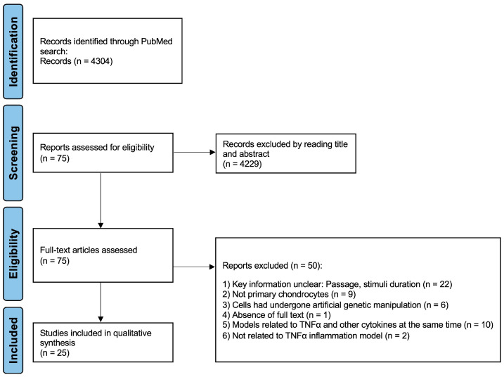
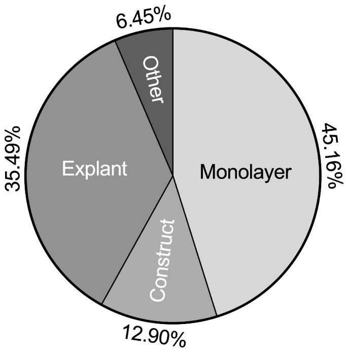
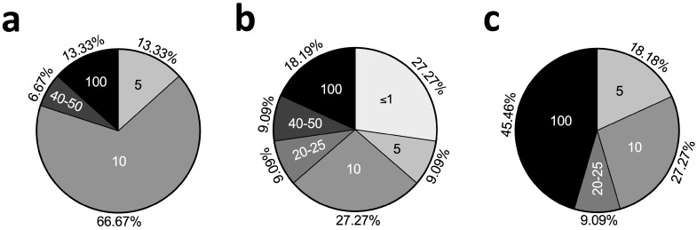
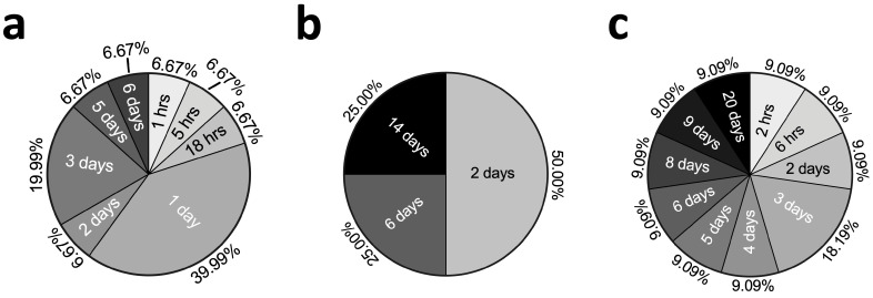
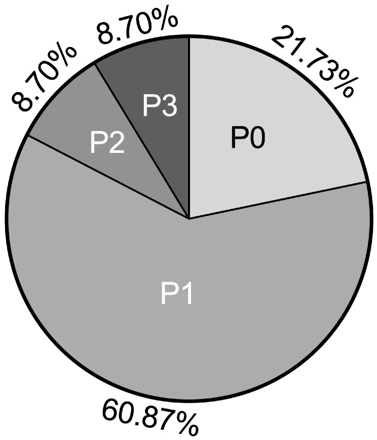
Tumor necrosis factor alpha (TNFα), as a key pro-inflammatory cytokine, plays a central role in joint diseases. In recent years, numerous models of TNFα-induced cartilage inflammation have been developed. However, due to the significant differences between these models and the lack of consensus in their construction, it becomes difficult to compare the results of different studies. Therefore, we summarized and compared these models based on important parameters for model construction, such as cell source, cytokine concentration, stimulation time, mechanical stimulation, and more. We attempted to analyze the advantages and disadvantages of each model and provide a compilation of the analytical methods used in previous studies. Currently, TNFα chondrocyte inflammation models can be categorized into four main types: monolayer-based, construct-based, explant-based TNFα chondrocyte inflammation models, and miscellaneous TNFα chondrocyte inflammation models. The most commonly used models were the monolayer-based TNFα chondrocyte inflammation models (42.86% of cases), with 10 ng/mL TNFα being the most frequently used concentration. The most frequently used chondrocyte cell passage is passage 1 (50%). Human tissues were most frequently used in experiments (51.43%). Only five articles included models with mechanical stimulations. We observed variations in design conditions between different models. This systematic review provides the essential experimental characteristics of the available chondrocyte inflammation models with TNFα, and it provides a platform for better comparison between existing and new studies in this field. It is essential to perform further experiments to standardize each model and to find the most appropriate experimental parameters.
Keywords: TNFα; cartilage; chondrocyte; cytokine; inflammation model.
Conflict of interest statement
The authors declare no conflicts of interest. The funders had no role in the design of the study; in the collection, analyses, or interpretation of data; in the writing of the manuscript; or in the decision to publish the results.
Figures

Similar articles
-
Erratum: Eyestalk Ablation to Increase Ovarian Maturation in Mud Crabs.J Vis Exp. 2023 May 26;(195). doi: 10.3791/6561. J Vis Exp. 2023. PMID: 37235796
-
Gadolinium Magnetic Resonance Imaging.2023 Jul 3. In: StatPearls [Internet]. Treasure Island (FL): StatPearls Publishing; 2025 Jan–. 2023 Jul 3. In: StatPearls [Internet]. Treasure Island (FL): StatPearls Publishing; 2025 Jan–. PMID: 29494094 Free Books & Documents.
-
Assessing the comparative effects of interventions in COPD: a tutorial on network meta-analysis for clinicians.Respir Res. 2024 Dec 21;25(1):438. doi: 10.1186/s12931-024-03056-x. Respir Res. 2024. PMID: 39709425 Free PMC article. Review.
-
Depressing time: Waiting, melancholia, and the psychoanalytic practice of care.In: Kirtsoglou E, Simpson B, editors. The Time of Anthropology: Studies of Contemporary Chronopolitics. Abingdon: Routledge; 2020. Chapter 5. In: Kirtsoglou E, Simpson B, editors. The Time of Anthropology: Studies of Contemporary Chronopolitics. Abingdon: Routledge; 2020. Chapter 5. PMID: 36137063 Free Books & Documents. Review.
-
Prognostic models for predicting clinical disease progression, worsening and activity in people with multiple sclerosis.Cochrane Database Syst Rev. 2023 Sep 8;9(9):CD013606. doi: 10.1002/14651858.CD013606.pub2. Cochrane Database Syst Rev. 2023. PMID: 37681561 Free PMC article. Review.
References
-
- Jiang Y., Yu M., Hu X., Han L., Yang K., Ba H., Zhang Z., Yin B., Yang X.-P., Li Z., et al. STAT1 mediates transmembrane TNF-alpha-induced formation of death-inducing signaling complex and apoptotic signaling via TNFR1. Cell Death Differ. 2017;24:660–671. doi: 10.1038/cdd.2016.162. - DOI - PMC - PubMed
Publication types
MeSH terms
Substances
Grants and funding
LinkOut - more resources
Full Text Sources
