. 2024 Oct 10;19(10):e0311817.
doi: 10.1371/journal.pone.0311817. eCollection 2024.

Preventive and treatment efficiency of dendrosomal nano-curcumin against ISO-induced cardiac fibrosis in mouse model

Affiliations

Preventive and treatment efficiency of dendrosomal nano-curcumin against ISO-induced cardiac fibrosis in mouse model

Behnaz Beikzadeh et al. PLoS One. .

Abstract

Cardiac fibrosis (c-fibrosis) is a critical factor in cardiovascular diseases, leading to impaired cardiac function and heart failure. This study aims to optimize the isoproterenol (ISO)-induced c-fibrosis model and evaluate the therapeutic efficacy of dendrosomal nano-curcumin (DNC) in both in-vitro and in-vivo conditions. Also, we were looking for the differentially expressed genes following the c-fibrosis induction. At the in-vitro condition, primary cardiac fibroblasts were exclusively cultured on collagen-coated or polystyrene plates and, were treated with ISO for fibrosis induction and post-treated or co-treated with DNC. RT-qPCR and flow cytometry analysis indicated that DNC treatment attenuated the fibrotic effect of ISO treatment in these cells. At the in-vivo condition, our findings demonstrated that ISO treatment effectively induces cardiac (and pulmonary) fibrosis, characterized by pro-fibrotic and pro-inflammatory gene expression and IHC (α-SMA, COL1A1, and TGFβ). Interestingly, fibrosis symptoms were reduced following the pretreatment, co-treatment, or post-treatment of DNC with ISO. Additionally, the intensive RNAseq analysis suggested the COMP gene is differentially expressed following the c-fibrosis and our RT-qPCR analysis suggested it as a novel potential marker. Overall, our results promise the application of DNC as a potential preventive or therapy agent before and after heart challenges that lead to c-fibrosis.


Conflict of interest statement

The authors have declared that no competing interests exist.

Figures

Fig 1
Fig 1. Outline of the study protocol.
The mice model was injected subcutaneously for four sequential days, and then 3 mice were harvested at days 5, 7, 11, 18, 25, and 32 either for the RNA extraction or IHC analysis.
Fig 2
Fig 2. Effect of DNC on cardiac fibroblasts activated by ISO.
(A) The optimal DNC treatment dose (1μm) for cardiac fibroblast without significant cytotoxicity was determined using the MTT assay. (B) Effects of ISO (10μm) and DNC (1μm) on profibrotic and pro-inflammatory gene expression were determined using RT-qPCR. Additionally, the differences between cardiac fibroblasts cultured on polystyrene plates (untreated) versus collagen plates (4 kPa) were reported. ISO and DNC are applied through sequential treatment (ISO/DNC) or co-treatment (ISO+DNC). Graph indicate that co-treatment of (ISO+DNC) is more effective for attenuation of fibrosis induction. (C) Cell cycle distribution of cardiac fibroblasts cultured in a softwell plate, and polystyrene plate namely untreated, ISO, ISO/DNC and ISO+DNC was examined using flow cytometry. Again, co-treatment of ISO+DNC is more effectively affecting cell cycle progression, shown through reduced S phase and increased G1 cell populations. Data are presented as mean ± SEM (triplicate). The mean expression is shown as relative expression level for RT-qPCR. T Student’s t-test for MTT assay, One-way ANOVA test for RT-qPCR: ns: non-significance, *p<0.05, **p<0.01, ***p<0.001, ****p<0.0001.
Fig 3
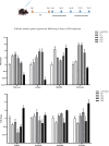
Fig 3. Successful induction of cardiac fibrosis and up-regulation of pro-fibrotic and pro-inflammatory genes following the protocol #4.
Detection of mRNA levels of pro-fibrotic (COL1A1, α-SMA, TGF-ß, SMAD2, SMAD3, and FURIN) and pro-inflammatory (MMP9) genes through RT-qPCR in the mice treated with ISO (140 mg/kg) for 4 days, and then harvested on D5, D7, D11, D18, D25, and D32 days. Two waves of gene expression upregulation is evident for most of the tested genes, following the ISO treatment. Data are presented as mean ± SEM vs. control (n  =  3). The mean expression is shown as a fold change. T Student’s t-test: *p<0.05, **p<0.01, ***p<0.001.
Fig 4
Fig 4. Verification of the induced cardiac fibrosis through histopathological staining and measuring the marker proteins level following protocol #4.
(A) The H&E staining shows the morphological alteration of the cardiomyocytes in the left ventricular, following the ISO induction. Black arrows show the irregular organization of cardiomyocytes, and yellow arrows indicate the irregular shape of nuclei without any organelles in the cytoplasm. (B) MTS staining shows the amount of cardiac fibrosis induction (identified by blue-colored tissues). (C) Quantitative analysis of the fibrotic area extent through evaluating the collagen surface area in the left ventricle of mice in the D5 (*p<0.05) and D32 (***p<0.001) groups, in comparison to the control group. (D-F) show the Immunohistochemistry (IHC) staining of COL1A1 (D), α-SMA (E), and TGF-ß (F) in mice’s left ventricular tissue sections on D5 and D32 after injection with ISO (140 mg/kg), compared to the control group. All these three marker proteins were upregulated/ accumulated on day 32nd, affirming the fibrosis induction. Nevertheless, COL1A1 (A) protein level has been drastically increased even on day 5th. Data are presented as the mean values ± SEM. Magnification (x400). H&E, hematoxylin and eosin; MTS, Masson’s trichrome staining.
Fig 5
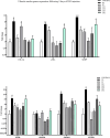
Fig 5. DNC modulates the expression of genes related to cardiac fibrosis.
The expression of profibrotic and pro-inflammatory genes was determined by RT-qPCR following the DNC (10 mg/kg) administration. In the first and second groups sacrificed on (A) D5 and (B) D32, the mice were pre-treated with DNC for 7 days, followed by co-treatment of ISO and DNC for 4 days (DNC/ISO+DNC). In the third group, the mice were treated with ISO for 4 days, followed by DNC post treatment for 7 days and sacrificed on D32 (B). In the fourth group, the mouse was treated with ISO for 4 days and then from D25-D31 injected with DNC and sacrificed on D32 (B). Marker genes expression results indicated that pretreatment of mice with DNC, reduces the risk of cardiac fibrosis following the ISO treatment. However, after the fibrosis induction, DNC treatment is less effective, up to the delay of DNC treatment. Data are presented as mean ± SEM. The mean expression is shown as a relative expression level. One-way ANOVA test: ns: non-significance, *p<0.05, **p<0.01, ***p<0.001.
Fig 6
Fig 6. Induction of pulmonary fibrosis based on the expression pattern of pro-fibrotic and pro-inflammatory genes.
RT-qPCR expression detection of pro-fibrotic (COL1A1, α-SMA, COL3A1, TGF-ß, SMAD2, SMAD3, and FURIN) and pro-inflammatory (MMP9) genes in the lung tissue samples prepared following the intraperitoneal-injection of 140 mg/kg ISO-chemical for 4 days and harvesting on D5, D7, D11, D18, D25, and D32. Data are presented as mean ± SEM vs. control (n  =  3). The mean expression was shown as a fold change. T Student’s t-test: *p<0.05, **p<0.01.
Fig 7
Fig 7. Shared DEGs between the 4 GEO datasets and enrichment analysis.
(A) 19 shared up-regulated and (B) 10 shared down-regulated genes were calculated with a Venn diagram. (C) Heat map of 29 shared genes according to their Log2 fold change in each project. (D) Molecular function, (E) biological process and (F) cellular component of shared genes.
Fig 8
Fig 8. Identifying the COMP gene as the first hub gene and confirming its increased expression in cardiac fibrosis.
PPI network of shared genes in (A) human and (B) mouse constructed by string. The top 10 hub genes for both (C) human and (D) mice; the COMP gene is the first one with a score of 3. (E) The expression of the COMP gene in cardiac fibroblast following ISO and DNC treatment with RT-qPCR. (F) The expression of the COMP gene in a mouse model of cardiac fibrosis and after the (G) DNC administration by RT-qPCR. Data are presented as mean ± SEM (n = 3). The mean expression is shown as the relative expression level and fold change. T Student’s t-test, One-way ANOVA test: ns: non-significance, *p<0.05, **p<0.0.

Similar articles

References

    1. Hall C, Gehmlich K, Denning C, Pavlovic D. Complex relationship between cardiac fibroblasts and cardiomyocytes in health and disease. Journal of the American Heart Association. 2021;10(5):e019338. doi: 10.1161/JAHA.120.019338 - DOI - PMC - PubMed
    1. Sarohi V, Chakraborty S, Basak T. Exploring the cardiac ECM during fibrosis: A new era with next-gen proteomics. Frontiers in Molecular Biosciences. 2022;9:1030226. doi: 10.3389/fmolb.2022.1030226 - DOI - PMC - PubMed
    1. Hinderer S, Schenke-Layland K. Cardiac fibrosis–a short review of causes and therapeutic strategies. Advanced drug delivery reviews. 2019;146:77–82. - PubMed
    1. Tian J, An X, Niu L. Myocardial fibrosis in congenital and pediatric heart disease. Experimental and therapeutic medicine. 2017;13(5):1660–1664. doi: 10.3892/etm.2017.4224 - DOI - PMC - PubMed
    1. Frangogiannis NG. Cardiac fibrosis. Cardiovascular research. 2021;117(6):1450–1488. doi: 10.1093/cvr/cvaa324 - DOI - PMC - PubMed

Grants and funding

This work is based upon research funded by Tarbiat Modares University and Iran National Science Foundation (INSF) under project No.4014277. The funders had no role in study design, data collection and analysis, decision to publish, or preparation of the manuscript.

LinkOut - more resources