Review
. 2024 Oct 10;5(1):46.
doi: 10.1186/s43556-024-00211-0.

Pathways regulating intestinal stem cells and potential therapeutic targets for radiation enteropathy

Affiliations
Review

Pathways regulating intestinal stem cells and potential therapeutic targets for radiation enteropathy

Si-Min Chen et al. Mol Biomed. .

Abstract

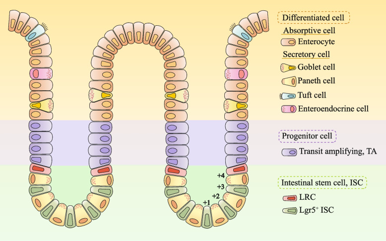
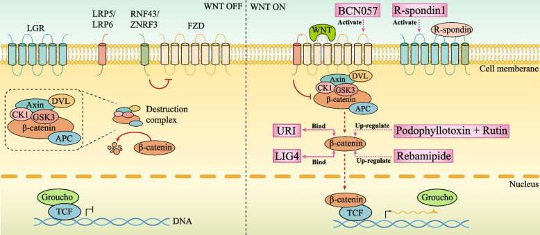
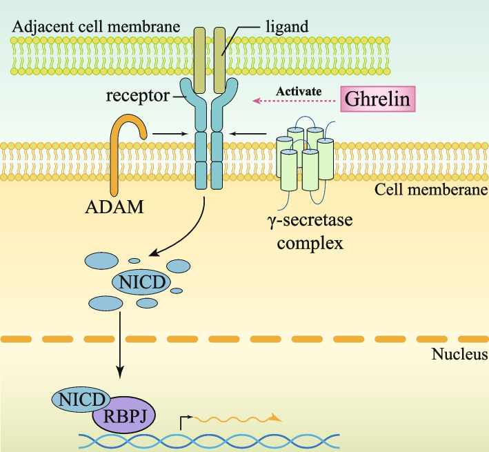
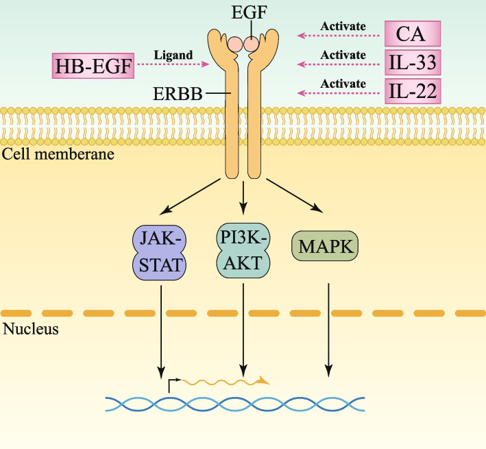
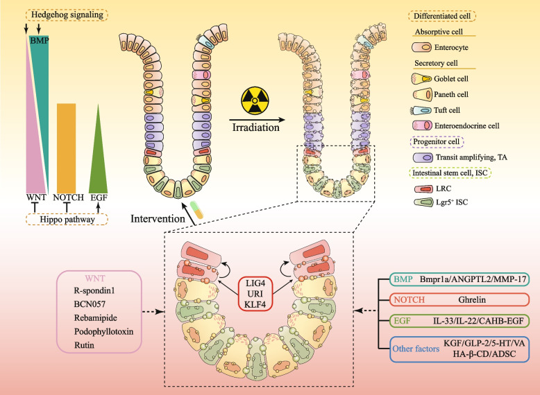
Radiotherapy is a pivotal intervention for cancer patients, significantly impacting their treatment outcomes and survival prospects. Nevertheless, in the course of treating those with abdominal, pelvic, or retroperitoneal malignant tumors, the procedure inadvertently exposes adjacent intestinal tissues to radiation, posing risks of radiation-induced enteropathy upon reaching threshold doses. Stem cells within the intestinal crypts, through their controlled proliferation and differentiation, support the critical functions of the intestinal epithelium, ensuring efficient nutrient absorption while upholding its protective barrier properties. Intestinal stem cells (ISCs) regulation is intricately orchestrated by diverse signaling pathways, among which are the WNT, BMP, NOTCH, EGF, Hippo, Hedgehog and NF-κB, each contributing to the complex control of these cells' behavior. Complementing these pathways are additional regulators such as nutrient metabolic states, and the intestinal microbiota, all of which contribute to the fine-tuning of ISCs behavior in the intestinal crypts. It is the harmonious interplay among these signaling cascades and modulating elements that preserves the homeostasis of intestinal epithelial cells (IECs), thereby ensuring the gut's overall health and function. This review delves into the molecular underpinnings of how stem cells respond in the context of radiation enteropathy, aiming to illuminate potential biological targets for therapeutic intervention. Furthermore, we have compiled a summary of several current treatment methodologies. By unraveling these mechanisms and treatment methods, we aspire to furnish a roadmap for the development of novel therapeutics, advancing our capabilities in mitigating radiation-induced intestinal damage.

Keywords: Biological targets; Intestinal stem cells; Radiation enteropathy; Signaling pathway; Treatment methods.


Conflict of interest statement

The authors declare that they have no competing interests.

Figures

Fig. 1
Fig. 1
Schematic diagram of small intestinal epithelial tissue structure. The main cell types of intestinal epithelial tissue are stem cells (ISCs), Progenitor cells and Differentiated cells. Label-retaining cells (LRC) and leucine-rich-repeat-containing G-protein-coupled receptor 5 (Lgr5+) ISC were intestinal stem cells and transit-amplifying cells (TAC) was progenitor cells. There are two types of differentiated cells: Absorptive cells and Secretory cells. Absorptive cells are Enterocytes; The secretory cells include Goblet cells, Paneth cells, Tuft cells and Enteroendocrine cells
Fig. 2
Fig. 2
WNT signalling pathways in the intestinal crypt and potential therapeutic targets. Sustained activation of the WNT signaling pathway is dependent on the presence of R-spondin. R-spondin binds to LGR family receptors and also binds to RNF43-ZNRF3, which in turn stabilizes FZD expression. Upon binding to FZD and LRP5-LRP6, the WNT pathway is activated. This activation leads to the activation of the intracellular scrambling protein Dvl (Dishevelled), a crucial component in the WNT signaling cascade. Dvl inhibits the phosphorylation of β-Catenin by disrupting the complex composed of Axin, adenomatous polyposis coli (APC), casein kinase 1 (CK1), and glycogen synthase 3 (GSK3). Accumulated β-catenin then enters the nucleus and promotes transcription by binding to T-cell factor (TCF) and displacing Groucho. The potential targets and their mechanisms of action have been marked with pink boxes
Fig. 3
Fig. 3
BMP signalling pathways in the intestinal crypt and potential therapeutic targets. BMP signaling pathway induces the dimerization of BMP type I and type II receptors (BMPR1 and BMPRII), resulting in the phosphorylation and subsequent dimerization of receptor-regulated mothers against decapentaplegic homologues (rSMADs). These rSMADs then form heterotrimeric complexes with common SMADs (cSMAD or SMAD4) and translocate to the nucleus. Within the nucleus, this complex regulates the expression of target genes. The potential targets and their mechanisms of action have been marked with pink boxes
Fig. 4
Fig. 4
NOTCH signalling pathways in the intestinal crypt and potential therapeutic targets. NOTCH signaling is initiated when NOTCH receptors bind to NOTCH ligands. The synthesis of NOTCH receptors takes place in the endoplasmic reticulum, followed by processing in the Golgi apparatus. Subsequently, the receptors are transported to the cell surface where they can bind and interact with NOTCH ligands present on adjacent cells. The transmembrane region of the receptor undergoes cleavage by ADAM (a disintegrin and metalloprotease), and then further cleavage occurs through γ-secretase protease. Once the receptor is cleaved, the NOTCH intracellular domain (NICD) is released and translocated to the nucleus where it binds to recombination signal binding protein for immunoglobulin kappa j region (RBPJ), thereby activating downstream genes. The potential targets and their mechanisms of action have been marked with pink boxes
Fig. 5
Fig. 5
EGF signalling pathways in the intestinal crypt and potential therapeutic targets. EGF: EGF stimulates the EGF receptor (ERBB), initiating the mitogen-activated protein kinase (MAPK) cascade. This cascade leads to the phosphorylation of various nuclear targets by ERK, which promotes cell proliferation and inhibits apoptosis. Simultaneously, EGF signaling enhances the interaction between phosphocarnosine 3-kinase (PI3K) and converts PIP2 to PIP3. Furthermore, the activated EGF receptor complex recruits Janus kinases (JAKs). The potential targets and their mechanisms of action have been marked with pink boxes
Fig. 6
Fig. 6
Potential intervention strategies for radiation enteropathy. The principal strategies of treating radiation enteropathy. Pharmacological intervention is up-left, hyperbaric oxygen therapy (HBOT) is down-left, endoscopic procedures is up-right, surgical management is down-right. Figure 6 was modified from Freepik (https://www.freepik.com/)
Fig. 7
Fig. 7
Summary Chart. Main regulatory pathways before intestinal radiation and potential intervention targets after radiation. URI: unconventional prefoldin RPB5 interactor, CA: centella asiatica, TCM: traditional chinese medicine, KGF: keratinocyte growth factor, GLP-2: glucagon-like peptide 2 analogue, 5-HT: 5-hydroxytryptamine, VA: valeric acid, KLF4: Krüppel-like factor 4, HA-β-CD: hyaluronic acid (HA) and β-cyclodextrin (HA-β-CD), ADSC: adipose-derived mesenchymal stem cells, IECs: intestinal epithelial cells, ISCs: intestinal stem cells

Similar articles

References

    1. Hauer-Jensen M, Denham JW, Andreyev HJ. Radiation enteropathy–pathogenesis, treatment and prevention. Nat Rev Gastroenterol Hepatol. 2014;11(8):470–9. 10.1038/nrgastro.2014.46. - PMC - PubMed
    1. Moraitis I, Guiu J, Rubert J. Gut microbiota controlling radiation-induced enteritis and intestinal regeneration. Trends Endocrinol Metab. 2023;34(8):489–501. 10.1016/j.tem.2023.05.006. - PubMed
    1. Kwak SY, Jang WI, Park S, Cho SS, Lee SB, Kim MJ, et al. Metallothionein 2 activation by pravastatin reinforces epithelial integrity and ameliorates radiation-induced enteropathy. EBioMedicine. 2021;73:103641. 10.1016/j.ebiom.2021.103641. - PMC - PubMed
    1. Gandle C, Dhingra S, Agarwal S. Radiation-induced enteritis. Clin Gastroenterol Hepatol. 2020;18(3):A39-a40. 10.1016/j.cgh.2018.11.060. - PubMed
    1. Hu B, Jin C, Li HB, Tong J, Ouyang X, Cetinbas NM, et al. The DNA-sensing AIM2 inflammasome controls radiation-induced cell death and tissue injury. Science. 2016;354(6313):765–8. 10.1126/science.aaf7532. - PMC - PubMed

Publication types

MeSH terms

LinkOut - more resources