. 2024 Nov 19;98(11):e0030024.
doi: 10.1128/jvi.00300-24. Epub 2024 Oct 9.

Retinoic acid-induced differentiation and oxidative stress inhibitors increase resistance of human neuroblastoma cells to La Crosse virus-induced cell death

Affiliations

Retinoic acid-induced differentiation and oxidative stress inhibitors increase resistance of human neuroblastoma cells to La Crosse virus-induced cell death

Paul F Policastro et al. J Virol. .

Abstract

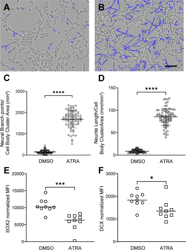
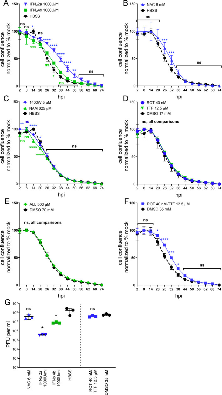
La Crosse Virus (LACV) encephalitis patients are at risk for long-term deficits in cognitive function due to neuronal apoptosis following virus infection. However, the specific etiology underlying neuronal damage remains elusive. In this study, we examined how differentiation and mitotic inhibition of neuroblastoma cells influence their susceptibility to LACV infection and cell death. Treatment of SH-SY5Y cells with retinoic acid induced a neuronal cell phenotype which was similarly susceptible to LACV infection as untreated cells but had significantly delayed virus-induced cell death. Protein and RNA transcript analysis showed that retinoic acid-treated cells had decreased oxidative stress responses to LACV infection compared to untreated cells. Modulation of oxidative stress in untreated cells with specific compounds also delayed cell death, without substantially impacting virus production. Thus, the oxidative stress response of neurons to virus infection may be a key component of neuronal susceptibility to virus-induced cell death.

Importance: Encephalitic viruses like La Crosse Virus (LACV) infect and kill neurons. Disease onset and progression is rapid meaning the time frame to treat patients before significant and long-lasting damage occurs is limited. Examining how neurons, the primary cells infected by LACV in the brain, resist virus-induced cell death can provide avenues for determining which pathways to target for effective treatments. In the current study, we studied how changing neuroblastoma growth and metabolism with retinoic acid treatment impacted their susceptibility to LACV-induced cell death. We utilized this information to test compounds for preventing death in these cells.

Keywords: La Crosse virus; all-trans retinoic acid; interferon; mitochondria; neuroblastoma; neurodifferentiation; oxidative stress.


Conflict of interest statement

The authors declare no conflict of interest.

Figures

Fig 1
Fig 1
ATRA treatment induces SH-SY5Y cell neurite formation and reduces SOX2 and DCX protein levels. Phase contrast image with neurite mask (BLUE) shows neurite extensions of (A) DMSO-treated cells and (B) ATRA-treated cells. Cells were analyzed after 8 days of treatment. Scale bar = 50 µm. Comparison of (C) neural branch points and (D) neurite length per cell body cluster area of images (n = 78) and analyzed by unpaired t tests: ****, <0.0001 P value. (E, F) MFI of gated cells for (E) SOX2 and (F) DCX in DMSO- and ATRA-treated cells (n = 9, 3 experiments with three replicates) analyzed by unpaired t test: P values: *, ≤0.05; ***, ≤0.001.
Fig 2
Fig 2
ATRA treatment delays LACV-induced SH-SY5Y cell death. SH-SY5Y cells treated for 8 days with DMSO or ATRA were infected with MOIs of (A–D) 0.01 or (E and F) 1.0 of LACV. (A, C, E) Cells were monitored for cell viability by MTT assay. Data were calculated relative to mock-inoculated controls for each treatment at each time point. (B, D, F) PFU per milliliter of supernatants from inoculated cells in A, C, and E, respectively. (B) DMSO-treated cell supernatants at 0 hpi include a value of <1 PFU per milliliter for one of three biological replicates. Cell survival and PFU data are representative of four similar experiments. Statistical analysis was calculated as (A, C–F) two-way ANOVA with Šídák’s multiple comparisons test or (B) one-way ANOVA, Šídák’s multiple comparisons test. n = 3 for all experiments. P values: *, ≤0.05; **, ≤0.01; ***, ≤0.001; and ****, ≤0.0001, ns, not significant.
Fig 3
Fig 3
SH-SY5Y cell ATRA treatment length increases resistance to LACV-induced cell death. Cells were cultured in DMSO or ATRA treatment conditions for different lengths of time prior to inoculation with LACV (MOI 0.01) or mock extract. (A, C, E) Cell viability as measured by MTT conversion for cells pretreated (A) 0 or 2 days, (C) 4 or 6 days, or (E) 8 or 10 days. Paired DMSO-ATRA values at each dilution were tested by 2-way ANOVA, Šídák’s multiple comparisons test, n = 3, all tests. (B, D, F) PFU per millileter from cell supernatants at 0 and 72 hpi for A, C, and E, respectively. Eight days DMSO 0 hpi includes value of <1 PFU per milliliter for one of three biological replicates. Statistical analysis was completed with a one-way ANOVA and Šídák’s multiple comparison test, n = 3. P values: *, ≤0.05; **, ≤0.01; ***, ≤0.001; and ****, ≤0.0001. ns, not significant. P value symbols are the same color as compared values.
Fig 4
Fig 4
Lower rate of infection and Caspase 3 activity is detected in ATRA-treated cells. (A) DMSO- or (B) ATRA-treated SH-SY5Y cells inoculated with LACV at 0.01 MOI and fixed at 24 hpi were stained for F-actin (magenta) with phalloidin, and antibodies to LACV (green) and active caspase-3 (blue). Scale bar = 50 µM (C) Percent of LACV-infected cells in equivalent fields from DMSO- and ATRA-treated cells, analyzed by Welch’s t test: P value: **** <0.0001. (D) Caspase-3/7 Green Dye was used to evaluate cell death in DMSO- and ATRA-treated cells (D), and statistics were calculated by two-way ANOVA and Šídák’s multiple comparison test. P values are denoted as *, ≤0.05; **, ≤0.01; ns, not significant. Colors are used to show P value differences between mock and LACV inoculations in DMSO (black asterisks) and ATRA (blue asterisks) cells or between DMSO and ATRA treatments following LACV (green asterisks) or mock inoculation (red asterisks).
Fig 5
Fig 5
DMSO-treated cells show greater oxidative stress transcript response to LACV than ATRA-treated cells. (A and B) DMSO- and ATRA-treated cells were inoculated with LACV (MOI 0.01) or mock lysate. At 36 hpi, RNA was isolated and processed for RT-qPCR analysis of oxidative stress-related transcripts. Data are plotted as log2 fold change relative to P value. Only transcripts with P values < 10−2 are labeled. (A) DMSO-treated, LACV-infected cells compared to DMSO-treated, mock-infected cells. (B) ATRA-treated, LACV-infected cells compared to ATRA-treated, mock-infected cells. P value analyses were multiple unpaired t tests, with P value stack analysis by the two-stage, step-up method of Benjamin, Krieger, and Yekutieli, desired false discovery rate (Q) of 1% (P value threshold for discovery was (A) <0.00285 and (B) <0.000367.
Fig 6
Fig 6
IFN and a combination of ROT and TTF reduce LACV-induced cell death. DMSO-treated cells inoculated LACV (MOI 0.01) or mock lysates were treated with indicated compounds or vehicle controls. (A–F) Wells were monitored for cell confluence at 6 h intervals, starting 2 hpi and ending at 74 hpi. Data are calculated as a percent of mock sample for each compound. Biological replicates per data point, (A, B, D, E) n = 3 (C, F) n = 2. Statistical analysis was calculated with a 2-way ANOVA and Šídák’s multiple comparison test. P values: *, ≤0.05; **, ≤0.01; ***, ≤0.001; and ****, ≤0.0001. ns, not significant. (G) PFU/mL in supernatants at 74 hpi for compounds with P values < 0.01 relative to vehicle control. Statistical analysis was completed by ordinary one-way ANOVA with Šídák’s multiple comparison test relative to HBSS control (left of line) or by unpaired t test relative to DMSO 35 mM control (right of line); n = 3, all tests. Significant P values are denoted as *, ≤0.05.

Similar articles

References

    1. Haddow AD, Bixler D, Odoi A. 2011. The spatial epidemiology and clinical features of reported cases of La Crosse virus infection in West Virginia from 2003 to 2007. BMC Infect Dis 11:29–37. doi:10.1186/1471-2334-11-29 - DOI - PMC - PubMed
    1. Gaensbauer JT, Lindsey NP, Messacar K, Staples JE, Fischer M. 2014. Neuroinvasive arboviral disease in the United States: 2003 to 2012. Pediatrics 134:e642–50. doi:10.1542/peds.2014-0498 - DOI - PMC - PubMed
    1. Vahey GM, Lindsey NP, Staples JE, Hills SL. 2021. La Crosse virus disease in the United States, 2003-2019. Am J Trop Med Hyg 105:807–812. doi:10.4269/ajtmh.21-0294 - DOI - PMC - PubMed
    1. Day CA, Odoi A, Trout Fryxell R. 2023. Geographically persistent clusters of La Crosse virus disease in the appalachian region of the United States from 2003 to 2021. PLoS Negl Trop Dis 17:e0011065. doi:10.1371/journal.pntd.0011065 - DOI - PMC - PubMed
    1. Rust RS, Thompson WH, Matthews CG, Beaty BJ, Chun RW. 1999. La Crosse and other forms of California encephalitis. J Child Neurol 14:1–14. doi:10.1177/088307389901400101 - DOI - PubMed

LinkOut - more resources