. 2024 Oct 29;9(10):e0078323.
doi: 10.1128/msphere.00783-23. Epub 2024 Oct 9.

A fur plucking model to study herpes simplex virus reactivation and recurrent disease

Affiliations

A fur plucking model to study herpes simplex virus reactivation and recurrent disease

Drake T Philip et al. mSphere. .

Abstract

Herpes simplex viruses (HSV-1 and HSV-2) most commonly cause ulcerative epithelial lesions (cold sores and genital herpes). Importantly, HSV establishes life-long persistent (latent) infection in peripheral neurons. Reactivation from latency produces recurrent epithelial lesions, which constitute the greatest burden of HSV disease in people. The mechanisms that regulate latency and reactivation remain incompletely understood, in part due to limitations in the animal models available for studying HSV reactivation. We have developed a simple and tractable model to induce HSV-1 and HSV-2 reactivation from latency to cause recurrent skin disease. We infected C57BL/6 mice with HSV-1 (strains NS, F, SC16, 17syn+) or HSV-2 (strain 333) on flank skin depilated by manual plucking. After at least 35 days post-infection (dpi), we replucked the fur from the infected flank and observed recurrent lesions in the same dermatome as the primary infection. We detected HSV DNA in dermatome skin through 4 days post-replucking and observed viral antigen and reporter signal in skin lesions by histology, consistent with viral replication following reactivation. In addition to C57BL/6 mice, we were able to produce reactivation in Balb/c and SKH-1 mice. We found that shaving the ipsilateral flank or plucking the contralateral flank did not induce recurrent skin lesions, suggesting that fur plucking is a specific stimulus that induces HSV reactivation. Furthermore, we were able to induce multiple rounds of plucking-induced recurrent disease, providing a model to investigate the lifelong nature of HSV infection. This new model provides a tractable system for studying pathogenic mechanisms of and therapeutic interventions against HSV reactivation and recurrent disease.

Importance: Herpes simplex viruses (HSV-1 and HSV-2) have infected over half of the US adult population to cause a lifelong, persistent infection; however, our understanding of the mechanisms that govern HSV reactivation and recurrent disease is incomplete. This is in part due to limitations in the animal models used to study recurrent disease, which are laborious and inefficient in mice. To address this technical gap, we developed a mouse model in which fur plucking after flank skin infection is sufficient to induce episodes of HSV reactivation and recurrent disease. Our work provides a model for the field to investigate the pathogenic mechanisms of HSV and immune responses during recurrent disease and provides an opportunity to investigate the neurobiology of HSV infection.

Keywords: dorsal root ganglia; herpes simplex virus; latency; reactivation; skin infection.


Conflict of interest statement

The authors declare no conflict of interest.

Figures

Fig 1
Fig 1
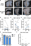
Plucking induces recurrent skin lesions in mice previously infected with HSV-1. WT (C57BL/6) mice were depilated by plucking 1 day prior to inoculation with 106 FFU of HSV-1 strain NS. After 35 days, mice were replucked and evaluated for recurrent skin disease. Blue boxes denote the dermatome area of interest. (A) Representative serial photos of the same mouse during acute disease (6 dpi), immediately post-replucking (R0), and 2 days post-replucking (R2). (B) Skin lesion areas for individual mice were measured using ImageJ from photographs during acute disease (A6) or 2, 4, or 6 days post-replucking. Blue stars indicate the lesion areas of the mouse depicted in (A). (C) Dermatome skin was harvested at the indicated times after replucking, and viral loads were measured by qPCR. The limit of detection and number of mice per group are indicated. (D) Frequency of HSV-1 positive skin by qPCR.
Fig 2
Fig 2
Ipsilateral shaving or contralateral plucking is not sufficient to cause HSV-1 recurrent skin disease. WT mice were depilated by plucking 1 day prior to inoculation with 106 FFU of HSV-1 strain NS. After 35 days, mice were either replucked on their ipsilateral flank, shaved on their ipsilateral flank, or plucked on their contralateral flank (plus shaved on their ipsilateral flank to allow skin lesions to be observed). Mice were then evaluated at R2 for skin lesions and viral loads. (A) Representative serial photos of mice during acute disease (6 dpi) and recurrent disease (R2). Blue boxes denote the dermatome area of interest. Blue star indicates the lesion area of the mouse depicted in (A). (B) Skin lesion areas were measured from photographs at R2 using ImageJ. (C) Frequency of recurrent disease observed at R2. (D) Dermatome skin was harvested at R2, and viral loads were measured by qPCR. The limit of detection and number of mice per group are indicated.
Fig 3
Fig 3
Diverse mouse lines are susceptible to plucking-induced recurrent skin lesions. BALB/c mice and SKH-1 mice were inoculated with 105 or 103 FFU, respectively, of HSV-1 strain NS. After 35 days, BALB/c mice were replucked, and SKH-1 mice were tape stripped to remove fine hairs, and recurrent skin lesions were evaluated. (A) Representative serial photos of mice during acute disease (6 dpi) and recurrent disease (R2). Blue boxes denote the dermatome area of interest. (B) Skin lesion areas were measured from photographs at R2 using ImageJ. (C) Frequency of recurrent disease observed at R2. (D) Dermatome skin was harvested from BALB/c mice (n = 12) at R2, and viral loads were measured by qPCR. The limit of detection is indicated. Stars indicate the lesion areas and skin viral loads of mice depicted in (A).
Fig 4
Fig 4
Multiple strains of HSV-1 can be reactivated by fur plucking. WT mice were depilated by plucking 1 day prior to inoculation with 105 FFU of HSV-1 strain F, 106 FFU of HSV-1 strain SC16, or 106 FFU of HSV-1 strain 17syn+. After 50–75 days, mice were replucked on their ipsilateral flank and then evaluated at R2 for disease and viral loads. (A) Representative serial photos of the same mouse during acute disease (6 dpi, A6) and recurrent disease (R2). Blue boxes denote the dermatome area of interest. (B–D) Paired skin lesion areas were measured using ImageJ from photographs during acute and recurrent disease. Blue stars indicate the lesion areas of the mice depicted in (A). (E) Frequency of recurrent disease observed at R2. (F) Dermatome skin was harvested at R2 from mice infected with HSV-1 strain SC16 and 17syn+, and viral loads were measured by qPCR (n = 10 mice per group). The limit of detection is indicated.
Fig 5
Fig 5
Fur plucking induces HSV-2 recurrent skin disease. WT mice were depilated by plucking 1 day prior to inoculation with 500 FFU of HSV-2 strain 333. After 35 days, mice were replucked and evaluated for recurrent skin disease. (A) Representative serial photos of the same mouse during acute disease (6 dpi), immediately post-replucking (R0), and 2 days post-replucking (R2). Blue boxes denote the dermatome area of interest. (B) Paired skin lesion areas were measured using ImageJ from photographs during acute and recurrent disease. Blue stars indicate the lesion areas of the mouse depicted in (A).
Fig 6
Fig 6
HSV-1 infection in the skin epithelium is evident after fur plucking. Stopf/f-tdTomato mice were depilated by plucking 1 day prior to inoculation with 106 FFU of HSV-1 expressing Cre recombinase (SC16-Cre). After 35 days, mice were replucked, and skin was harvested for histology at R0 (n = 5) or R2 (n = 3). WT mice were depilated by plucking 1 day prior to inoculation with 106 FFU of HSV-1 strain NS. Mice were either harvested at A6 (6 dpi) (n = 5) or reactivated 35 days later and harvested at R2 (n = 3) for skin histology. Slides were processed for IHC with an anti-HSV-1 antibody and imaged in brightfield (IHC) or fluorescent (tdTomato) channels. Images are oriented with epithelium upward and muscle downward. (A) Black dashed ovals indicate HSV-1 lesions in the skin epithelium with corresponding immune infiltrate. White oval notes a region of autofluorescence due to red blood cells (RBCs) in the lower dermis. (B) Magnified image from panel A (red and white boxes). Black arrows indicate faint HSV-1 antigen staining. White arrows indicate corresponding regions of tdTomato fluorescence in the lesion skin epithelium. Scale bars indicate 100 µM.
Fig 7
Fig 7
Serial plucking can induce multiple rounds of recurrent HSV-1 disease. WT mice were depilated by plucking 1 day prior to inoculation with 106 FFU of HSV-1 strain NS. After 35 days, mice were replucked, evaluated for recurrent skin disease, and fur was allowed to grow back. Then, 35 days after replucking, mice were replucked again and evaluated for recurrent skin disease. (A) Representative serial photos of the same mouse during acute disease (6 dpi, A6), immediately post-replucking (R0), 2 days post-replucking (R2), immediately following the second replucking (RR0), and 2 days after the second replucking (RR2). Blue boxes denote the dermatome area of interest. (B) Skin lesion areas for individual mice were measured using ImageJ from photographs at A6, R2, and RR2. Blue stars indicate the lesion areas of the mouse depicted in (A).

Update of

Similar articles

References

    1. Bradley H, Markowitz LE, Gibson T, McQuillan GM. 2014. Seroprevalence of herpes simplex virus types 1 and 2--United States, 1999-2010. J Infect Dis 209:325–333. doi:10.1093/infdis/jit458 - DOI - PubMed
    1. Kollias CM, Huneke RB, Wigdahl B, Jennings SR. 2015. Animal models of herpes simplex virus immunity and pathogenesis. J Neurovirol 21:8–23. doi:10.1007/s13365-014-0302-2 - DOI - PubMed
    1. Marcocci ME, Napoletani G, Protto V, Kolesova O, Piacentini R, Li Puma DD, Lomonte P, Grassi C, Palamara AT, De Chiara G. 2020. Herpes simplex virus-1 in the brain: the dark side of a sneaky infection. Trends Microbiol 28:808–820. doi:10.1016/j.tim.2020.03.003 - DOI - PubMed
    1. Fellner A, White S, Rockwell E, Giandomenico D, Diaz MM, Weber DJ, Miller MB, Boyce RM. 2023. The clinical epidemiology, management, and outcomes of patients diagnosed with encephalitis in North Carolina, 2015-2020. J Clin Microbiol 61:e0073123. doi:10.1128/jcm.00731-23 - DOI - PMC - PubMed
    1. Zhang S-Y, Jouanguy E, Ugolini S, Smahi A, Elain G, Romero P, Segal D, Sancho-Shimizu V, Lorenzo L, Puel A, et al. . 2007. TLR3 deficiency in patients with herpes simplex encephalitis. Science 317:1522–1527. doi:10.1126/science.1139522 - DOI - PubMed

LinkOut - more resources