. 2024 Oct 5;21(1):245.
doi: 10.1186/s12985-024-02502-y.

Oncolytic activity of a coxsackievirus B3 strain in patient-derived cervical squamous cell carcinoma organoids and synergistic effect with paclitaxel

Affiliations

Oncolytic activity of a coxsackievirus B3 strain in patient-derived cervical squamous cell carcinoma organoids and synergistic effect with paclitaxel

Yanzhen Lin et al. Virol J. .

Abstract

Background: Cervical squamous cell carcinoma (CSCC) is a prevalent gynecological malignancy worldwide. Current treatments for CSCC can impact fertility and cause long-term complications, underscoring the need for new therapeutic strategies. Oncolytic virotherapy has emerged as a promising option for cancer treatment. Previous research has demonstrated the oncolytic activity of the coxsackievirus B3 strain 2035 A (CVB3/2035A) against various tumor types. This study aims to evaluate the clinical viability of CVB3/2035A for CSCC treatment, focusing on its oncolytic effect in patient-derived CSCC organoids.

Methods: The oncolytic effects of CVB3/2035A were investigated using human CSCC cell lines in vitro and mouse xenograft models in vivo. Preliminary tests for tumor-selectivity were conducted on patient-derived CSCC tissue samples and compared to normal cervical tissues ex vivo. Three patient-derived CSCC organoid lines were developed and treated with CVB3/2035A alone and in combination with paclitaxel. Both cytotoxicity and virus replication were evaluated in vitro.

Results: CVB3/2035A exhibited significant cytotoxic effects in human CSCC cell lines and xenograft mouse models. The virus selectively induced oncolysis in patient-derived CSCC tissue samples while sparing normal cervical tissues ex vivo. In patient-derived CSCC organoids, which retained the immunohistological characteristics of the original tumors, CVB3/2035A also demonstrated significant cytotoxic effects and efficient replication, as evidenced by increased viral titers and presence of viral nucleic acids and proteins. Notably, the combination of CVB3/2035A and paclitaxel resulted in enhanced cytotoxicity and viral replication.

Conclusions: CVB3/2035A showed oncolytic activity in CSCC cell lines, xenografts, and patient-derived tissue cultures and organoids. Furthermore, the virus exhibited synergistic anti-tumor effects with paclitaxel against CSCC. These results suggest CVB3/2035A could serve as an alternative or adjunct to current CSCC chemotherapy regimens.

Keywords: Cervical squamous cell carcinoma; Coxsackievirus B3; Oncolytic virus; Organoid; Virotherapy.


Conflict of interest statement

The authors declare no competing interests.

Figures
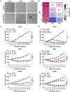
Fig. 1
Fig. 1
Oncolytic activity of CVB3/2035A in human CSCC cell lines and cell line-derived xenografts. (A) CSCC cell monolayers were either mock-infected or infected with CVB3/2035A at an MOI of 1. Photomicrographs were taken at 72 h post-infection. Scale bar, 100 μm. (B) CSCC cells were infected with CVB3/2035A at an MOI of 10, 1, 0.1, and 0.01 for 72 h. Cell viability was assessed using the CCK8 assay. (C) Nude mice bearing C33A, SiHa, and CaSki xenografts were treated i.t. or i.v. with five consecutive doses of CVB3/2035A (1 × 108 TCID50 per dose). An equivalent volume of PBS was administered i.t. as a control (Mock). Tumor growth was monitored over a 20-day period. (D) The combination of CVB3/2035A and paclitaxel improved efficacy in CSCC CDX models. Nude mice with C33A, SiHa, and CaSki xenografts were either mock-treated (PBS i.t. + PTX diluting solution i.p.) or treated with CVB3/2035A (1 × 108 TCID50 i.t. + PTX diluting solution i.p.), PTX (5 mg/kg i.p. + PBS i.t.), or a combination (1 × 108 TCID50 CVB3/2035A i.t. + 5 mg/kg PTX i.p.). Data are presented as means ± SEM. Statistical analysis was performed using a two-way ANOVA (n = 5). Black arrows indicate treatments; ns, not significant; *p < 0.05; ****p < 0.0001
Fig. 2
Fig. 2
Oncolytic effect of CVB3/2035A on patient-derived CSCC samples ex vivo. Four CSCC samples (Tumor 1–4) and three normal cervical samples (Normal 1–3) were divided into approximately 1-mm3 pieces and treated with CVB3/2035A (1 × 107 TCID50/well) or left untreated (n = 3 per group). After 72 h, (A) tissue viability was assessed using a modified HDRA method, and (B) replication of CVB3/2035A in both tumor and normal tissue samples was quantified by qRT-PCR. Statistical analysis was performed by comparing each tumor sample’s value to the mean value of all three normal samples using one-way ANOVA. Data are presented as means ± SEM. **p < 0.01; ***p < 0.001; ****p < 0.0001
Fig. 3
Fig. 3
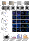
Generation of CSCC Organoids. (A) Schematic illustration of the derivation of CSCC organoids from patient samples. (B) Representative brightfield microscopy images of CSCC organoids post-seeding (exemplified by CSCC-2; see Fig. S4 for more examples). Scale bar, 25 μm. (C) Measurements of the growth diameters of CSCC organoids at days 1, 7, and 11. Horizontal lines and error bars represent the mean diameter ± SEM for five organoids (n = 5). (D) Immunohistological features of CSCC organoids derived from patients, compared to original tumors (exemplified by CSCC-2, see Fig. S5 for more examples). Representative immunofluorescence images display markers Ki67, p53, PanCK, PAX8, and CAR (all in red), with nuclei counterstained using DAPI (blue) and cell membranes with phalloidin (green). Inset images from the left panels are magnified three-fold in the right panels for enhanced detail. Scale bar, 50 μm. (E) Proportion of immunofluorescence staining-positive cells in primary tumors and organoids (n = 3). Statistical significance was assessed using two-way ANOVA. T, tumor; O, organoid; ns, not significant; *p < 0.05; ***p < 0.001; ****p < 0.0001; ns, not significant
Fig. 4
Fig. 4
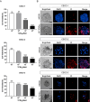
Cytotoxic effects of CVB3/2035A on CSCC organoids. (A) CSCC organoids were exposed to 10-fold serial dilutions of CVB3/2035A and incubated at 37 ℃ for 72 h. Cell viability was evaluated using the Cell Titer-Glo 3D assay, with results presented as mean ± SEM (n = 4). Statistical analysis between CVB3/2035A-infected groups and the control group were assessed using One-way ANOVA followed by Tukey’s test. ***p < 0.001; ****p < 0.0001; ns, not significant. (B) Representative images of CSCC organoids 72 h post-infection with 1 × 107 TCID50/well CVB3/2035A. Dead cells were visualized with PI staining (red), while nuclei were counterstained with DAPI (blue). Scale bar, 50 μm
Fig. 5
Fig. 5
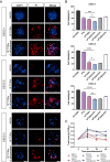
Replication of CVB3/2035A in CSCC organoids. (A) Growth kinetics of CVB3/2035A in CSCC organoid lines. Following infection with 10-fold serial dilutions of CVB3/2035A (ranging from 1 × 108 to 1 × 105 TCID50/well), supernatant samples were collected at 4, 12, 24, and 48 hpi for viral titration by the TCID50 assay. Statistical analysis of peak titers compared to the titers at 4 hpi was performed using two-way ANOVA. *p < 0.05; **p < 0.01. (B) Immunofluorescence analysis of CVB3/2035A infection in CSCC organoid lines. CSCC organoids, infected with 1 × 107 TCID50/well of CVB3/2035A, were collected at 72 hpi and subjected to immunofluorescent staining for double-stranded RNA (J2) and the CVB3 VP1 protein (both in red). Nuclei were counterstained with DAPI (blue). Scale bar, 50 μm
Fig. 6
Fig. 6
Combined treatment of CVB3/2035A and paclitaxel for CSCC organoids. (A) Representative images of CSCC organoids 72 h post-infection with 1 × 107 TCID50/well CVB3/2035A, with or without 2 µM PTX. Dead cells were visualized with PI staining (red), while nuclei were counterstained with DAPI (blue). Scale bar, 50 μm. (B) CSCC organoid lines were treated with 2 µM PTX, 1 × 107 TCID50/well CVB3/2035A, or 2 µM PTX plus 10-fold diluted CVB3/2035A (ranging from 1 × 107 to 1 × 105 TCID50/well). Cell viability was evaluated using the Cell Titer-Glo 3D assay, with results presented as mean ± SEM. Statistical analysis was performed using one-way ANOVA followed by Tukey’s test. *p < 0.05; **p < 0.01; ***p < 0.001; ****p < 0.0001. (C) Comparison of viral growth kinetics between groups treated with 2 µM PTX plus 1 × 107 TCID50/well CVB3/2035A and those treated with 1 × 107 TCID50/well CVB3/2035A alone. Data are represented as mean ± SEM, statistical analysis for virus peak titer was performed using Student’s t-test

Similar articles

References

    1. Sung H, Ferlay J, Siegel RL, Laversanne M, Soerjomataram I, Jemal A, et al. Global Cancer statistics 2020: GLOBOCAN estimates of incidence and Mortality Worldwide for 36 cancers in 185 countries. CA Cancer J Clin. 2021;71(3):209–49. - PubMed
    1. Lõhmussaar K, Oka R, Espejo Valle-Inclan J, Smits MHH, Wardak H, Korving J, et al. Patient-derived organoids model cervical tissue dynamics and viral oncogenesis in cervical cancer. Cell Stem Cell. 2021;28(8):1380–e966. - PubMed
    1. Cohen PA, Jhingran A, Oaknin A, Denny L. Cervical cancer. Lancet. 2019;393(10167):169–82. - PubMed
    1. Lin D, Shen Y, Liang T. Oncolytic virotherapy: basic principles, recent advances and future directions. Signal Transduct Target Ther. 2023;8(1):156. - PMC - PubMed
    1. Shalhout SZ, Miller DM, Emerick KS, Kaufman HL. Therapy with oncolytic viruses: progress and challenges. Nat Rev Clin Oncol. 2023;20(3):160–77. - PubMed

MeSH terms