. 2024 Oct 3;20(10):e1011424.
doi: 10.1371/journal.pgen.1011424. eCollection 2024 Oct.

The middle domain of Hsp104 can ensure substrates are functional after processing

Affiliations

The middle domain of Hsp104 can ensure substrates are functional after processing

Hannah E Buchholz et al. PLoS Genet. .

Abstract

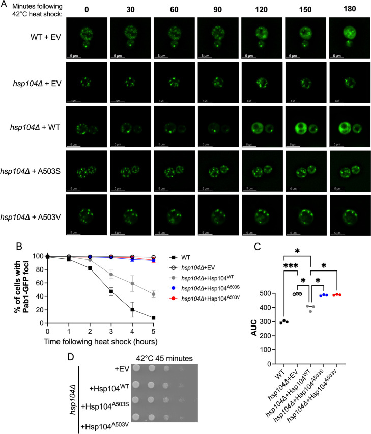
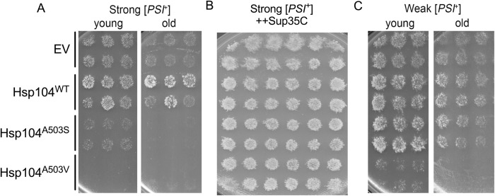
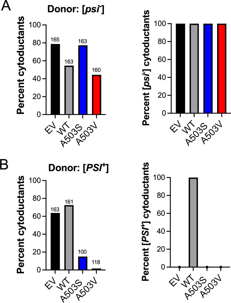
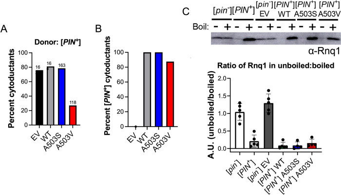
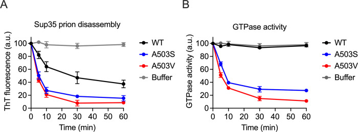
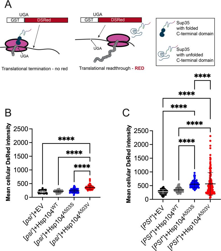
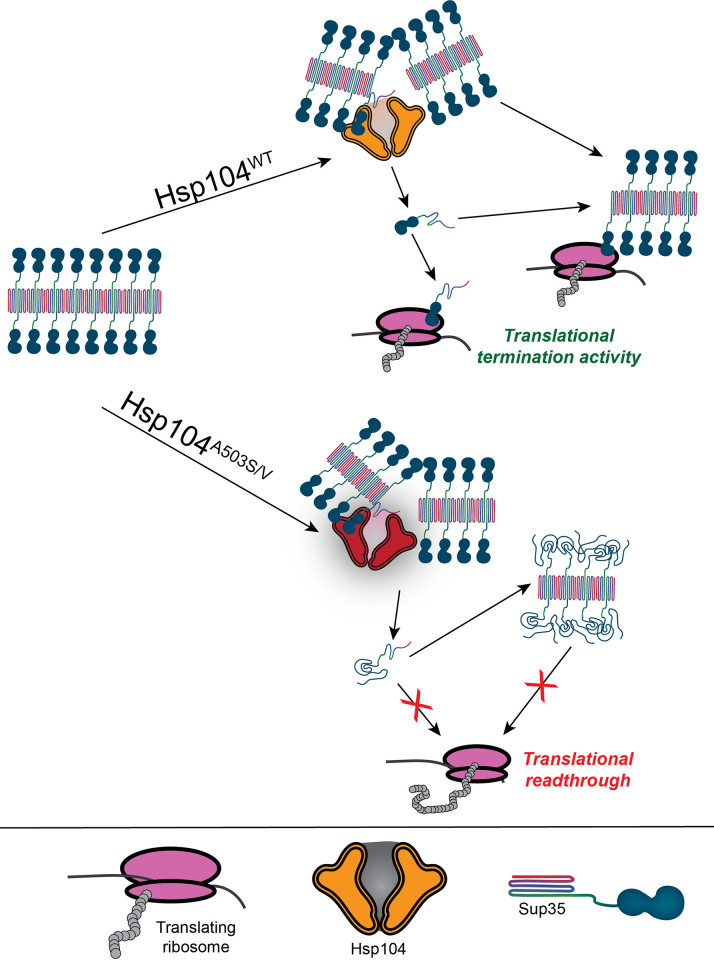
Molecular chaperones play a central role in protein disaggregation. However, the molecular determinants that regulate this process are poorly understood. Hsp104 is an AAA+ ATPase that disassembles stress granules and amyloids in yeast through collaboration with Hsp70 and Hsp40. In vitro studies show that Hsp104 processes different types of protein aggregates by partially translocating or threading polypeptides through the central pore of the hexamer. However, it is unclear how Hsp104 processing influences client protein function in vivo. The middle domain (MD) of Hsp104 regulates ATPase activity and interactions with Hsp70. Here, we tested how MD variants, Hsp104A503S and Hsp104A503V, process different protein aggregates. We establish that engineered MD variants fail to resolve stress granules but retain prion fragmentation activity required for prion propagation. Using the Sup35 prion protein, our in vitro and in vivo data indicate that the MD variants can disassemble Sup35 aggregates, but the disaggregated protein has reduced GTPase and translation termination activity. These results suggest that the middle domain can play a role in sensing certain substrates and plays an essential role in ensuring the processed protein is functional.


Conflict of interest statement

The authors have declared that no competing interests exist.

Figures

Fig 1
Fig 1. Hsp104A503S and Hsp104A503V exhibit reduced stress granule disassembly as the only source of Hsp104.
A) WT and hsp104Δ strains, transformed with a plasmid containing the stress granule marker, poly-A binding protein, fused to GFP (Pab1-GFP) and the indicated HSE plasmids. Strains were heat shocked at 42°C for 45 minutes. Shown are representative cells from indicated strains undergoing stress granule disassembly following acute heat stress. All images were taken at the same magnification (630X) and scale bars are all equivalent to 5μm. B) Images were taken at the indicated time points following heat shock, and the number of cells with Pab1-GFP foci were quantified. C) Area under the curve (AUC) was quantified from B to determine statistical differences between strains. Analysis using Brown-Forsythe ANOVA with a Games-Howell’s multiple comparisons test showed the indicated significance bwteen strains (*p>0.03; ***p≤0.0009). D) hsp104Δ strains, as in A, were subjected to 42°C heat shock for 45 minutes and plated on SD-Ura.
Fig 2
Fig 2. Hsp104A503S is toxic in strong [PSI+] but not weak [PSI+].
A) Left Galactose-inducible Hsp104 plasmids were transformed into a wildtype strong [PSI+][pin-] strain. Left, six transformants of each indicated strain were first pronged onto media containing 2% dextrose media lacking uracil and grown for 2 days (young). Strains were then velveted onto 2% galactose media lacking uracil to induce plasmid expression. The image of toxicity shown in the figure is observed after three passages on 2% galactose media. Right The original transformants plated on 2% dextrose were incubated for one week (old) before being velveted onto 2% galactose media lacking uracil to induce plasmid expression. The image of toxicity shown is after two passages on 2% galactose media. B) Galactose-inducible Hsp104 plasmids as well as a galactose-inducible Sup35C plasmid were transformed into a wildtype strong [PSI+][pin-] strain. Twelve transformants of each strain were pronged onto media containing 2% dextrose media lacking uracil and tryptophan and grown for two days, then velveted onto 2% galactose media lacking uracil and tryptophan to induce plasmid expression. Viability shown is after two passages on 2% galactose media. C) Same as A, but in a wildtype weak [PSI+][pin-] strain.
Fig 3
Fig 3. Hsp104A503S is sufficient to cure weak [PSI+] similarly to Hsp104WT.
A) Wildtype weak [PSI+][pin-] strains with indicated HSE plasmids were plated on SD-Ura to assess toxicity and on YPD to assess [PSI+] curing by a colony color assay. Shown is a representative of three trials. B) Fresh transformants of a weak [PSI+] strain with indicated plasmids were grown in liquid SD-Ura media and plated for single colony onto YPD at the indicated time points. Curing was determined by colony color 7 days after the cells were plated. Three trials were conducted, 50–150 colonies were assessed for each trial. Two-way ANOVA with Tukey’s multiple comparisons tests detected significant differences between Hsp104WT and Hsp104A503V at 48 hours p = 0.0118. C) Weak [PSI+] strains with indicated galactose-inducible plasmids were grown in 2% raffinose or 2% galactose media lacking uracil to an OD600 of 0.8–1.0 (approximately 20 hours) and plated on YPD to visually assess [PSI+] curing by colony color. The labeling indicates whether the strains were initially grown in either raffinose (no induction) or galactose (induction) prior to plating on YPD to assess color. E) Strains in C were also plated for single colony and scored for the presence of [PSI+]. At least three trials were conducted and approximately 800–1,500 colonies were assessed per trial. (*p = 0.027 t-test with Welch’s correction).
Fig 4
Fig 4. Hsp104A503S or Hsp104A503V as the only source of Hsp104 cannot maintain [PSI+].
A) Left panel, cytoduction was performed from a donor strain lacking prions ([psi-][pin-]) to a hsp104Δ recipient strain containing the indicated HSE plasmid. Definitive cytoductants were identified based upon acquisition of [RHO+] phenotype, and cytoductant selective markers. Numbers above bars represent the total sample number of potential cytoductants that were screened whereas the bar indicates the percentage of definitive cytoductants obtained. Right panel, definitive cytoductants were assayed for the [PSI+] state. B) Left panel, the percentage of definitive cytoductants obtained from a strong [PSI+] donor. Right panel, quantification of cytoductants that were able to retain [PSI+].
Fig 5
Fig 5. Hsp104A503S and Hsp104A503V retain [PIN+] prion fragmentation activity and do not change the soluble pool of Rnq1.
A) The percentage of definitive cytoductants obtained from a [psi-] high [PIN+] donor. B) Quantification of cytoductants that were able to retain [PIN+]. C) Well trap assay used to determine the amount of SDS-sensitive soluble Rnq1 protein in the indicated [PIN+] cytoductants obtained from Fig 4C. Western blot (top) and quantification of four independent experiments (bottom) are shown.
Fig 6
Fig 6. Hsp104A503S and Hsp104A503V disassemble Sup35 fibrils into non-functional protein in vitro.
A) Preformed full-length Sup35 prions (1 μM monomer) were incubated with GroELtrap (2 μM), Ssa1 (1μM), Sis1 (1μM), Sse1 (0.1μM), and either buffer, Hsp104WT, Hsp104A503S, or Hsp104A503V (1μM) and monitored over 60 minutes. Sup35 fibril integrity was measured with Thioflavin-T (ThT) fluorescence. Values represent means±SEM (n = 3). B) The GTPase activity of full-length Sup35 from A was assessed by measuring the GTPase activity associated with the C-terminal domain of Sup35 over time. Values represent means±SEM (n = 3).
Fig 7
Fig 7. Hsp104A503S and Hsp104A503V increase stop codon readthrough.
A) Schematic of indirectly assaying Sup35 function by quantifying fluorescent intensity of DsRed, which contains a UGA codon between GST and DsRed. In the presence of functional Sup35, translation termination at the UGA codon results in a truncated protein. In the presence of non-functional Sup35, translational readthrough of UGA will result in a GST-DsRed fusion protein. B) A [psi-][pin-] strain integrated with a GST(UGA)DsRed reporter was transformed with the indicated galactose-inducible Hsp104 variants. Strains were grown on galactose-containing media for two days to drive plasmid expression. Cells were picked directly from plates and subjected to fluorescent microscopy. Mean DsRed pixel intensity was measured in individual cells and normalized to the mean DsRed pixel intensity of the field background. Bars represent mean±SD Approximately 25–250 cells were counted for each of three trials for a minimum total of 275 cells. C) Same as in B, but in a [PSI+][pin-] strain. A Brown-Forsythe and Welch’s ANOVA test with Games-Howell’s multiple comparisons test was used to compare strains (****p≤0.0001).
Fig 8
Fig 8. Contributions of the Hsp104 middle domain in prion processing.
Sup35 contains a prion domain (shown as rainbow core), a linker thought to bind to Hsp104, and a functional GTPase C-terminal domain (shown as an attached blue shape). Within the prion aggregate, the C-terminal domain is accessible on the surface and retains translation termination activity. The A503 within the middle domain plays a role in ensuring that the extracted monomer retains functional activity, allowing the functional Sup35 protein to remain a monomer or join the prion aggregate, both of which can participate in translational termination. Mutations in the middle domain may lead to complete unfolding of Sup35 monomers (bottom), resulting in monomers that are nonfunctional. These nonfunctional proteins may stay as monomers, join the prion aggregate (as shown), form amorphous aggregates, or could be degraded.

Similar articles

References

    1. Shorter J. The mammalian disaggregase machinery: Hsp110 synergizes with Hsp70 and Hsp40 to catalyze protein disaggregation and reactivation in a cell-free system. PLoS One. 2011;6(10):e26319. Epub 20111014. doi: 10.1371/journal.pone.0026319 ; PubMed Central PMCID: PMC3194798. - DOI - PMC - PubMed
    1. Rampelt H, Kirstein-Miles J, Nillegoda NB, Chi K, Scholz SR, Morimoto RI, et al.. Metazoan Hsp70 machines use Hsp110 to power protein disaggregation. EMBO J. 2012;31(21):4221–35. Epub 20120918. doi: 10.1038/emboj.2012.264 ; PubMed Central PMCID: PMC3492728. - DOI - PMC - PubMed
    1. Nillegoda NB, Kirstein J, Szlachcic A, Berynskyy M, Stank A, Stengel F, et al.. Crucial HSP70 co-chaperone complex unlocks metazoan protein disaggregation. Nature. 2015;524(7564):247–51. Epub 20150805. doi: 10.1038/nature14884 ; PubMed Central PMCID: PMC4830470. - DOI - PMC - PubMed
    1. Labbadia J, Morimoto RI. The biology of proteostasis in aging and disease. Annual review of biochemistry. 2015;84:435–64. Epub 2015/03/19. doi: 10.1146/annurev-biochem-060614-033955 ; PubMed Central PMCID: PMC4539002. - DOI - PMC - PubMed
    1. Klaips CL, Jayaraj GG, Hartl FU. Pathways of cellular proteostasis in aging and disease. J Cell Biol. 2018;217(1):51–63. doi: 10.1083/jcb.201709072 ; PubMed Central PMCID: PMC5748993. - DOI - PMC - PubMed

MeSH terms

LinkOut - more resources