. 2024 Oct 3;14(1):22939.
doi: 10.1038/s41598-024-73339-2.

Development of novel canine phage display-derived neutralizing monoclonal antibody fragments against rabies virus from immunized dogs

Affiliations

Development of novel canine phage display-derived neutralizing monoclonal antibody fragments against rabies virus from immunized dogs

Apidsada Chorpunkul et al. Sci Rep. .

Abstract

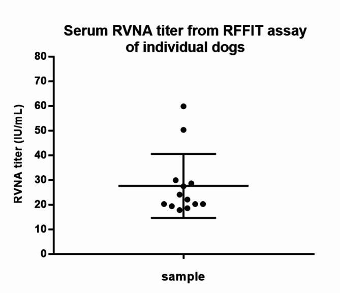
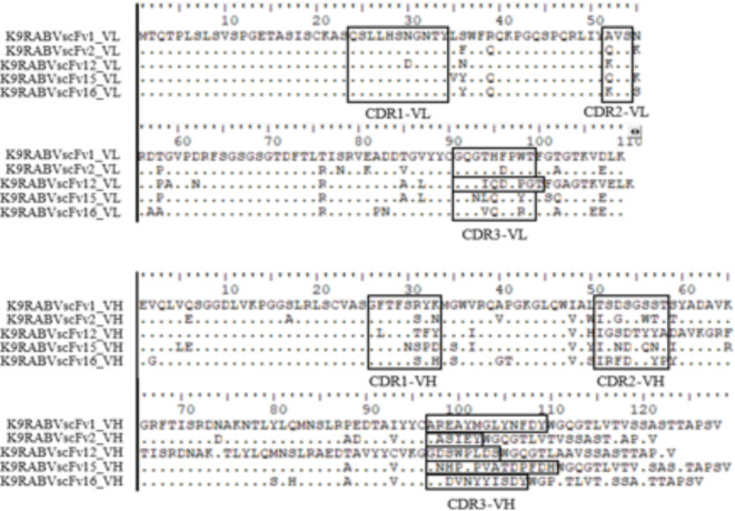
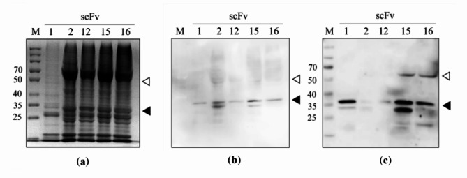
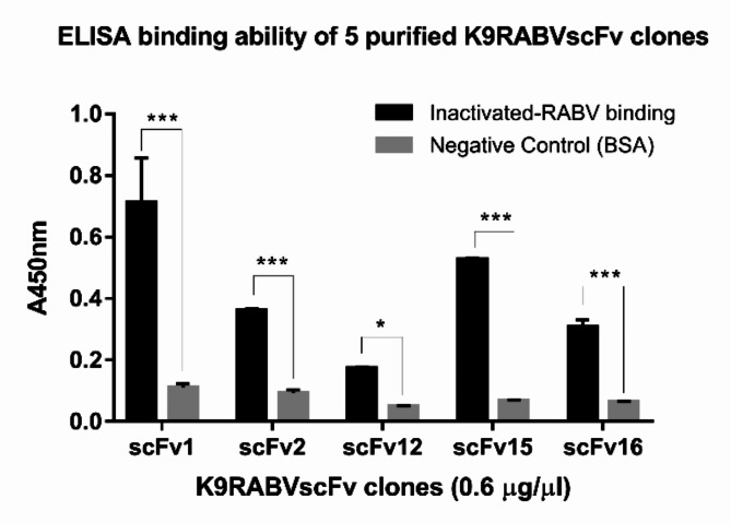
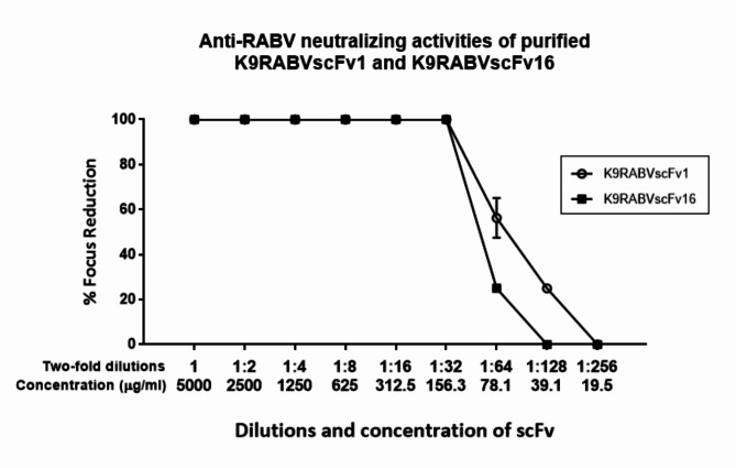
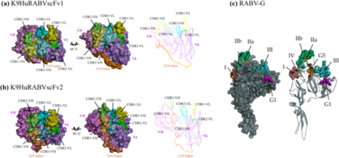
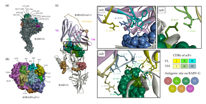
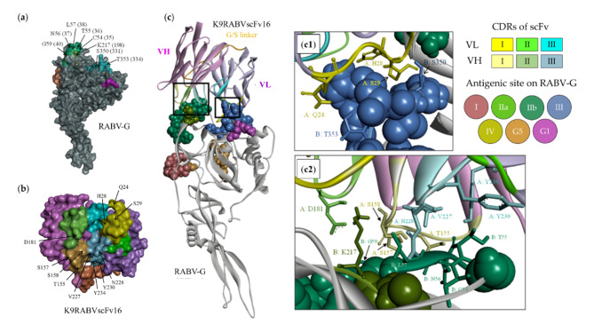
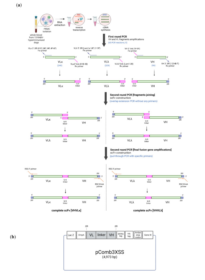
Animal rabies is a potentially fatal infectious disease in mammals, especially dogs. Currently, the number of rabies cases in pet dogs is increasing in several regions of Thailand. However, no passive postexposure prophylaxis (PEP) has been developed to combat rabies infection in animals. As monoclonal antibodies (MAbs) are promising biological therapies for postinfection, we developed a canine-neutralizing MAb against rabies virus (RABV) via the single-chain variable fragment (scFv) platform. Immunized phage-displaying scFv libraries were constructed from PBMCs via the pComb3XSS system. Diverse canine VHVLκ and VHVLλ libraries containing 2.4 × 10⁸ and 1.3 × 10⁶ clones, respectively, were constructed. Five unique clones that show binding affinity with the RABV glycoprotein were then selected, of which K9RABVscFv1 and K9RABVscFv16 showed rapid fluorescent foci inhibition test (RFFIT) neutralizing titers above the human protective level of 0.5 IU/ml. Finally, in silico docking predictions revealed that the residues on the CDRs of these neutralizing clones interact mainly with similar antigenic sites II and III on the RABV glycoprotein. These candidates may be used to develop complete anti-RABV MAbs as a novel PEP protocol in pet dogs and other animals.


Conflict of interest statement

The authors declare no competing interests.

Figures

Fig. 1
Fig. 1
Serum VNA titer against rabies virus in dogs via the RFFIT assay. All 13 serum samples presented neutralizing antibody levels greater than 0.5 IU/ml. The average titers ranged from 20 to 30 IU/ml. Two outliers presented levels greater than 50 IU/ml.
Fig. 2
Fig. 2
VH, VLλ, and VLκ amplification via conventional PCR. (a) VLκ amplicons were amplified via sets of forward primers (CSCK1-F, 24-F, 34-F, 4-F, 5-F, and 6-F; numbers 1, 24, 34, 4, 5, and 6 in the figure). Four reverse primers were used: CSCJK1-B, CSCJK2-B, CSCJK3-B, and CSCJK4-B. (b) VLλ amplicons were amplified via sets of forward primers (CSCLam1a-F–CSCLam13-F; numbers 1a, 1b, 1c, 1d, and 2–13 in the figure). CSCJLam1-B and CSCJLam2-B served as reverse primers. The size of the VLκ and VLλ PCR products was 350 bp. (c) VH amplicons were amplified via the forward primer (CSCVH1-F–CSCVH9-F; numbers 1–9 in the figure) and the CSCG1234-B reverse primer. The size of the VH PCR products was 450 bp. (d) Purified variable chain fragments. After the antibody fragments were amplified via PCR, the corrected PCR products were purified via gel extraction (Lane 1, purified VH; Lane 2, purified VLλ; and Lane 3, purified VLκ). The PCR products were run on 1.2% agarose gels.
Fig. 3
Fig. 3
Colony PCR of 16 randomly selected clones from the (a) VHVLκ scFv library and (b) VHVLλ scFv library. The inserted scFv antibodies were amplified via second-round PCR primers (RSC-F and RSC-B(AA) primers), and the size of the PCR product was 850–900 bp, as observed on a 1% agarose gel. M refers to the 100 bp DNA ladder.
Fig. 4
Fig. 4
BstNI analysis of the scFv libraries. (a) Fingerprint BstNI digestion of 15 randomly selected clones from the VHVLκ library. (b) Fifteen randomly selected clones from the VHVLλ library.
Fig. 5
Fig. 5
Input and output phage titers (CFU/ml) of the (a) VHVLҡ and (b) VHVLλ libraries. Four selection rounds were performed per target antigen. The results of the affinity selection process are shown in the scatter plots. (a1) (b1) rG binding selection. (a2) (b2) RABV binding selection. The Y-axis shows the power of ten logarithmic scales, the left side indicates the input titers, and the right side indicates the output titers. The gray and black dots indicate the input and output phage titers of each round, respectively.
Fig. 6
Fig. 6
ELISA absorbance signal at 450 nm from soluble fractions of all 37 clones. Each sample consisted of four wells: expression control, RABV, rG, and negative control (SM). (a) Clone numbers 1–19. (b) Clone numbers 20–37. (c) Clone numbers 38–58. Differences between the absorbance levels of the target antigens and SM binding were assessed via independent t tests. The asterisks indicate significant differences as follows: P value ≤ 0.05 (*), ≤ 0.01 (**), and ≤ 0.001 (***). The symbol “#” refers to an excluded sample whose signal from the negative control was greater than or equal to that of the scFv samples. A secondary anti-HA-conjugated HRP antibody bound to the scFv was detected using a TMB substrate. The error bars indicate the SDs of two replicates.
Fig. 7
Fig. 7
Amino acid sequence alignments for the five selected K9RABVscFv clones. The complementarity-determining regions (CDRs) of the VL (top) and VH (below) fragments are in black-bordered rectangles.
Fig. 8
Fig. 8
SDS‒PAGE and Western blot analysis of the five purified, concentrated, and soluble K9RABVscFv clones. The actual scFv molecular weight was approximately 30–37 kDa (black arrow), and a 60-kDa (white arrow) dimeric form of scFv was observed in four clones. (a) SDS‒PAGE analysis of 5 K9RABVscFv (b) Anti-6XHIS tag-conjugated HRP secondary monoclonal antibody (clone HIS-1, Sigma‒Aldrich, UK) at a concentration of 1:10,000 bound to the scFv antibody in the WB assay and detected by ECL reagents (Cytiva Amersham). (c) All five scFv antibodies incorporated with primary antibody 1:5,000 dilution of unconjugated goat anti-human IgG [F(ab’)2] (Invitrogen; Thermo Fisher Scientific) and a 1:5,000 dilution of HRP-conjugated rabbit anti-goat IgG (H + L) (Invitrogen; Thermo Fisher Scientific) as a secondary antibody. The secondary antibodies were detected via enhanced chemiluminescence (ECL) reagents (Cytiva Amersham). The original Western blot images are shown in Fig. S3. A Spectra Multicolor Broad Range Protein Ladder (Thermo Scientific) was used in panels (a) and (c).
Fig. 9
Fig. 9
ELISA binding absorbance signal at 450 nm of five purified soluble K9RABVscFv clones (0.6 µg/µl; total 60 µg/well). BSA was used as a negative control. Asterisks indicate significant differences at P values ≤ 0.05 (*), ≤ 0.01 (**), and ≤ 0.001 (***). A TMB substrate was used for detection. The error bars indicate the SDs of duplicate samples.
Fig. 10
Fig. 10
Percent neutralization of K9RABVscFv1 (5.35 mg/ml) and K9RABVscFv16 (5.88 mg/ml) interpreted from the RFFIT results. Twofold dilutions of both antibodies were conducted from 1:2 to 1:256. A total of 8 fields per well were observed. Percent neutralization was calculated from the number of negative fields.
Fig. 11
Fig. 11
Three-dimensional (3D) tertiary structure prediction of the K9RABVscFv1 and K9RABVscFv16 antibodies and the rabies virus glycoprotein (RABV-G). PDBID no. 1 × 9q, 8dy2.1. A and the monomer form of PDBID no. 7u9g were used as templates for K9RABVscFv1, K9RABVscFv16, and RABV-G protein structure simulations. The light purple and pink clusters represent the VL and VH chains, respectively. The orange fragment was a glycine/serine linker (G/S linker). CDR1–3 on the VH and VL chains are shown in several colors. Surface display and line-ribbon style of (a) K9RABVscFv1, (b) K9RABVscFv16, and (c) RABV-G (SQ382). The SWISS model online server (https://swissmodel.expasy.org/) was used to construct protein structure homology models, which were decorated with BIOVIA Discovery Studio Visualizer 2021.
Fig. 12
Fig. 12
The most likely binding conformation between the CDR1–3 area on K9RABVscFv1 and the antigenic sites on RABV-G was predicted by the HADDOCK 2.4 web server. (a) Surface display structures of RABV-G with residues on antigenic sites that interact with (b) residues on the CDR of K9RABVscFv1. The numbers in brackets “()” in the RABV-G labels refer to the positions without 19 amino acids in the signal sequence. (c) Interactions between the residues on the CDR of K9RABVscFv1 and the antigenic sites of RABV-G on the basis of the results in Table 3. The sticks display the amino acid residues on the CDR (chain A) that interact with the residues on the antigenic sites of RABV-G (chain B). Significant interactions are shown in panels (c1), (c2), and (c3). The VH chain, VL chain, G/S linker, CDR1–3 VH, CDR1–3 VL, antigenic major sites I, II, III, IV, and G5, and minor site G1 (or “a”) are shown in different colors.
Fig. 13
Fig. 13
The most likely binding conformation between the CDR1–3 area on K9RABVscFv16 and the antigenic sites on RABV-G was predicted by the HADDOCK 2.4 web server. (a) Surface display structures of RABV-G with residues on antigenic sites that interact with (b) residues on the CDR of K9RABVscFv1. The numbers in brackets “()” in the RABV-G labels refer to the positions without 19 amino acids in the signal sequence. (c) Interactions between the residues on the CDR of K9RABVscFv1 and the antigenic sites of RABV-G on the basis of the results in Table 3. The sticks display the amino acid residues on the CDR (A chain) that interact with the residues on the antigenic sites of RABV-G (B chain). Significant interactions are shown in panels (c1) and (c2). The VH chain, VL chain, G/S linker, CDR1–3 VH, CDR1–3 VL, antigenic major sites I, II, III, IV, and G5, and minor site G1 (or “a”) are shown in different colors.
Fig. 14
Fig. 14
Schematic representation of canine variable-chain fragment amplification and scFv construction via the pComb3XSS phage display system. (a) Step of PCR amplification of variable fragments and scFv construction. (b) Structure of pComb3XSS harboring the scFv fragment.

Similar articles

References

    1. Thanapongtharm, W., Suwanpakdee, S., Chumkaeo, A., Gilbert, M. & Wiratsudakul, A. Current characteristics of animal rabies cases in Thailand and relevant risk factors identified by a spatial modeling approach. PLoS Negl. Trop. Dis.15, e0009980. 10.1371/journal.pntd.0009980 (2021). - PMC - PubMed
    1. Niezgoda, M., Hanlon, C. A. & Rupprecht, C. E. Animal rabies. Rabies 163–218 (2003).
    1. Dietzschold, B., Tollis, M., Lafon, M., Wunner, W. H. & Koprowski, H. Mechanisms of Rabies virus neutralization by glycoprotein-specific monoclonal antibodies. Virology161, 29–36 (1987). - PubMed
    1. Callaway, H. M. et al. Structure of the Rabies virus glycoprotein trimer bound to a prefusion-specific neutralizing antibody. Sci. Adv.8, eabp9151. 10.1126/sciadv.abp9151 (2022). - PMC - PubMed
    1. Flamand, V. et al. Vaccination with tumor-antigen-pulsed dendritic cells induces in vivo resistance to a B cell lymphoma. Adv. Exp. Med. Biol.329, 611–616. 10.1007/978-1-4615-2930-9_102 (1993). - PubMed