

doi: 10.1038/s41467-024-52918-x.
The molecular dissection of TRIM25's RNA-binding mechanism provides key insights into its antiviral activity
Affiliations
- PMID: 39353916
- PMCID: PMC11445558
- DOI: 10.1038/s41467-024-52918-x
Item in Clipboard
The molecular dissection of TRIM25's RNA-binding mechanism provides key insights into its antiviral activity
Nat Commun.
.
Abstract
TRIM25 is an RNA-binding ubiquitin E3 ligase with central but poorly understood roles in the innate immune response to RNA viruses. The link between TRIM25's RNA binding and its role in innate immunity has not been established. Thus, we utilized a multitude of biophysical techniques to identify key RNA-binding residues of TRIM25 and developed an RNA-binding deficient mutant (TRIM25-m9). Using iCLIP2 in virus-infected and uninfected cells, we identified TRIM25's RNA sequence and structure specificity, that it binds specifically to viral RNA, and that the interaction with RNA is critical for its antiviral activity.
© 2024. The Author(s).
Conflict of interest statement
The authors declare no competing interests.
Figures

a Domain arrangement of TRIM25. b
1H-15N-HSQC spectra of 100 µM 15N-labelled TRIM25 PRY/SPRY domain free (dark blue) and in presence of different ratios of pre-let-7 RNA (1:1 light blue, 1:2 green and 1:3 yellow). c Significant CSPs from the titration of TRIM25 PRY/SPRY with pre-let-7 plotted on the structure (PDB: 6FLM) indicate two binding sites (binding site 1 coloured red, binding site 2 coloured blue). The structure shown was generated using UCSF ChimeraX. d Histograms of chemical shift perturbations (CSPs) for TRIM25-PRY/SPRY upon binding to full-length pre-let-7 (top), pre-let-7-loop (middle) and pre-let-7-stem (bottom). The continuous line shows the average CSP and the dashed line indicates the average plus one standard deviation. Residues for which the CSPs are more than one standard deviation above the average are shown in open bars. Significantly affected regions are indicated in the protein sequence by red and blue horizontal bars. The RNA is coloured according to where it binds in the PRY/SPRY domain (binding site 1 in red, binding site 2 in blue). Source data are provided as a Source Data file. e Representative ITC binding isotherm for TRIM25 PRY/SPRY:pre-let-7 complex (n = 4). The value shown in the figure is the average of all replicates and its standard deviation. f Representative ITC binding isotherm for TRIM25 PRY/SPRY triple mutant (m3): pre-let-7 complex (n = 3). All experimental setups and results of ITC measurements including replicates can be found in Supplementary Table 1.
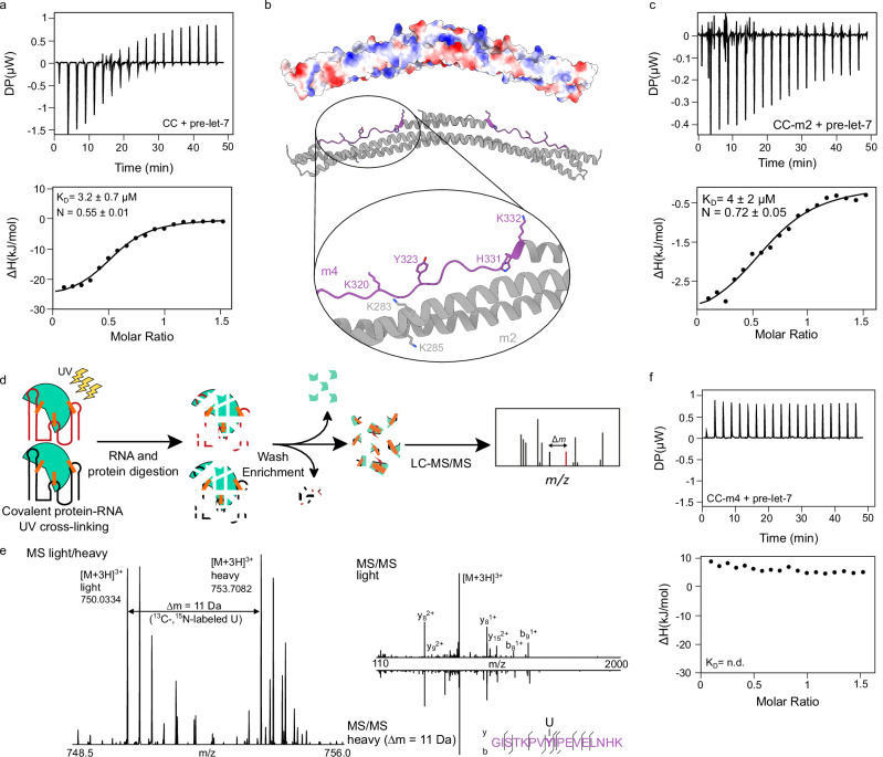
a Representative ITC binding isotherm for TRIM25 CC:pre-let-7 complex (n = 3). The value shown in the figure is the average of all replicates and its standard deviation. b The surface potential representation of the CC dimer (PDB:4LTB) indicates a positively charged surface. The enlargement of this interface shows potential RNA binding residues. Mutation of residues K283 and K285, shown in grey, leads to a TRIM25-CC mutant we term m2. In purple, the peptide detected by CLIR-MS/MS (see below) is highlighted including the four potential RNA binding residues. Mutation of these four residues leads to a TRIM25-CC mutant we term m4. The structure shown was generated using UCSF ChimeraX. c Representative ITC binding isotherm for TRIM25 CC-m2 with pre-let-7 complex (n = 3). d Schematic representation of the CLIR-MS/MS method. The RNA binding protein (grey) is crosslinked to an equimolar mixture of unlabelled (black) and uniformly 13C/15N-labelled RNA (red). e MS spectrum of the TRIM25-derived peptide GISTKPVYIPEVELNHK crosslinked to a single U nucleotide (see methods). f Representative ITC binding isotherm for TRIM25 CC-m4:pre-let-7 complex (n = 3). All experimental setups and ITC measurements including replicates are listed in Supplementary Table 1.

a Representative ITC binding isotherm for TRIM25 CC-PRY/SPRY (n = 3). The value shown is the average of all replicates and its standard deviation. b
1H,15N-HSQC spectra comparing CC-PRY/SPRY in the absence (black) and presence (red) of pre-let-7. The strong signal loss of peaks corresponding to residues in structured regions of the PRY/SPRY domain upon addition of equimolar amounts of pre-let-7 indicates that the RNA keeps the PRY/SPRY domain at the CC interface leading to joint tumbling and thus increased transverse relaxation and line broadening beyond detection. c Proposed mechanisms of RNA-induced conformational change. CC and PRY/SPRY of TRIM25 interact only transiently in the absence of RNA. Binding of stem-loop RNA stabilizes the interaction between the two domains. CC-PRY/SPRY dimer structure (PDB: 6FLN) shown as a surface representation with two binding sites in the PRY/SPRY domain (binding site 1 coloured red and binding site 2 coloured blue) and the binding site in the CC domain (coloured purple). The structure shown was generated using UCSF ChimeraX. In a second possibility, the stem-loop RNA is sandwiched between the CC and PRY/SPRY domains, which do not interact with each other. d SAXS curves and pairwise distance distributions for free TRIM25 CC-PRY/SPRY (black) and its complex with pre-let-7 RNA (red). The distance distribution of the free protein has two maxima, indicating independent tumbling of the CC and PRY/SPRY domains, whereas the distribution for the RNA-bound complex is much narrower and contains only one peak, indicating a conformational change towards a more compact form. e CC-PRY/SPRY dimer structure (PDB: 6FLN) in cartoon representation showing all RNA binding sites in the PRY/SPRY domain (binding site 1, red and binding site 2, blue), and CC domain (purple) and residues mutated to obtain the TRIM25 CC-PRY/SPRY m9 mutant. f Representative ITC binding isotherm for the TRIM25 CCPRY/SPRY-m9:pre-let-7 complex (n = 3). All experimental setups and ITC measurements including replicates are provided in Supplementary Table 1.
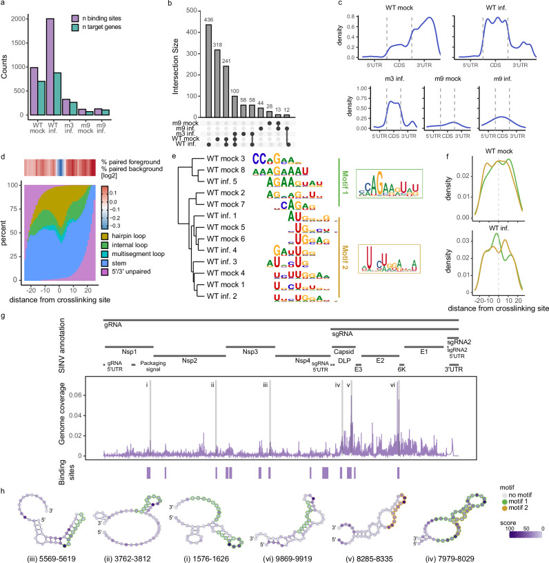
iCLIP2 was applied to HEK293 TRIM25 KO Flip-In expressing TRIM25-WT or the different TRIM25 mutants (m3 or m9) and infected with SINV and harvested 9 hour-post-infection, hpi (inf.) or mock-infected (mock). a Bar plots showing the number of cellular RNA binding sites (purple) and target gene (green) counts identified for the different samples. b Intersection plot comparing the different cellular targets RNAs identified in the different samples. c Density plot showing the distribution of binding sites across 5’ UTRs, CDSs and 3’ UTRs on cellular target RNAs for the different samples. d Percentage of paired and unpaired sequences across the binding site for the mock WT sample (see Supplementary Fig. 4f for TRIM25-WT infected sample). e Motifs enriched in the cellular RNA binding sites of the WT mock and SINV infected samples can be clustered into two prominent classes, an AGAA motif (motif 1) and a UGG motif (motif 2). f Density plot showing the distribution of the sequence motifs across the binding site for the TRIM25-WT mock and infected samples (green for motif 1 and orange for motif 2). The dotted line indicates the peak in the crosslinking signal. g Binding profile of TRIM25-WT on SINV RNA. Significant binding sites (p < 0.01) are indicated as blue boxes underneath the binding site density plot. h Secondary structure of the binding sites using SHAPE data. The colour of the nucleotides indicates the cross-linking density at each nucleotide position. The line surrounding the nucleotide indicates the presence of the identified motifs in the RNA structure (green for motif 1 and orange for motif 2).
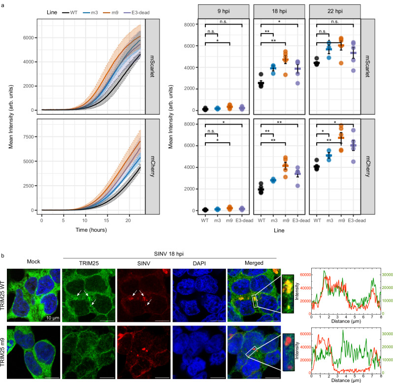
a Red fluorescence signal in TRIM25 WT (black) and mutant lines (m3 in blue, m9 in orange and E3-dead in purple) infected with SINV-mCherry (left panel) or SINV-nsp3-mScarlet (right panel). Fluorescence was measured every 15 min in a plate reader with atmospheric control (5% CO2 and 37 °C). The fluorescence is represented as mean ± SD of six independent infections in three biological replicates. Statistical differences are based on a two-tailed homoscedastic t-test (***p < 0.001; **p < 0.01; *p < 0.05). No adjustments were made for multiple comparisons. b Localization analysis by immunofluorescence and smFISH of TRIM25 (green) and SINV RNA (red). Nuclei are labelled with DAPI. Green and red fluorescence profiles for regions of interest are displayed on the right.
Similar articles
-
Depressing time: Waiting, melancholia, and the psychoanalytic practice of care.In: Kirtsoglou E, Simpson B, editors. The Time of Anthropology: Studies of Contemporary Chronopolitics. Abingdon: Routledge; 2020. Chapter 5. In: Kirtsoglou E, Simpson B, editors. The Time of Anthropology: Studies of Contemporary Chronopolitics. Abingdon: Routledge; 2020. Chapter 5. PMID: 36137063 Free Books & Documents. Review.
-
TRIM25 predominately associates with anti-viral stress granules.Nat Commun. 2024 May 15;15(1):4127. doi: 10.1038/s41467-024-48596-4. Nat Commun. 2024. PMID: 38750080 Free PMC article.
-
Functional anatomy of zinc finger antiviral protein complexes.Nat Commun. 2024 Dec 30;15(1):10834. doi: 10.1038/s41467-024-55192-z. Nat Commun. 2024. PMID: 39738020 Free PMC article.
-
Unlocking data: Decision-maker perspectives on cross-sectoral data sharing and linkage as part of a whole-systems approach to public health policy and practice.Public Health Res (Southampt). 2024 Nov 20:1-30. doi: 10.3310/KYTW2173. Online ahead of print. Public Health Res (Southampt). 2024. PMID: 39582242
-
Antioxidants for female subfertility.Cochrane Database Syst Rev. 2017 Jul 28;7(7):CD007807. doi: 10.1002/14651858.CD007807.pub3. Cochrane Database Syst Rev. 2017. Update in: Cochrane Database Syst Rev. 2020 Aug 27;8:CD007807. doi: 10.1002/14651858.CD007807.pub4. PMID: 28752910 Free PMC article. Updated. Review.
References
MeSH terms
Substances
Grants and funding
- BB/T002751/1/RCUK | Biotechnology and Biological Sciences Research Council (BBSRC)
- MR/R021562/1/RCUK | Medical Research Council (MRC)
- n.a./Joachim Herz Stiftung (Joachim Herz Foundation)
- BB/M011224/1/RCUK | Biotechnology and Biological Sciences Research Council (BBSRC)
- HE7291/8-1/Deutsche Forschungsgemeinschaft (German Research Foundation)
- WT_/Wellcome Trust/United Kingdom
- MC_UU_00034/5/MRC_/Medical Research Council/United Kingdom
- CC2075/WT_/Wellcome Trust/United Kingdom
- HE7291/1-1/Deutsche Forschungsgemeinschaft (German Research Foundation)
- CC2075/ARC_/Arthritis Research UK/United Kingdom
- MC_UU_00034/2/RCUK | Medical Research Council (MRC)
- 51NF40-182880/Schweizerischer Nationalfonds zur Förderung der Wissenschaftlichen Forschung (Swiss National Science Foundation)
- 101001634/EC | EU Framework Programme for Research and Innovation H2020 | H2020 Priority Excellent Science | H2020 European Research Council (H2020 Excellent Science - European Research Council)
LinkOut - more resources
Full Text Sources
