. 2024 Sep 30:13:RP94898.
doi: 10.7554/eLife.94898.

An arms race between 5'ppp-RNA virus and its alternative recognition receptor MDA5 in RIG-I-lost teleost fish

Affiliations

An arms race between 5'ppp-RNA virus and its alternative recognition receptor MDA5 in RIG-I-lost teleost fish

Shang Geng et al. Elife. .

Abstract

The incessant arms race between viruses and hosts has led to numerous evolutionary innovations that shape life's evolution. During this process, the interactions between viral receptors and viruses have garnered significant interest since viral receptors are cell surface proteins exploited by viruses to initiate infection. Our study sheds light on the arms race between the MDA5 receptor and 5'ppp-RNA virus in a lower vertebrate fish, Miichthys miiuy. Firstly, the frequent and independent loss events of RIG-I in vertebrates prompted us to search for alternative immune substitutes, with homology-dependent genetic compensation response (HDGCR) being the main pathway. Our further analysis suggested that MDA5 of M. miiuy and Gallus gallus, the homolog of RIG-I, can replace RIG-I in recognizing 5'ppp-RNA virus, which may lead to redundancy of RIG-I and loss from the species genome during evolution. Secondly, as an adversarial strategy, 5'ppp-RNA SCRV can utilize the m6A methylation mechanism to degrade MDA5 and weaken its antiviral immune ability, thus promoting its own replication and immune evasion. In summary, our study provides a snapshot into the interaction and coevolution between vertebrate and virus, offering valuable perspectives on the ecological and evolutionary factors that contribute to the diversity of the immune system.

Keywords: 5'ppp-RNA; MDA5; RIG-I; chicken; evolutionary biology; genetic compensation response; immune evasion; immunology; inflammation; pattern recognition receptors.

Plain language summary

Before the immune system can eliminate a bacterium, virus or other type of pathogen, it needs to be able to recognize these foreign elements. To achieve this, cells in the immune system have proteins called pattern recognition receptors (PRRs) which can identify distinct molecular features of certain pathogens. One specific group of PRRs is a family of retinoic acid-induced RIG-I-like receptors (RLRs), which help immune cells detect different types of viruses. Members of this family recognize distinct motifs on the genetic material of viruses known as RNA. For instance, RIG-I recognizes a marker known as 5’ppp on the end of single-stranded RNA molecules, whereas MDA5 recognizes long strands of double-stranded RNA. Many vertebrates – including various mammals, birds, and fish – lost the RIG-I receptor over the course of evolution. However, Geng et al. predicted that some animals lacking the RIG-I receptor may still be able to activate an immune response against viruses that contain the 5’ppp-RNA motif. To investigate this possibility, Geng et al. studied chickens and miiuy croakers (a type of ray-finned fish) which no longer have a RIG-I receptor. They found that both animals can still sense and eliminate two 5’ppp-RNA viruses called VSV and SCRV. Further experiments revealed that these two viruses are detected by a modified MDA5 receptor that had evolved to bind to 5’-ppp and activate the antiviral response. Viruses are also continuously evolving new ways to escape the immune system. This led Geng et al. to investigate whether SCRV, which causes serious harm to marine fish, has evolved a way to evade the MDA5 protection mechanism. Using miiuy croakers as a model, they found that SCRV causes the transcripts that produce the MDA5 protein to contain more molecules of m6a. This molecular tag degrades the transcript, leading to lower levels of MDA5, reducing the antiviral response against SCRV. The findings of Geng et al. offer valuable perspectives on how the immune system adapts over the course of evolution, and highlight the diversity of antiviral responses in vertebrates. Chickens and miiuy croakers are commonly farmed animals, and further work investigating how viruses invade these species could prevent illnesses from spreading and having a negative impact on the economy.


Conflict of interest statement

SG, XL, WZ, TX No competing interests declared

Figures

Figure 1.
Figure 1.. A genetic compensation response model for RIG loss.
(A) Loss of RIG-I in vertebrates. Each branch tip represents one species. Red lines show lineages where loss of RIG-I. The red circle indicates the occurrence of an independent loss event. Branch lengths scale in millions of years (MYA). (B) Comparative analysis of gene synteny of RIG-I in vertebrate genomes. (C and D) SCRV-RNA and VSV-RNA were extracted from SCRV and VSV virus and dephosphorylated them with Calf Intestinal Alkaline Phosphatase (CIAP). Then, MKC cells were transfected with SCRV-RNA and SCRV-RNA +CIAP, and DF-1 cells were transfected with VSV-RNA and VSV-RNA +CIAP. IRF3 dimerization was analyzed by native gel electrophoresis. (E) MKC cells were transfected with different concentrations of SCRV-RNA and SCRV-RNA +CIAP, then the expression of IFN-1 was detected by qRT-PCR. As a control, the expression level of IFN-1 in MKC cells transfected with the same volume of water was set to ‘1’. (F) Predicted protein structures of MDA5 and LGP2 of M. miiuy and RIG-I of H. sapiens. (G) The expression of MDA5 and LGP2 plasmids of M. miiuy were detected by western Blot. (H) MKC cells were co-transfected with MDA5 plasmids and LGP2 plasmids for 24 hr and then treated with SCRV (MOI = 5) for 24 hr. The expression of IFN-1 was determined by qPCR. (I) MKC cells were co-transfected with MDA5 plasmids and si-LGP2 for 24 hr and then treated with SCRV (MOI = 5) for 24 hr. The expression of LGP2 and IFN-1 was determined by qPCR. (J) A genetic compensation response model for RIG loss. All data presented as the means ± SE from three independent triplicated experiments. **, p<0.01, as determined by Student’s t test.
Figure 2.
Figure 2.. MDA5 promotes host antiviral innate immunity.
(A) Silence efficiency of si-MDA5 measured by qRT-PCR and Western blotting. Two siRNAs of MDA5 were transfected into MPC cells for 48 hr, respectively. (B) MKC cells were transfected with MDA5 plasmids for 48 hr, then the overexpression efficiency was detected through MDA5 endogenous antibodies. (C) Subcellular localization of MDA5 in MKCs by immunofluorescence. (D) MDA5 knockdown suppresses NF-κB, IRF3, IRF7, and IFN-1 signaling. MPC cells were transfected with pRL-TK Renilla luciferase plasmid, luciferase reporter genes, together with MDA5 expression plasmid. At 48 hr post-transaction, the luciferase activity was measured and normalized to renilla luciferase activity. (E) Knockdown of MDA5 attenuates the expression of antiviral genes. MPC cells were transfected with si-Ctrl or si-MDA5 for 24 hr and then treated with SCRV (MOI = 5) or poly(I:C)-HMW for 24 hr. The expression of IFN-1, Mx1, ISG15, and Viperin were determined by qPCR. (F and G) Overexpression of MDA5 promotes the expression of antiviral genes. MKC cells were transfected with vector or MDA5 expression plasmids for 24 hr and then treated with SCRV (MOI = 5) or poly (I:C)-HMW for 24 hr. The expression of MDA5 (F), and antiviral genes, including IFN-1, Mx1, ISG15, and Viperin (G) were determined by qPCR. (H and J) MPC cells transfected with si-Ctrl or si-MDA5 and MKC cells transfected with pcDNA3.1 vector or MDA5 plasmid 24 hr and then treated with SCRV at the dose indicated for 48 hr. Then, cell monolayers were fixed with 4% paraformaldehyde and stained with 1% crystal violet. (I and K) MDA5 inhibits SCRV replication. MPC cells were transfected with si-Ctrl or si-MDA5 and MKC cells were transfected with pcDNA3.1 vector or MDA5 expression plasmid for 24 hr, then infected with SCRV (MOI = 5) for 12, 24, or 48 hr. The qPCR analysis was conducted for SCRV-M and SCRV-G RNA levels. (L and M) MDA5 immunoprecipitates with MAVS (L) and STING (M). EPC cells (~1 × 10⁷) were co-transfected with MAVS-Myc or STING-Myc and MDA5-Flag expression plasmids for 24 hr, followed by immunoprecipitation (IP) with anti-Myc. (N and O) MKC cells were transfected with MDA5 plasmids and STING plasmids (N) or si-STING (O) for 24 hr and then treated with SCRV (MOI = 5) for 24 hr. The expression of IFN-1 was determined by qPCR. (P and Q) MKC cells were transfected with si-MDA5 for 24 hr and then transfected with SCRV-RNA for 24 hr, then the IRF3 dimerization and IFN-1 levels were analyzed by native gel electrophoresis (P) and qPCR (Q), respectively. All data presented as the means ± SE from three independent triplicated experiments. **, p<0.01; *, p<0.05, as determined by Student’s t test.
Figure 3.
Figure 3.. RD domain is required for MDA5 to recognize SCRV RNA.
(A) The association of MDA5 proteins with SCRV. (B) Schematics and expression of MDA5 and truncated MDA5 (MDA5-△RD). (C) The association of MDA5 and MDA5-△RD proteins with SCRV. (D) MKC cells transfected with pcDNA3.1 vector, MDA5, or MDA5-△RD plasmids for 24 hr and then treated with SCRV at the dose indicated for 48 hr. Then, cell monolayers were fixed with 4% paraformaldehyde and stained with 1% crystal violet. (E) MKC cells were transfected with pcDNA3.1 vector, MDA5 or MDA5-△RD expression plasmids for 24 hr, then infected with SCRV for 24 hr, then the qPCR analysis was conducted for SCRV-M and SCRV-G RNA levels. (F–I) Expression of IFN-1 (F), Mx1 (G), ISG15 (H), and Viperin (I) in Mock-infected, SCRV-infected, and poly(I:C)-HMW-stimulated MKC cells transfected with pcDNA3.1 vector, MDA5, or MDA5-△RD expression plasmids. All data presented as the means ± SE from three independent triplicated experiments. **, p<0.01, as determined by Student’s t test.
Figure 4.
Figure 4.. MDA5 recognizes 5’ppp-RNA in M. miiuy and G. gallus.
(A) Schematic representation of 5’ppp-SCRV and dsRNA. The product of in vitro transcription runs as a single product degraded by RNase I. (B) Pull-down of biotinylated or non-biotinylated dsRNA and 5’ppp-SCRV by mmiMDA5. MKC cells were transfected with mmiMDA5-Flag plasmid, and the input and immunoprecipitated MDA5 proteins were analyzed by Western blot. (C) MKC cells were transfected with 5’ppp-SCRV, 5’OH-SCRV (5’ppp-SCRV dephosphorylated by CIAP), or 5’pppGG-SCRV (5’ppp-SCRV capped by m7G cap analog), then IRF3 dimerization was analyzed by native gel electrophoresis. (D) WT and si-MDA5 MKC cells were transfected with 5’ppp-SCRV and poly(I:C)-HMW, then IRF3 dimerization was analyzed by native gel electrophoresis. (E) The cytoplasmic fraction of MKC cells transfected with Flag-tagged mmiMDA5 was incubated with biotinylated 5’ppp-SCRV, 5’OH-SCRV, or 5’pppGG-SCRV. RNA-protein complexes were pulled down using streptavidin affinity beads. Input and pull-down samples were analyzed by SDS-PAGE and immunoblotting using anti-Flag antibody. (F) Purity of recombinant mmiMDA5 and mmiMDA5-△RD were determined by SDS-PAGE and immunoblotting using anti-His antibody. (G) EMSA of 5’ppp-SCRV with mmiMDA5. For binding competition, indicated non-biotinylated RNAs (50-fold molar excess over the probe) were included. The asterisk marks the binding bands between mmiMDA5 protein and different RNA probes. (H) EMSA of 5’ppp-SCRV with mmiMDA5 or mmiMDA5-△RD. The asterisk marks the binding bands between mmiMDA5 or mmiMDA5-△RD proteins and 5’ppp-SCRV RNA probes. (I) Schematic representation of 5’ppp-VSV. The product of in vitro transcription runs as a single product degraded by RNase I. (J) Pull-down of dsRNA and 5’ppp-VSV by ggaMDA5. DF-1 cells were transfected with ggaMDA5-Flag plasmids, and the input and immunoprecipitated MDA5 proteins were analyzed by Western blot. (K) DF-1 cells were transfected with 5’ppp-VSV, 5’OH-VSV (5’ppp-VSV dephosphorylated by CIAP), or 5’pppGG-VSV (5’ppp-VSV capped by m7G cap analog), then IRF3 dimerization was analyzed by native gel electrophoresis. (L) The cytoplasmic fraction of DF-1 cells transfected with Flag-tagged ggaMDA5 was incubated with biotinylated 5’ppp-VSA, 5’OH-VSV, or 5’pppGG-RNA. RNA-protein complexes were pulled down using streptavidin affinity beads. Input and pull-down samples were analyzed by SDS-PAGE and immunoblotting using anti-Flag antibody. (M) EMSA of 5’ppp-RNA with ggaMDA5. For binding competition, indicated non-biotinylated RNAs (50-fold molar excess over the probe) were included. The asterisk marks the binding bands between ggaMDA5 protein and different RNA probes.
Figure 5.
Figure 5.. Increased m6A modification and expression of MDA5 upon SCRV infection.
(A) Schematic of the MeRIP-seq protocol used to identify differential m6A methylation following infection of M. miiuy spleen tissues with SCRV. (B) Circos plots showing differentially m6A-methylated peaks identified from normal and SCRV-infected spleen tissues of M. miiuy. The green and red lines on the innermost ring represented the m6A peaks identified from normal and SCRV-infected spleen tissues of M. miiuy, respectively. The orange lines represent the log2 fold change values for each differentially m6A peak. The blue spots represent the fold change of gene expression. Chromosomes were shown on the outermost ring, the ruler on which represented the physical distance is in millions of bases (Mb). (C) The m6A abundance in MDA5 mRNA transcripts detected by MeRIP-seq, the m6A peak of MDA5 is circled in the green box. (D) The exon1 sequence of MDA5 was submitted to the SRAMP website, and then the predicted m6A site was displayed. Four predicted methylation sites located on the exon1 of MDA5 were marked by red box, then we designed two m6A specific primers (MDA5-m6A-1 and MDA5-m6A-2). (E) m6A abundance on MDA5 exon1 detected by MeRIP-qPCR and emiquantitative PCR in MKC cells. (F) MeRIP-qPCR analysis of relative m6A level of MDA5 in Mock, SCRV-infected, and poly(I:C)-HMW-stimulated MKC cells. (G) Relative luciferase activity of pmirGLO-MDA5-exon1 firefly luciferase reporters in Mock and SCRV-infected (MOI = 5) MKC cells that treated with DMSO or Cycloleucine (CL, 20 mM). (H) mRNA levels of MDA5 in spleen and kidney tissues measured by qRT-PCR at indicated time after SCRV infection (MOI = 5). (I and J) mRNA and protein levels of MDA5 in MKC cells measured by qRT-PCR and Western blotting at indicated time after SCRV infection (MOI = 5) (I) and poly(I:C)-HMW stimulation (J). All data presented as the means ± SE from three independent triplicated experiments. **, p<0.01, as determined by Student’s t test (E-G) and one-way ANOVA with Dunnett’s multiple comparisons test (H-J).
Figure 6.
Figure 6.. m6A-modification weakens MDA5 mRNA stability and antiviral ability.
(A) Silence efficiency of si-METTL3 and si-METTL14 measured. Three siRNAs of METTL3 and METTL14 were transfected into MPC cells for 48 hr, respectively. (B) METTL3 immunoprecipitates with METTL14. EPC cells (1×10⁷) were co-transfected with METTL3-Myc and METTL14-Flag expression plasmids for 24 hr, followed by immunoprecipitation (IP) with anti-Myc. (C) METTL3 and METTL14 significantly increased the m6A content. MPCs were transfected with si-Ctrl or si-METTL3 or si-METTL14 and MKCs were transfected with vector or METTL3 or METTL14 plasmids for 48 hr, then the m6A level was measured by colorimetry. (D) METTL3 and METTL14 overexpressed MKC cells seeded in 48-well plates overnight were treated with SCRV at the dose indicated for 48 hr. Then, cell monolayers were fixed with 4% paraformaldehyde and stained with 1% crystal violet. (E) MKC cells were transfected with pcDNA3.1 vector and METTL3 or METTL14 expression plasmid for 24 hr, then infected with SCRV (MOI = 5) for 24 hr. The qPCR analysis was conducted for SCRV-M and SCRV-G RNA levels. (F) The m6A level alteration of MDA5 upon METTL3 or METTL14 knockdown or overexpression was examined by MeRIP-qPCR. MPC cells were transfected with si-Ctrl or si-METT3 and MKC cells were transfected with vector or METTL3 or METTL14 plasmids for 48 hr. (G and H) MPC cells were transfected with si-Ctrl, si-METTL3 or METTL14 and MKC cells were transfected with vector, METTL3, or METTL14 plasmids for 48 hr, the expression of MDA5 was detected by qRT-PCR and western blotting. (I) MPC cells were transfected with si-Ctrl, si-METTL3, or si-METTL14, and MKC cells were transfected with vector, METTL3, or METTL14 plasmids, then 5 µg/ml actinomycin D was added to the cells for 0 hr, 2 hr, and 4 hr. The half-life of MDA5 was analyzed by qRT-PCR. (J) MKC cells were transfected with vector or METTL3&14 plasmids for 24 hr and then treated with Cycloleucine (CL) for 24 hr in a final concentration of 20 mM. The expression of MDA5 was detected by qRT-PCR and western blotting. (K) MKC cells were transfected with vector or METTL3&14 plasmids for 24 hr and then treated with CL for 24 hr at 20 mM, then 5 µg/ml actinomycin D was added to the cells for 0 hr, 2 hr, and 4 hr. The half-life of MDA5 was analyzed by qRT-PCR. (L) MKC cells were transfected with vector or METTL3&14 plasmids for 24 hr and stimulated with SCRV (MOI = 5) for 24 hr, then the expression of MDA5 was detected by qRT-PCR and western blotting. (M) MKC cells seeded in 48-well plates overnight were transfected with MDA5 or MDA5 + METTL3&14 plasmids for 48 hr, then the expression of MDA5 was detected by qRT-PCR. (N) MKC cells seeded in 48-well plates overnight were transfected with MDA5 or MDA5 + METTL3&14 plasmids were treated with SCRV at the dose indicated for 48 hr. Then, cell monolayers were fixed with 4% paraformaldehyde and stained with 1% crystal violet. (O) MKC cells were transfected with MDA5 or MDA5 + METTL3&14 plasmids for 24 hr, then infected with SCRV (MOI = 5) for 24 hr. The qPCR analysis was conducted for SCRV-M and SCRV-G RNA levels. All data presented as the means ± SE from three independent triplicated experiments. **, p<0.01; *, p<0.05, as determined by Student’s t test.
Figure 6—figure supplement 1.
Figure 6—figure supplement 1.. The regulation of m6A writers (METTL3&14) to the MDA5-exon1.
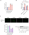
(A) Relative luciferase activity of pmirGLO-MDA5-exon1 firefly luciferase reporters in MPC cells transfected with si-Ctrl, si-METTL3 or si-METTL14. (B) Schematic diagram of m6A site mutation on exon1 of MDA5. ‘A’ in the predicted methylation motif was replaced by ‘U’. (C) Relative luciferase activity of wild-type or mutant pmirGLO-MDA5-exon1 firefly luciferase reporter in MPC cells transfected with si-Ctrl or si-METTL3&14. (D) METTL3 and METTL14 could downregulate GFP expression of mVenus-MDA5-exon1. EPC cells were co-transfected with the mVenus-MDA5-exon1 and vector, METTL3 or METTL14 plasmids. At 48 h post-transfection, the fluorescence intensity and the GFP expression were evaluated by enzyme-labeled instrument and western blotting, respectively. All data presented as the means ± SE from three independent triplicated experiments. **, p<0.01, as determined by Student’s t test.
Figure 7.
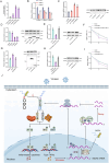
Figure 7.. Detailed m6A regulatory mechanism of MDA5.
(A) The binding relationship between YTHDF1, YTHDF2, or YTHDF3 and MDA5 mRNA was validated using a RIP assay. MKC cells were transfected with YTHDF1-Flag, YTHDF2-Flag, YTHDF3-Flag, or pcDNA3.1-Flag for 48 hr. (B) SiRNA silencing effect test of YTHDF1, YTHDF2, and YTHDF3. MPC cells were transfected with si-YTHDF1, si-YTHDF2, or si-YTHDF3 for 48 h. (C and D) MPC cells were transfected with si-YTHDF1, si-YTHDF2, si-YTHDF3 or si-Ctrl (C), and MKC cells were transfected with YTHDF1, YTHDF2, YTHDF3 or vector for 48 hr (D), then the expression of MDA5 was detected by qRT-PCR and western blotting. (E) Relative mRNA and protein levels of MDA5 in MKC cells after co-transfected with YTHDF2&3 plasmids and si-METTL3&14 by qPCR and western blot assays. (F) MKC cells were transfected with YTHDF2&3 plasmids and si-METTL3&14, then 5 µg/ml actinomycin D was added to the cells for 0 hr, 2 hr, and 4 hr. The half-life of MDA5 was analyzed by qRT-PCR. (G) MKC cells were transfected with vector, FTO, ALKBH5 plasmids for 48 hr, then the expression of MDA5 was detected by qRT-PCR and Western blotting. (H) Relative mRNA and protein levels of MDA5 in MKC cells after co-transfected with YTHDF2&3 plasmids and FTO plasmids by qPCR and western blotting. (I) MKC cells were transfected with YTHDF2&3 plasmids and FTO plasmids, then 5 µg/ml actinomycin D was added to the cells for 0 hr, 2 hr, and 4 hr. The half-life of MDA5 was analyzed by qRT-PCR. (J) Schematic diagram of arms race between MDA5 and 5’ppp-RNA virus in M. miiuy. All data presented as the means ± SE from three independent triplicated experiments. **, p<0.01; *, p<0.05, as determined by Student’s t test.
Figure 7—figure supplement 1.
Figure 7—figure supplement 1.. The regulation of YTHDF readers (YTHDF1&2&3) to the MDA5-exon1.
(A) Relative luciferase activity of pmirGLO-MDA5-exon1 firefly luciferase reporters in MPC cells transfected with si-Ctrl, si-YTHDF1, si-YTHDF2 or si-YTHDF3. (B) Relative luciferase activity of wild-type or mutant pmirGLO-MDA5-exon1 plasmids in MPC cells transfected with si-Ctrl or si-YTHDF2&3. (C) YTHDF2 and YTHDF3 could downregulate GFP expression of mVenus-MDA5-exon1. EPC cells were co-transfected with the mVenus-MDA5-exon1 and vector, YTHDF1, YTHDF2, or YTHDF3 plasmids. At 48 hr post-transfection, the fluorescence intensity and the GFP expression were evaluated by enzyme-labeled instrument and western blotting, respectively. All data presented as the means ± SE from three independent triplicated experiments. **, p<0.01, as determined by Student’s t test.

Update of

  • doi: 10.1101/2024.01.10.575132
  • doi: 10.7554/eLife.94898.1
  • doi: 10.7554/eLife.94898.2
  • doi: 10.7554/eLife.94898.3

Similar articles

Cited by

References

    1. Barber MRW, Aldridge JR, Webster RG, Magor KE. Association of RIG-I with innate immunity of ducks to influenza. PNAS. 2010;107:5913–5918. doi: 10.1073/pnas.1001755107. - DOI - PMC - PubMed
    1. Che RB, Sun YY, Sun DQ, Xu TJ. Characterization of the miiuy croaker (Miichthys miiuy) transcriptome and development of immune-relevant genes and molecular markers. PLOS ONE. 2014;9:e94046. doi: 10.1371/journal.pone.0094046. - DOI - PMC - PubMed
    1. Chen SN, Zou PF, Nie P. Retinoic acid-inducible gene I (RIG-I)-like receptors (RLRs) in fish: current knowledge and future perspectives. Immunology. 2017;151:16–25. doi: 10.1111/imm.12714. - DOI - PMC - PubMed
    1. Chen C, Chen H, Zhang Y, Thomas HR, Frank MH, He Y, Xia R. TBtools: an integrative toolkit developed for interactive analyses of big biological data. Molecular Plant. 2020;13:1194–1202. doi: 10.1016/j.molp.2020.06.009. - DOI - PubMed
    1. Chu Q, Xu T, Zheng W, Chang R, Zhang L. Long noncoding RNA MARL regulates antiviral responses through suppression miR-122-dependent MAVS downregulation in lower vertebrates. PLOS Pathogens. 2020;16:e1008670. doi: 10.1371/journal.ppat.1008670. - DOI - PMC - PubMed

MeSH terms

LinkOut - more resources