Review
. 2024 Sep 23;14(9):1208.
doi: 10.3390/life14091208.

Practical Use of Ultrasound in Modern Rheumatology-From A to Z

Affiliations
Review

Practical Use of Ultrasound in Modern Rheumatology-From A to Z

Tanya Sapundzhieva et al. Life (Basel). .

Abstract

During the past 20 years, the use of ultrasound (US) in rheumatology has increased tremendously, and has become a valuable tool in rheumatologists' hands, not only for assessment of musculoskeletal structures like joints and peri-articular tissues, but also for evaluation of nerves, vessels, lungs, and skin, as well as for increasing the accuracy in a number of US-guided aspirations and injections. The US is currently used as the imaging method of choice for establishing an early diagnosis, assessing disease activity, monitoring treatment efficacy, and assessing the remission state of inflammatory joint diseases. It is also used as a complementary tool for the assessment of patients with degenerative joint diseases like osteoarthritis, and in the detection of crystal deposits for establishing the diagnosis of metabolic arthropathies (gout, calcium pyrophosphate deposition disease). The US has an added value in the diagnostic process of polymyalgia rheumatica and giant-cell arteritis, and is currently included in the classification criteria. A novel use of US in the assessment of the skin and lung involvement in connective tissue diseases has the potential to replace more expensive and risky imaging modalities. This narrative review will take a close look at the most recent evidence-based data regarding the use of US in the big spectrum of rheumatic diseases.

Keywords: imaging; musculoskeletal; rheumatology; ultrasound.


Conflict of interest statement

The authors declare no conflict of interest.

Figures
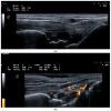
Figure 1
Figure 1
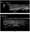
Synovitis. (A) A dorsal longitudinal scan of the wrist joint on Gray Scale Ultrasound (GSUS) in an RA patient—Grade 3 synovitis of the radiocarpal and intercarpal joints; (B) a dorsal longitudinal scan of the wrist joint on Power Doppler Ultrasound (PDUS) in an RA patient—Grade 2 synovitis; (C) a dorsal longitudinal scan of the second metacarpophalangeal (MCP) joint on PDUS in an RA patient—Grade 3 synovitis; (D) a dorsal longitudinal scan of the third proximal interphalangeal (PIP) joint on PDUS in an RA patient—Grade 3 synovitis; (E) a dorsal longitudinal scan of the first metatarsophalangeal (MTP) joint on GSUS—effusion and grade 3 synovitis.
Figure 1
Figure 1
Synovitis. (A) A dorsal longitudinal scan of the wrist joint on Gray Scale Ultrasound (GSUS) in an RA patient—Grade 3 synovitis of the radiocarpal and intercarpal joints; (B) a dorsal longitudinal scan of the wrist joint on Power Doppler Ultrasound (PDUS) in an RA patient—Grade 2 synovitis; (C) a dorsal longitudinal scan of the second metacarpophalangeal (MCP) joint on PDUS in an RA patient—Grade 3 synovitis; (D) a dorsal longitudinal scan of the third proximal interphalangeal (PIP) joint on PDUS in an RA patient—Grade 3 synovitis; (E) a dorsal longitudinal scan of the first metatarsophalangeal (MTP) joint on GSUS—effusion and grade 3 synovitis.
Figure 2
Figure 2
Paratenonitis—inflammation of the finger extensor tendon on PDUS. A dorsal longitudinal scan of the third PIP joint in a PsA patient.
Figure 3
Figure 3
Tenosynovitis. (A) Finger flexor tenosynovitis—a palmar longitudinal scan of the second PIP joint in a PsA patient—Grade 3 tenosynovitis of the finger flexor tendon on PDUS; (B) tenosynovitis of the VI extensor compartment of the wrist—extensor carpi ulnaris tedon—in an RA patient—ulnar longitudinal scan—grade 2 tenosynovitis on PDUS; (C) tenosynovitis of the VI extensor compartment of the wrist—extensor carpi ulnaris in an RA patient—ulnar transverse scan—grade 2 tenosynovitis on PDUS.
Figure 3
Figure 3
Tenosynovitis. (A) Finger flexor tenosynovitis—a palmar longitudinal scan of the second PIP joint in a PsA patient—Grade 3 tenosynovitis of the finger flexor tendon on PDUS; (B) tenosynovitis of the VI extensor compartment of the wrist—extensor carpi ulnaris tedon—in an RA patient—ulnar longitudinal scan—grade 2 tenosynovitis on PDUS; (C) tenosynovitis of the VI extensor compartment of the wrist—extensor carpi ulnaris in an RA patient—ulnar transverse scan—grade 2 tenosynovitis on PDUS.
Figure 4
Figure 4
Enthesitis. (A) Enthesitis of the enthesis of the Achilles tendon on the calcaneus in a patient with ankylosing spondylitis. A thickened hypoechoic enthesis, with loss of the normal fibrillar pattern, calcifications, enthesophytes, exhibiting a PD signal; (B) enthesitis of the common extensor tendon enthesis on the lateral epicondyle of the humerus—thickened and hypoechoic enthesis, loss of the normal fibrillar pattern, exhibiting a PD signal.
Figure 4
Figure 4
Enthesitis. (A) Enthesitis of the enthesis of the Achilles tendon on the calcaneus in a patient with ankylosing spondylitis. A thickened hypoechoic enthesis, with loss of the normal fibrillar pattern, calcifications, enthesophytes, exhibiting a PD signal; (B) enthesitis of the common extensor tendon enthesis on the lateral epicondyle of the humerus—thickened and hypoechoic enthesis, loss of the normal fibrillar pattern, exhibiting a PD signal.
Figure 5
Figure 5
A Baker’s cyst (semimembranosus-gastrocnemius bursa) of the knee joint—a posterior longitudinal scan of the medial tibio-femoral joint space shows a cyst with a heterogenous predominantly anechoic internal structure.
Figure 6
Figure 6
Double-contour sign (DCS) in a gout patient. (A) A dorsal longitudinal scan of the second MCP joint in a gout patient. Grade 3 synovitis and DCS of the hyaline cartilage of the metacarpal head; (B) a plantar longitudinal scan of the second MTP joint in a gout patient. DCS of the hyaline cartilage of the metatarsal head.
Figure 7
Figure 7
Calcium pyrophosphate deposition disease (CPPD). (A) A posterior longitudinal scan of the tibio-femoral joint in a CPPD patient. A linear hyperechoic calcification within the femoral hyaline cartilage. (B) A posterior transverse scan of the tibio-femoral joint in a CPPD patient. A linear hyperechoic calcification within the femoral hyaline cartilage. (C) A posterior scan of the gleno-humeral joint in a CPPD patient. A hyperechoic calcification (between the cursors) in the fibrocartilage of the labrum.
Figure 7
Figure 7
Calcium pyrophosphate deposition disease (CPPD). (A) A posterior longitudinal scan of the tibio-femoral joint in a CPPD patient. A linear hyperechoic calcification within the femoral hyaline cartilage. (B) A posterior transverse scan of the tibio-femoral joint in a CPPD patient. A linear hyperechoic calcification within the femoral hyaline cartilage. (C) A posterior scan of the gleno-humeral joint in a CPPD patient. A hyperechoic calcification (between the cursors) in the fibrocartilage of the labrum.
Figure 8
Figure 8
Lymphoma of the parotid gland in a patient with Sjogren Syndrome. (A) Parotid gland lymphoma on GSUS—a mass with a heterogenous internal structure, a lobulated appearance, and irregular, poorly defined borders; (B) parotid gland lymphoma on PDUS.

Similar articles

References

    1. McDonald D.G., Leopold G.R. Ultrasound B-scanning in the differentiation of Baker’s cyst and thrombophlebitis. Br. J. Radiol. 1972;45:729–732. doi: 10.1259/0007-1285-45-538-729. - DOI - PubMed
    1. Cooperberg P.L., Tsang I., Truelove L., Knickerbocker W.J. Gray scale ultrasound in the evaluation of rheumatoid arthritis of the knee. Radiology. 1978;126:759–763. doi: 10.1148/126.3.759. - DOI - PubMed
    1. Sapundzhieva T.L., Karalilova R., Batalov A. Musculoskeletal Ultrasound in Rheumatology—New Horizons. Folia Medica. 2020;62:7–16. doi: 10.3897/folmed.62.e47772. - DOI - PubMed
    1. Kaeley G.S., Bakewell C., Deodhar A. The importance of ultrasound in identifying and differentiating patients with early inflammatory arthritis: A narrative review. Arthritis Res. Ther. 2020;22:1. doi: 10.1186/s13075-019-2050-4. - DOI - PMC - PubMed
    1. Saku A., Furuta S., Kato M., Furuya H., Suzuki K., Fukuta M., Suehiro K., Makita S., Tamachi T., Ikeda K., et al. Experience of musculoskeletal ultrasound scanning improves physicians’. physical examination skills in assessment of synovitis. Clin. Rheumatol. 2020;39:1091–1099. doi: 10.1007/s10067-020-04960-5. - DOI - PubMed

Grants and funding

This research received no external funding.

LinkOut - more resources