. 2024 Sep 3;13(9):27.
doi: 10.1167/tvst.13.9.27.

Pharmacological Activation and Transgenic Overexpression of SIRT1 Attenuate Traumatic Optic Neuropathy Induced by Blunt Head Impact

Affiliations

Pharmacological Activation and Transgenic Overexpression of SIRT1 Attenuate Traumatic Optic Neuropathy Induced by Blunt Head Impact

Alex Kwok et al. Transl Vis Sci Technol. .

Abstract

Purpose: Resveratrol (RSV) is a nutraceutical compound known for its therapeutic potential in neurodegenerative and metabolic diseases. RSV promotes survival signals in retinal ganglion cells (RGCs) through activation of SIRT1, an NAD+-dependent deacetylase. RSV and SIRT1 reduce RGC loss induced by direct optic nerve injury, but effects in indirect models of traumatic optic neuropathy remain unknown and are examined in this study.

Methods: An electromagnetic stereotaxic impactor device was used to impart five traumatic skull impacts with an inter-concussion interval of 48 hours to wild type (WT) and SIRT1 knock in (KI) C57BL/6J mice overexpressing the SIRT1 gene. A cohort of WT mice also received intranasal administration of RSV (16 mg/kg) throughout the experimental period. Loss of righting reflex (RR), optokinetic response (OKR) scores, and immunolabeled RGC count are determined to assess optic neuropathy in this model of traumatic brain injury (TBI).

Results: TBI significantly decreases RGC survival and decreases OKR scores compared with control uninjured mice. Either RSV administration in WT mice, or SIRT1 overexpression in SIRT1 KI mice, significantly increases RGC survival and improves OKR scores. RR time increases after the first few impacts in all groups of mice subjected to TBI, demonstrating that RSV and SIRT1 overexpression are able to attenuate optic neuropathy following similar degrees of TBI.

Conclusions: Intranasal RSV is effective in preserving visual function in WT mice following TBI. Constitutive overexpression of SIRT1 recapitulates the neuroprotective effect of RSV.

Translational relevance: Results support future exploration of RSV as a potential therapy for indirect traumatic optic neuropathy.


Conflict of interest statement

Disclosure: A. Kwok, None; B. Chaqour, None; R.S. Khan, None; P. Aravand, None; K. Dine, None; A.G. Ross, None; K.S. Shindler, None

Figures

Figure 1.
Figure 1.
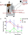
Effects of TBI and RSV treatment on RR time. (A) Schematic representation of the stereotaxic impactor device used to induce TBI. (B) Effects of RSV on the RR time in WT mice. RR time was determined in isoflurane-anesthetized mice of the control (no TBI, n = 3) mice, TBI (n = 6), and TBI + RSV (n = 3) groups. Mice of the latter 2 groups received, each, a single impact on days 0, 2, 4, 6, and 8 on their heads. These two groups also received intranasal administration of PBS and RSV (16 mg/kg daily), respectively, throughout the duration of the experiment. RR time was defined as the time from placement in the supine position until the animals turned all four paws and stood upright. Data are means ± SEM. RR times were compared by repeated measures ANOVA (**P < 0.001).
Figure 2.
Figure 2.
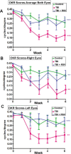
Intranasal administration of RSV attenuates visual function loss following TBI. (A) OKR scores averaged between left and right eyes show a significant (***P < 0.001) decrease over a 6-week time course in TBI mice (n = 6) compared with control (no TBI, n = 3) mice. Daily intranasal RSV administration significantly attenuated vision loss in TBI + RSV mice (n = 3, ***P < 0.001) compared with TBI mice. There was no significant difference in visual function between TBI + RSV and control no TBI mice. (B) OKR scores of the left eye show a significant (**P < 0.01) progressive decrease in TBI mice compared with control mice. RSV significantly attenuated vision loss in TBI + RSV mice (***P < 0.001) compared with TBI mice. There was no significant difference in visual function between TBI + RSV and control no TBI mice. (C) OKR scores of the right eye show a significant (**P < 0.01) progressive decrease in OKR scores in TBI mice as compared with control mice. RSV significantly attenuated vision loss in TBI + RSV mice (**P < 0.01) compared with TBI mice. There was no significant difference in visual function between TBI + RSV and control mice. OKR scores of all panels were compared by repeated measures ANOVA. Data from one of three representative experiments are shown.
Figure 3.
Figure 3.
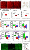
Pharmacologic activation of SIRT1 attenuates RGC loss. (A) Representative images of Brn3a-labeled flat-mounted retinas of mice from control (no TBI), TBI, and TBI + RSV groups. (B) Diagram shows 12 standardized fields imaged in each retina. (CE) Aggregate RGC counts across all retinal regions of both eyes, and right and left eyes separately. Average RGC count in both eyes dropped significantly (*P < 0.05) in the TBI group (n = 6) compared with controls (n = 3). TBI + RSV mice (n = 3) showed a significant (*P < 0.05) improvement in the number of RGCs compared with TBI mice. The right eyes of TBI mice demonstrated significantly (**P < 0.01) reduced numbers of RGCs compared with control mice. RSV treatment induced a nonsignificant trend toward improved RGC numbers. The left eyes of TBI mice demonstrated a significantly (***P < 0.001) reduced number of RGCs compared with control mice. TBI + RSV mice showed a significant (*P < 0.05) improvement in the number of RGCs compared with TBI mice. (F, G) RGC counts in central, mid-peripheral, and peripheral retina. RSV significantly attenuated RGC numbers in TBI + RSV mice (**P < 0.01) mice compared with TBI mice in the central retina of the left eyes. RSV significantly attenuated RGC numbers in TBI + RSV mice compared with TBI mice in the mid-peripheral and central retinas of the right eyes. (H) Representative images of neurofilament-labeled sections of optic nerves of mice from control (no TBI), TBI, and TBI + RSV groups. (I) TBI mice demonstrated significantly (**P < 0.01) lower mean fluorescence signal values of neurofilament staining compared with control mice. TBI + RSV mice showed a significant (**P < 0.01) improvement in mean fluorescence signal values of neurofilament staining compared with TBI mice. RGC counts and neurofilament staining were compared by 1-way ANOVA. Data from one of three representative experiments are shown.
Figure 4.
Figure 4.
SIRT1 overexpression in TBI mice did not affect RR time but improved visual function. (A) The RR time did not differ significantly between WT TBI and SIRT1 KI TBI mice, and both were higher than control mice. (BD) When averaged between both eyes, or measured separately in right and left eyes, OKR scores compared by repeated measures ANOVA show a significant (***P < 0.001) progressive decrease over a 6-week time course in TBI mice (n = 4) compared with control (n = 4) mice. SIRT1 overexpression significantly attenuated vision loss in SIRT1 KI TBI mice (n = 4, ***P < 0.001) compared with WT TBI mice. There was no significant difference in visual function between SIRT1 KI TBI and control WT mice. Data from one of three representative experiments are shown.
Figure 5.
Figure 5.
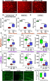
SIRT1 overexpression attenuates RGC loss in mice subjected to TBI. (A) Representative images of Brn3a-labeled flat-mounted retinas of mice from control (no TBI), TBI, and SIRT1 KI TBI mouse groups. (B) TBI mice (n = 4) demonstrated a significantly (***P < 0.001) reduced number of RGCs (averaged across all retinal regions and averaged between both eyes) compared with control mice (n = 4). SIRT1 KI TBI (n = 4) mice showed a significant (*P < 0.05) improvement in the number of RGCs compared with TBI mice. (C) The right eyes of TBI mice demonstrated a significantly (***P < 0.001) reduced number of RGCs compared with the right eyes of control mice. SIRT1 overexpression significantly (*P < 0.05) preserved the number of RGCs compared with TBI mice. (D) The left eyes of TBI mice demonstrated a significantly (***P < 0.001) reduced the number of RGCs compared with control mice. SIRT1 overexpression significantly (*P < 0.05) preserved the number of RGCs compared with TBI mice. (E, F) RGC counts in central, mid-peripheral, and peripheral retina of control, TBI and SIRT1 KI TBI mice. SIRT1 overexpression significantly increased RGC numbers in the SIRT1 KI TBI mice compared to TBI mice in the central, mid-peripheral, and peripheral retina of both the right and left eyes (***P < 0.001; **P < 0.01; and *P < 0.05). (G) Representative images of neurofilament-labeled sections of optic nerves of mice from control (no TBI), TBI, and SIRT1 KI TBI mouse groups. (H) TBI mice demonstrated significantly (*P < 0.05) lower mean fluorescence signal values of neurofilament staining compared with control mice. SIRT1 KI TBI mice showed a significant (*P < 0.05) improvement in mean fluorescence values of neurofilament staining compared with TBI mice. RGC counts and neurofilament staining were compared by 1-way ANOVA. Data from one of three representative experiments are shown.

Similar articles

References

    1. Levin LA, Beck RW, Joseph MP, Seiff S, Kraker R.. The treatment of traumatic optic neuropathy: the International Optic Nerve Trauma Study. Ophthalmology. 1999; 106: 1268–1277. - PubMed
    1. Bastakis GG, Ktena N, Karagogeos D, Savvaki M.. Models and treatments for traumatic optic neuropathy and demyelinating optic neuritis. Dev Neurobiol. 2019; 79: 819–836. - PubMed
    1. Levin LA. Axonal loss and neuroprotection in optic neuropathies. Can J Ophthalmol. 2007; 42: 403–408. - PubMed
    1. Ropposch T, Steger B, Meco C, et al. .. The effect of steroids in combination with optic nerve decompression surgery in traumatic optic neuropathy. Laryngoscope. 2013; 123: 1082–1086. - PubMed
    1. Yu-Wai-Man P, Griffiths PG.. Steroids for traumatic optic neuropathy. Cochrane Database Syst Rev. 2011; 1: CD006032. - PubMed

MeSH terms