Review
. 2024 Sep 21;13(18):1591.
doi: 10.3390/cells13181591.

The Nucleolus and Its Interactions with Viral Proteins Required for Successful Infection

Affiliations
Review

The Nucleolus and Its Interactions with Viral Proteins Required for Successful Infection

José Manuel Ulloa-Aguilar et al. Cells. .

Abstract

Nuclear bodies are structures in eukaryotic cells that lack a plasma membrane and are considered protein condensates, DNA, or RNA molecules. Known nuclear bodies include the nucleolus, Cajal bodies, and promyelocytic leukemia nuclear bodies. These bodies are involved in the concentration, exclusion, sequestration, assembly, modification, and recycling of specific components involved in the regulation of ribosome biogenesis, RNA transcription, and RNA processing. Additionally, nuclear bodies have been shown to participate in cellular processes such as the regulation of transcription of the cell cycle, mitosis, apoptosis, and the cellular stress response. The dynamics and functions of these bodies depend on the state of the cell. It is now known that both DNA and RNA viruses can direct their proteins to nuclear bodies, causing alterations in their composition, dynamics, and functions. Although many of these mechanisms are still under investigation, it is well known that the interaction between viral and nuclear body proteins is necessary for the success of the viral infection cycle. In this review, we concisely describe the interaction between viral and nuclear body proteins. Furthermore, we focus on the role of the nucleolus in RNA virus infections. Finally, we discuss the possible implications of the interaction of viral proteins on cellular transcription and the formation/degradation of non-coding RNAs.

Keywords: Cajal bodies; non-coding RNAs; nuclear bodies; nucleolus; promyelocytic leukemia nuclear bodies; ribosome biogenesis; transcription.


Conflict of interest statement

The authors declare no conflicts of interest.

Figures

Figure 1
Figure 1
Subnuclear structures and their interaction with viruses.
Figure 2
Figure 2
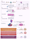
The ole of non-coding RNAs during viral infections. (A) RNA molecules play various roles in cells, and they are broadly classified into two categories: coding RNA and non-coding RNA. Non-coding RNAs do not code for proteins but have crucial roles in regulating gene expression and maintaining cellular functions. Here is an overview of the main types of ncRNA: microRNA (miRNA), small interfering RNA (siRNA), piwi-Interacting RNA (piRNA), small nuclear RNA (snRNA), small nucleolar RNA (snoRNA), long non-coding RNA (lncRNA), circular RNA (circRNA), and long intergenic non-coding RNA (lincRNA). (B) miRNAs are small, non-coding, single-stranded RNAs ~23 nt (ranging from 19 to 25 nt). The majority of mammalian miRNAs genes are located in intergenic regions or in antisense orientation and are transcribed by RNA polymerase II (Pol II) as primary miRNA transcripts (pri-miRNAs). (C) pri-miRNAs are capped, polyadenylated, and contain a local stem–loop structure that encodes miRNA sequences in the arm of the stem. This stem–loop structure is cleaved by the nuclear RNase III type enzyme Drosha in a process known as ‘cropping’. In the nucleus, the RNA hairpin structure is excised by the RNAse III-like enzyme Drosha and its co-factor DGCR8 to form the precursor miRNA (pre-miRNA). (D) pre-miRNA is translocated to the cytosol by exportin5, where it is processed by the Dicer protein complex, resulting in an miRNA duplex (miRNA/miRNA*), which is made up of a guide chain (miRNA) and a passenger chain (miR-NA*). (E) The miRNA/miRNA* is then loaded into the Argonaute (AGO), promoting the expulsion and degradation of the miRNA and the formation of the RNA-induced silencing complex (RISC). The RISC recognizes the targeted mRNA through base-pairing with miRNA. (F) miRNAs function as key regulators of gene expression in many different cellular pathways and systems, including immune response. So, several viruses with the purpose of carrying out an efficient replication or a persistent infection are able to modify their biogenesis, such as in the case of HIV. In this sense, several studies report an increase in miRNAs that facilitate its replication while inhibiting the Dicer–TRBP–PACT complex.
Figure 3
Figure 3
(A) Atorvastatin and Ivermectin: Both drugs are known to disrupt the nuclear–cytoplasmic transport of proteins. Specifically, they impair the trafficking of viral proteins, which is crucial for the assembly and maturation of viral particles. This action has been noted in viruses like Dengue and Zika virus (ZIKV), where effective viral replication relies on the proper localization of viral proteins within the host cell. (B) 5-Fluorouracil (5-FU): A pyrimidine analog that interferes with nucleotide metabolism and RNA function. 5-FU targets nucleolar structures, disrupting the organization and function of the nucleolus. This disruption impairs the transport and processing of rRNA and other molecules, crucial for ribosome assembly and function. (C) Quarfloxin (CX-3543): Its main mechanism is the inhibition of the interaction between the nucleolar protein nucleophosmin (NPM1) and DNA containing regions rich in G-quadruplexes, secondary structures present in promoter regions of ribosomal DNA. This drug destabilizes ribosome assembly by blocking the transcription of ribosomal RNA, which reduces protein production in the cell. (D) Leptomycin B: Inhibits CRM1 (also known as exportin 1), a key protein in the nuclear export of proteins and RNAs. By blocking the nuclear export of viral proteins and RNAs, Leptomycin B effectively prevents the replication of various viruses, including HIV and Influenza. This inhibition disrupts the life cycle of these viruses, which rely on the export of viral components for replication and assembly. Selinexor: Another inhibitor of CRM1/exportin 1, like Leptomycin B. Used in the treatment of certain cancers and viral infections, Selinexor blocks the nuclear export of viral and cellular components, thereby disrupting viral replication and cancer cell proliferation by affecting cellular stress responses and apoptotic pathways. (E) Cisplatin: Forms covalent adducts with DNA, including ribosomal DNA (rDNA), and proteins within the nucleolus. These adducts create steric hindrances that prevent the proper assembly and function of nucleolar components. This action blocks the synthesis and maturation of rRNA, thereby hindering viral access to the nucleolar machinery necessary for replication. (F) CDK inhibitors: Target cyclin-dependent kinases (CDKs), which are critical regulators of cell cycle progression and nucleolar function. These inhibitors disrupt the nucleolar scaffold, leading to nucleolar dissolution. This disruption affects rRNA transcription and processing, impairing the nucleolus’s ability to produce ribosomes, which are necessary for protein synthesis, including viral proteins. (G) Camptothecin and Doxorubicin: Inhibit RNA polymerase I (Pol I), which is responsible for the transcription of rRNA genes. These drugs reduce the synthesis of rRNA, leading to decreased ribosome production. Since ribosomes are essential for the translation of viral proteins, their reduced availability impairs viral replication. BMH-21: Exerts its action by binding to DNA in rRNA gene regions, which leads to inhibition of RNA polymerase I and degradation of the enzyme. This inhibition specifically affects cells with a high rate of rRNA synthesis, such as tumor cells, without severely impacting normal cells.

Similar articles

References

    1. Kvansakul M. Viral Infection and Apoptosis. Viruses. 2017;9:356. doi: 10.3390/v9120356. - DOI - PMC - PubMed
    1. Stern-Ginossar N., Thompson S.R., Mathews M.B., Mohr I. Translational Control in Virus-Infected Cells. Cold Spring Harb. Perspect. Biol. 2019;11:a033001. doi: 10.1101/cshperspect.a033001. - DOI - PMC - PubMed
    1. Rawlinson S.M., Moseley G.W. The Nucleolar Interface of RNA Viruses. Cell. Microbiol. 2015;17:1108–1120. doi: 10.1111/cmi.12465. - DOI - PubMed
    1. Rawlinson S.M., Zhao T., Rozario A.M., Rootes C.L., McMillan P.J., Purcell A.W., Woon A., Marsh G.A., Lieu K.G., Wang L.-F., et al. Viral Regulation of Host Cell Biology by Hijacking of the Nucleolar DNA-Damage Response. Nat. Commun. 2018;9:3057. doi: 10.1038/s41467-018-05354-7. - DOI - PMC - PubMed
    1. Rattay S., Hufbauer M., Hoboth P., Sztacho M., Akgül B. Viruses and Phospholipids: Friends and Foes during Infection. J Med. Virol. 2023;95:e28658. doi: 10.1002/jmv.28658. - DOI - PubMed

Grants and funding

This research received no external funding.

LinkOut - more resources