. 2024 Sep 11:15:1465952.
doi: 10.3389/fimmu.2024.1465952. eCollection 2024.

Establishment and characterization of an immortalized red river hog blood-derived macrophage cell line

Affiliations

Establishment and characterization of an immortalized red river hog blood-derived macrophage cell line

Takato Takenouchi et al. Front Immunol. .

Abstract

Red river hogs (RRHs) (Potamochoerus porcus), a wild species of Suidae living in Africa with a major distribution in the Guinean and Congolian forests, are natural reservoirs of African swine fever virus (ASFV) and typically are asymptomatic. Since blood and tissue macrophages of suid animals are target cell lineages of ASFV, RRH-derived macrophages are expected to play an important role in suppressing disease development in infected individuals. In the present study, we successfully isolated RRH-derived blood macrophages using co-culture techniques of RRH blood cells with porcine kidney-derived feeder cells and immortalized them by transferring SV40 large T antigen and porcine telomerase reverse transcriptase genes. The newly established macrophage cell line of the RRH-derived blood cell origin (RZJ/IBM) exhibited an Iba1-, CD172a-, and CD203a-positive typical macrophage-like phenotype and up-regulated the phosphorylation of nuclear factor-κB p65 subunit and p38 mitogen-activated protein kinase in response to the bacterial cell wall components, lipopolysaccharide (LPS) and muramyl dipeptide. In addition, RZJ/IBM cells produced the precursor form of interleukin (IL)-1β and IL-18 upon a stimulation with LPS, leading to the conversion of IL-18, but not IL-1β, into the mature form. Time-lapse live cell imaging with pHrodo dye-conjugated Escherichia coli BioParticles demonstrated the phagocytotic activity of RZJ/IBM cells. It is important to note that RZJ/IBM cells are clearly susceptible to ASFV infection and support viral replication in vitro. Therefore, the RZJ/IBM cell line provides a unique model for investigating the pathogenesis of ASFV.

Keywords: African swine fever virus; immortalization; in vitro model; macrophages; red river hog.


Conflict of interest statement

The authors declare that the research was conducted in the absence of any commercial or financial relationships that could be construed as a potential conflict of interest. The author(s) declared that they were an editorial board member of Frontiers, at the time of submission. This had no impact on the peer review process and the final decision.

Figures

Figure 1
Figure 1
Proliferation and isolation of primary RBMs. RBMs proliferated under the mixed culture conditions of RRH whole blood with primary porcine kidney feeder cells (A, arrows). The macrophage-like morphology of isolated RBMs was observed under a phase-contrast microscope (B). RBMs were seeded on 8-well chamber slides and cultured for 1 day. Cells were then fixed using 4% paraformaldehyde phosphate buffer solution and immunostained with specific antibodies against cell markers of macrophages (brown) (C). No specific staining was observed when cells were treated without primary antibodies (NC: negative control in C). All nuclei were counterstained with hematoxylin (blue) (C). Images are representative of two independent experiments.
Figure 2
Figure 2
Establishment and characterization of RZJ/IBM cells. The morphology of RZJ/IBM cells was observed under a phase-contrast microscope (A). They were seeded on 8-well chamber slides and cultured for 1 day. Cells were then fixed using 4% paraformaldehyde phosphate buffer solution and immunostained with specific antibodies against the cell markers of macrophages (brown) (B). No specific staining was observed when cells were treated without primary antibodies (NC: negative control in B). The cumulative population doublings of RZJ/IBM cells were plotted against the duration of the culture period (in days) (C). The PCR products generated from the SV40LT (128 bp) and pTERT (143 bp) genes were detected by a genomic DNA PCR analysis (D).
Figure 3
Figure 3
Enhanced expression of CD169 and MHC-II in LPS-treated RZJ/IBM cells. RZJ/IBM cells were treated with (lower panels) or without 1 μg/mL LPS (upper panels) for 1 day, and then immunostained with specific antibodies against cell markers of macrophages (brown) (A). All nuclei were counterstained with hematoxylin (blue) (A). Cells were also reacted with mouse monoclonal anti-MHC-II (blue line), anti-CD203a (green line), or anti-CD169 (red line) antibodies before being labeled with Alexa Fluor 488-conjugated anti-mouse IgG antibodies (Alexa 488) (B). Cells treated with Alexa 488 alone were used as a negative control (NC) (black line) (B, C). Flow cytometry histograms are representative of three independent experiments (B), and MFI data are expressed as mean ± SEM values (C).
Figure 4
Figure 4
Up-regulated expression of CD163 in DEX-treated RZJ/IBM cells. RZJ/IBM cells were treated with (lower panels) or without 100 ng/mL DEX (upper panels) for 3 days, and then immunostained with specific antibodies against cell markers of macrophages (brown) (A). All nuclei were counterstained with hematoxylin (blue) (A). Cells were also reacted with mouse monoclonal anti-MHC-II (blue line), anti-CD203a (green line), or anti-CD163 (red line) antibodies before being labeled with Alexa Fluor 488-conjugated anti-mouse IgG antibodies (Alexa 488) (B). Cells treated with Alexa 488 alone were used as a negative control (NC) (black line) (B, C). Flow cytometry histograms are representative of three independent experiments (B), and MFI data are expressed as mean ± SEM values (C).
Figure 5
Figure 5
LPS-induced increases in the expression of inflammatory cytokine and interferon β mRNAs in RZJ/IBM cells. Total RNA was recovered from RZJ/IBM cells treated with or without 1 μg/mL LPS, and RNA-seq experiments were performed independently three times. The transcripts per million (TPM) values of the genes indicated were expressed as mean ± SEM values. The TPM value of GAPDH was used as an internal control.
Figure 6
Figure 6
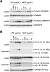
Phosphorylation of p65 NF-κB and p38 MAPK, and the production of IL-1β and IL-18 in RZJ/IBM cells in response to LPS or MDP. The treatment with LPS or MDP induced the phosphorylation of NF-κB p65 and p38 MAPK in a dose-dependent manner (A, first and third panels). The equivalent protein loading of these molecules was confirmed by immunoblotting with anti-NF-κB p65 or anti-p38 MAPK antibodies (A, second and fourth panels). The dose-dependent production of pro-IL-1β and pro-IL-18 was also detected in cell lysates (B, first and third panels) or culture supernatants (B, second and fourth panels) from RZJ/IBM cells that had been stimulated with LPS for 3 days. The secretion of mIL-18 from LPS-treated RZJ/IBM cells into the culture supernatant was also detected (B, fourth panel), whereas that of mIL-1β was not (B, second panel). MDP exerted a negligible effect on the production of pro-IL-1β (B, first panel) and pro-IL-18 (B, third panel). GAPDH was used as an internal control (B, fifth panel). Blots are representative of three independent experiments.
Figure 7
Figure 7
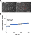
Phagocytotic activity of RZJ/IBM cells. RZJ/IBM cells were treated with pHrodo-labeled E. coli BioParticles and monitored via the time-lapse fluorescence imaging of live cells (A). The mean intensity of fluorescence emitted by pHrodo was plotted against the duration of the culture period (in minutes) (B). The experiments were performed independently three times and data are expressed as mean ± SEM values (B).
Figure 8
Figure 8
CPE and HAD assays of ASFV-inoculated RZJ/IBM and IPKM cells. The presence of CPE was detected by the disruption of monolayer cell sheets caused by viral infection and examined by microscopy. HAD assays were performed using porcine red blood cells, and rosette formation was examined by microscopy. CPE and rosette formation were detected in IPKM cells inoculated with the virulent ASFV field isolates Armenia07, Kenya05/Tk-1, Espana75, and Vero cell-adapted isolate Lisbon60V (MOI = 0.1) at 2 dpi (A). Rosette formation, but not CPE, was observed in RZJ/IBM cells inoculated with Armenia07, Kenya05/Tk-1, and Espana75 (MOI = 0.1) at 3 dpi (B). CPE and rosette formation were both detected in Lisbon60V-inoculated RZJ/IBM cells (MOI = 0.1) at 3 dpi relative to the findings obtained with mock-infected control cells (B).
Figure 9
Figure 9
Comparison of ASFV production in RZJ/IBM and IPKM cell cultures. Cell cultures were infected with Armina07, Kenya05/Tk-1, Espana75, or Lisbon60V isolates (MOI = 0.01). Culture supernatant samples were collected at the indicated timepoints. Viral production in the RZJ/IBM (closed circles) and IPKM (open triangles) cell cultures was estimated by titration experiments with IPKM cells. Data represent the mean and standard deviation of three experiments. Asterisks indicate significant differences in viral production between the RZJ/IBM and IPKM cell cultures (**p < 0.001, *p < 0.05).

Similar articles

References

    1. Oura CA, Powell PP, Anderson E, Parkhouse RM. The pathogenesis of African swine fever in the resistant bushpig. J Gen Virol. (1998) 79:1439–43. doi: 10.1099/0022-1317-79-6-1439 - DOI - PubMed
    1. Gaudreault NN, Madden DW, Wilson WC, Trujillo JD, Richt JA. African swine fever virus: An emerging DNA arbovirus. Front Vet Sci. (2020) 7:215. doi: 10.3389/fvets.2020.00215 - DOI - PMC - PubMed
    1. Gordon S, Plüddemann A. Tissue macrophages: heterogeneity and functions. BMC Biol. (2017) 15:53. doi: 10.1186/s12915-017-0392-4 - DOI - PMC - PubMed
    1. Finlay BB, McFadden G. Anti-immunology: evasion of the host immune system by bacterial and viral pathogens. Cell. (2006) 124:767–82. doi: 10.1016/j.cell.2006.01.034 - DOI - PubMed
    1. Bordet E, Maisonnasse P, Renson P, Bouguyon E, Crisci E, Tiret M, et al. . Porcine Alveolar Macrophage-like cells are pro-inflammatory Pulmonary Intravascular Macrophages that produce large titers of Porcine Reproductive and Respiratory Syndrome Virus. Sci Rep. (2018) 8:10172. doi: 10.1038/s41598-018-28234-y - DOI - PMC - PubMed

Grants and funding

The author(s) declare financial support was received for the research, authorship, and/or publication of this article. The present study was conducted as part of the research project on “Regulatory research projects for food safety, animal health and plant protection (JPJ008617. 20319736)” funded by the Ministry of Agriculture, Forestry and Fisheries of Japan.

LinkOut - more resources