. 2024 Sep 11:12:1460061.
doi: 10.3389/fcell.2024.1460061. eCollection 2024.

Fast and quantitative mitophagy assessment by flow cytometry using the mito-QC reporter

Affiliations

Fast and quantitative mitophagy assessment by flow cytometry using the mito-QC reporter

Juan Ignacio Jiménez-Loygorri et al. Front Cell Dev Biol. .

Abstract

Mitochondrial quality control is finely tuned by mitophagy, the selective degradation of mitochondria through autophagy, and mitochondrial biogenesis. Removal of damaged mitochondria is essential to preserve cellular bioenergetics and prevent detrimental events such as sustained mitoROS production, pro-apoptotic cytochrome c release or mtDNA leakage. The array of tools available to study mitophagy is very limited but in constant development. Almost a decade ago, we developed a method to assess mitophagy flux using MitoTracker Deep Red in combination with lysosomal inhibitors. Now, using the novel tandem-fluorescence reporter mito-QC (mCherry-GFP-FIS1101-152) that allows to differentiate between healthy mitochondria (mCherry+GFP+) and mitolysosomes (mCherry+GFP-), we have developed a robust and quantitative method to assess mitophagy by flow cytometry. This approach has been validated in ARPE-19 cells using PINK1/Parkin-dependent (CCCP) and PINK1/Parkin-independent (DFP) positive controls and complementary techniques. Furthermore, we show that the mito-QC reporter can be multiplexed, especially if using spectral flow cytometry, to simultaneously study other cellular parameters such as viability or ROS production. Using this technique, we evaluated and characterized two prospective mitophagy inducers and further dissected their mechanism of action. Finally, using mito-QC reporter mice, we developed a protocol to measure mitophagy levels in the retina ex vivo. This novel methodology will propel mitophagy research forward and accelerate the discovery of novel mitophagy modulators.

Keywords: FACS; Fisetin; SI; autophagy; mitochondria; phenanthroline; retina.


Conflict of interest statement

The authors declare that the research was conducted in the absence of any commercial or financial relationships that could be construed as a potential conflict of interest.

Figures

FIGURE 1
FIGURE 1
Mitophagy assessment in ARPE-19 cells by traditional flow cytometry using the mito-QC reporter. (A) Basis of the mito-QC reporter, whereby mitochondria are tagged with the chimeric protein mCherry-GFP-FIS1101–152. Upon mitophagy induction, mitochondria will be delivered to the lysosome where acidic pH will quench GFP and mitolysosomes will be identified as mCherry-only puncta. (B) ARPE-19 cells stably expressing the mito-QC reporter treated with 25 μM CCCP or 1 mM DFP for the indicated timepoints. (C) Quantification of % of mitophagyhigh population as defined in Supplementary Figure S1. (D) Representative histogram of FITC-GFP mean fluorescence intensity (MFI), equivalent to mitochondrial mass, in cells from (C). (E) Quantification of mitochondrial mass as shown in (D). Scale bar, 15 μm. All data are expressed as the mean ± s.e.m. Dots represent independent experiments. P values were calculated using two-way ANOVA with Dunnett’s post hoc test. Diagrams were created using BioRender.
FIGURE 2
FIGURE 2
Mitophagy assessment in organotypic ex vivo retina culture. (A) Neuroretinas from C57BL/6J mice expressing the mito-QC reporter were dissected and cultured ex vivo for 6 h in defined medium in the present of 25 μM CCCP or 1 mM DFP. Samples were dissociated into a single-cell suspension and analyzed by FC. (B) Representative images of the photoreceptor-containing outer nuclear layer (ONL). (C) Quantification of the % of mitophagyhigh cells in neuroretina ex vivo culture as defined in Supplementary Figure S3. Scale bar, 25 μm. All data are expressed as the mean ± s.e.m. Dots represent individual mice. P values were calculated using one-way ANOVA with Dunnet’s post hoc test. Diagrams were created using BioRender.
FIGURE 3
FIGURE 3
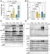
Phenanthroline and Fisetin are potent mitophagy inducers. (A) ARPE-19 mito-QC cells were treated with 10 μM Fisetin (Fis) or 50 μM Phenanthroline (Phen) for the indicated timepoints, quantification of % of mitophagyhigh population is shown. (B) Quantification of mitochondrial mass (GFP MFI). (C) Immunobloting against proteins involved in receptor-mediated mitophagy (HIF-1α, BNIP3L/NIX, BNIP3) and mitochondrial markers (TOMM20, TOMM40). Vinculin was used as a loading control. (D) Immunoblotting against proteins involved in PINK1/Parkin-dependent mitophagy (phospho-UbiquitinSer65, PINK1, Parkin, OPTN, SQSTM1/p62, NDP52). Vinculin was used as a loading control. All data are expressed as the mean ± s.e.m. Dots represent independent experiments. P values were calculated using two-way ANOVA with Dunnett’s post hoc test.
FIGURE 4
FIGURE 4
Oxidative stress reduces cell viability, differentially modulates mitophagy and stimulates ROS production. ARPE-19 mito-QC cells were treated with 20 mM sodium iodate (SI) or 750 μM H2O2 for 24 h. (A) Simultaneous measurement of viability by nuclear exclusion assay using DAPI in SI-treated cells. Viable population does not incorporate the dye and are identified as DAPI. (B) Quantification of % of mitophagyhigh population in SI-treated cells. (C) Quantification of mitochondrial mass (GFP MFI) in SI-treated cells. (D) Simultaneous measurement of reactive oxygen species (ROS) using CellROX Deep Red dye in SI-treated cells. (E) Simultaneous measurement of viability using ViaDye Red in H2O2-treated cells. Viable population does not incorporate the dye and are identified as ViaDye. (F) Quantification of % of mitophagyhigh population in H2O2-treated cells. (G) Quantification of mitochondrial mass (GFP MFI) in H2O2-treated cells. (H) Simultaneous measurement of mitochondrial ROS using MitoSOX Red dye in H2O2-treated cells. All data are expressed as the mean ± s.e.m. Dots represent independent experiments. P values were calculated using one-way ANOVA with Dunnet’s post hoc test.

Similar articles

References

    1. Allen G. F., Toth R., James J., Ganley I. G. (2013). Loss of iron triggers PINK1/Parkin-independent mitophagy. EMBO Rep. 14 (12), 1127–1135. 10.1038/embor.2013.168 - DOI - PMC - PubMed
    1. Chan C. M., Huang D. Y., Sekar P., Hsu S. H., Lin W. W. (2019). Reactive oxygen species-dependent mitochondrial dynamics and autophagy confer protective effects in retinal pigment epithelial cells against sodium iodate-induced cell death. J. Biomed. Sci. 26 (1), 40. 10.1186/s12929-019-0531-z - DOI - PMC - PubMed
    1. Chiu S. C., Huang S. Y., Tsai Y. C., Chen S. P., Pang C. Y., Lien C. F., et al. (2012). Poly (ADP-ribose) polymerase plays an important role in intermittent hypoxia-induced cell death in rat cerebellar granule cells. J. Biomed. Sci. 19 (1), 29. 10.1186/1423-0127-19-29 - DOI - PMC - PubMed
    1. Chowers G., Cohen M., Marks-Ohana D., Stika S., Eijzenberg A., Banin E., et al. (2017). Course of sodium iodate-induced retinal degeneration in albino and pigmented mice. Invest Ophthalmol. Vis. Sci. 58 (4), 2239–2249. 10.1167/iovs.16-21255 - DOI - PubMed
    1. Ding H., Li Y., Chen S., Wen Y., Zhang S., Luo E., et al. (2022). Fisetin ameliorates cognitive impairment by activating mitophagy and suppressing neuroinflammation in rats with sepsis-associated encephalopathy. CNS Neurosci. Ther. 28 (2), 247–258. 10.1111/cns.13765 - DOI - PMC - PubMed

Grants and funding

The author(s) declare that financial support was received for the research, authorship, and/or publication of this article. Research in the PB lab is supported by grants 310030_215271 Swiss National Science Foundation (SNSF) and PID2021-126864NB I00 from MCIN, Spain. JJ-L holds a FPI fellowship from MCIN (PRE2019-088222). CJ-G is supported by the University of Fribourg.

LinkOut - more resources