Observational Study
. 2024 Sep 24;9(18):e171029.
doi: 10.1172/jci.insight.171029.

T cell responses and clinical symptoms among infants with congenital cytomegalovirus infection

Affiliations
Observational Study

T cell responses and clinical symptoms among infants with congenital cytomegalovirus infection

Alexandra K Medoro et al. JCI Insight. .

Abstract

BACKGROUNDCongenital cytomegalovirus (cCMV) infection can cause developmental impairment and sensorineural hearing loss (SNHL). To determine the relationship between immune responses to cCMV infection and neurologic sequelae, T cell responses were compared for their connection to clinical symptoms at birth and neurodevelopmental outcomes.METHODSThirty cCMV-infected and 15 uninfected infants were enrolled in a single-center prospective observational case-control study. T cell pp65-specific cytokine responses; CD57, CD28, and PD-1 expression; and memory subsets were compared.RESULTSInfected neonates (73% symptomatic at birth) lacked pp65-specific cytokine-secreting T cells, with elevated frequencies of CD57+, CD28-, and PD-1+CD8+ T cells and effector memory subsets. Though frequencies overlapped between cCMV symptom groups, asymptomatic infants had higher frequencies of CD57+PD-1+CD8+ T cells. Neonates with subsequent developmental delay lacked detectable CMV-specific T cell responses, with patterns resembling those of uninfected infants. Two children with progressive SNHL had high frequencies of PD-1+CD8+ T cells over the first year compared with children without progressive SNHL.CONCLUSIONSimilar to published reports, neonatal viral antigen-specific cytokine-secreting T cell responses were not detected, but overall patterns indicate that globally differentiated memory CD8+ T cell populations were induced by cCMV infection, with higher frequencies of terminally differentiated PD-1+CD8+ T cells potentially associated with asymptomatic infection. In this cohort, a lack of in utero T cell differentiation was associated with developmental delay, and high frequencies of PD-1+CD8+ T cells persisted only in children with progressive SNHL. Further work is needed to define the specificity of these T cells and their mechanistic connection to these outcomes.FUNDINGThis study was funded through an intramural research award at Nationwide Children's Hospital, the Pediatric Infectious Disease Society Fellowship Award funded by Stanley and Susan Plotkin and Sanofi Pasteur, the Abigail Wexner Research Institute at Nationwide Children's Hospital, and the Pichichero Family Foundation Vaccines for Children Initiative Research Award from the Pediatric Infectious Diseases Society Foundation.

Keywords: Immunology; Infectious disease; Neurodevelopment; T cells.


Conflict of interest statement

Conflict of interest: The authors have declared that no conflict of interest exists.

Figures

Figure 1
Figure 1. CMV pp65-specific T cell responses in cCMV-infected neonates.
PBMCs from cCMV-infected infants (n = 21) or uninfected infants (n = 5) at ≤60 days of age and CMV-seropositive adults (n = 10) were unstimulated or stimulated with CMV pp65 peptide pool and analyzed by intracellular cytokine staining and flow cytometry. Representative flow plots (A and C) and aggregated data (B and D) are shown for frequencies of CD4+ (A and B) and CD8+ (C and D) T cells expressing IFN-γ, MIP-1β, TNF-α, and IL-2 after pp65 stimulation (background subtracted from unstimulated frequencies) for CMV-uninfected infants, cCMV-infected infants, and CMV-seropositive (CMV+) adults. Groups were compared using the Kruskal-Wallis test (B and D) with Dunn’s multiple comparisons. *P < 0.05; **P < 0.01; ***P < 0.001; ns, not significant (P > 0.05).
Figure 2
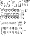
Figure 2. Global CD8+ T cell differentiation and memory in cCMV-infected infants categorized by clinical symptoms at birth.
PBMCs of cCMV-infected (n = 18) or uninfected infants (n = 4) at ≤60 days of age were analyzed by flow cytometry for frequencies of global populations of CD8+ T cells expressing CD57, CD28, PD-1, CD45RO, CCR7, and CD45RA. Uninfected and cCMV-infected groups (A and B) and cCMV-infected individuals grouped by clinical symptoms at birth (CF) were compared. Sx + CNS, symptomatic with CNS findings (n = 7); Sx no CNS, symptomatic without CNS findings (n = 5); eoSNHL, early onset sensorineural hearing loss (n = 2); Asx, asymptomatic (n = 6). (A) Frequencies of CD57+, CD28, or PD-1+CD8+ T cells between infants with cCMV infection (n = 18) and uninfected infants (n = 4). (B) Frequencies of naive (CCR7+/CD45RA+), central memory (Tcm; CCR7+/CD45RA), effector memory (Tem; CCR7/CD45RA), and effector memory expressing RA (Temra; CCR7/CD45RA+) subsets for infants with cCMV (n = 18) and uninfected infants (n = 5). (C) Serum viral loads of infants according to clinical symptom group. (D) Representative flow plots of CD8+ T cells stained for CD57, CD28, and PD-1 for an uninfected infant (stained and unstained to show gating) and then for cCMV-infected infants (stained only) from each clinical symptom group, with aggregated data shown in graphs. (E) Representative flow plots of memory markers CCR7/CD45RA shown for an uninfected infant and cCMV-infected infants from each clinical symptom group, with graph showing proportions of memory subsets by group. (F) Representative flow plots and graphs showing frequencies of PD-1+CD8+ T cells coexpressing either CD45RO or CD57. Comparisons were made using the Mann-Whitney U test (A, B, and D), Kruskal-Wallis test (C and F) with Dunn’s multiple comparisons, or 2-way ANOVA (E). *P < 0.05; **P < 0.01; ns, not significant (P > 0.05).
Figure 3
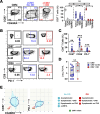
Figure 3. Association of T cell phenotypes with developmental delay.
The neonatal T cell phenotypes (≤60 days of age) of cCMV-infected infants with developmental delay (DD, n = 4) were compared with those with normal development (No DD, n = 14) and uninfected controls (n = 5). (A) CD8+ T cell memory subset distribution and (B and C) frequencies of CD57+, CD28, or PD-1+CD8+ T cells were compared between groups. (D) Plasma viral loads were compared between infants with and without DD. (E) Correlation plots show frequencies of CD45RO+ and either CD57+ or PD-1+CD8+ T cells by clinical symptom group (shown as shapes connected by lines) and outcomes (red, DD; blue, no DD). Ellipses show 95% CIs for DD/no DD groups. Comparisons were made using Mann-Whitney U tests (D), Kruskal-Wallis tests with Dunn’s comparisons (A and C), and SD ellipses (E) as appropriate. *P < 0.05; **P < 0.01; ns, not significant (P > 0.05).
Figure 4
Figure 4. Association of T cell phenotypes with SNHL.
The cCMV-infected infants with SNHL (n = 9) were compared with those with normal hearing (n = 14) and to uninfected controls (n = 5). (A) For infants with samples available at ≤60 days of age, frequencies of CD57+, CD28, or PD-1+CD8+ T cells were compared among those with (n = 6) and without (n = 12) SNHL and among uninfected controls (n = 5). (B) For all infants with longitudinal samples available for analysis, frequencies of PD-1+ and CD57+ CD8+ T cells are shown over the first 365 days of age for those with no SNHL (n = 14), nonprogressive SNHL (n = 6), and progressive SNHL (n = 3), with 95% CIs shown as shaded areas. (C) Plasma viral loads at ≤60 days of age were compared between cCMV-infected groups with and without SNHL. Comparisons were made using Mann-Whitney U tests (C), Kruskal-Wallis tests with Dunn’s comparisons (A), and linear-mixed models (B). *P < 0.05; **P < 0.01; ns, not significant (P > 0.05).
Figure 5
Figure 5. CMV antigen-specific T cell responses during the second year of age.
PBMCs from cCMV-infected infants (n = 5) and uninfected controls (n = 3) at 12–18 months age were analyzed by flow cytometry in comparison with CMV-seropositive adults (n = 10). (A) CMV pp65-specific CD4+ and CD8+ T cell responses are shown for uninfected (n = 3), cCMV IFN-γ+ responder (n = 2), and cCMV IFN-γ nonresponder (n = 3) infants as representative flow plots and graphs. (B) Representative flow plots and graphs of CD57+, CD28, or PD-1+CD8+ T cells are shown for uninfected children (n = 2; 1 infant had insufficient PBMCs for staining) and for cCMV responder and nonresponder infants. (C) CD8+ T cell polyfunctionality of cCMV responders was compared with that of CMV-seropositive adults using SPICE software and the polyfunctionality index (PI). (D) CCR7/CD45RA memory subset distribution of CMV pp65+IFN-γ+ CD4+ and CD8+ T cells is shown for cCMV-infected infants and CMV-seropositive adults as representative flow plots and graphs. Comparison were made using Mann Whitney U test (C). ***P < 0.001.
Figure 6
Figure 6. Consort diagram.
Participant enrollment and experimental flow.

Similar articles

References

    1. Fowler KB, Boppana SB. Congenital cytomegalovirus (CMV) infection and hearing deficit. J Clin Virol. 2006;35(2):226–231. doi: 10.1016/j.jcv.2005.09.016. - DOI - PubMed
    1. Nance WE, et al. Importance of congenital cytomegalovirus infections as a cause for pre-lingual hearing loss. J Clin Virol. 2006;35(2):221–225. doi: 10.1016/j.jcv.2005.09.017. - DOI - PubMed
    1. Dollard SC, et al. New estimates of the prevalence of neurological and sensory sequelae and mortality associated with congenital cytomegalovirus infection. Rev Med Virol. 2007;17(5):355–363. doi: 10.1002/rmv.544. - DOI - PubMed
    1. Ross SA, et al. CMV Infections. In: Elzouki AY, et al, eds. Textbook of Clinical Pediatrics. Springer; 2012:1145–1161.
    1. Fowler KB, et al. Progressive and fluctuating sensorineural hearing loss in children with asymptomatic congenital cytomegalovirus infection. J Pediatr. 1997;130(4):624–630. doi: 10.1016/S0022-3476(97)70248-8. - DOI - PubMed

Publication types

MeSH terms