[Preprint]. 2024 Sep 15:2024.09.15.613133.
doi: 10.1101/2024.09.15.613133.

Light-triggered protease-mediated release of actin-bound cargo from synthetic cells

Affiliations

Light-triggered protease-mediated release of actin-bound cargo from synthetic cells

Mousumi Akter et al. bioRxiv. .

Abstract

Synthetic cells offer a versatile platform for addressing biomedical and environmental challenges, due to their modular design and capability to mimic cellular processes such as biosensing, intercellular communication, and metabolism. Constructing synthetic cells capable of stimuli-responsive secretion is vital for applications in targeted drug delivery and biosensor development. Previous attempts at engineering secretion for synthetic cells have been confined to non-specific cargo release via membrane pores, limiting the spatiotemporal precision and specificity necessary for selective secretion. Here, we designed and constructed a protein-based platform termed TEV Protease-mediated Releasable Actin-binding protein (TRAP) for selective, rapid, and triggerable secretion in synthetic cells. TRAP is designed to bind tightly to reconstituted actin networks and is proteolytically released from bound actin, followed by secretion via cell-penetrating peptide membrane translocation. We demonstrated TRAP's efficacy in facilitating light-activated secretion of both fluorescent and luminescent proteins. By equipping synthetic cells with a controlled secretion mechanism, TRAP paves the way for the development of stimuli-responsive biomaterials, versatile synthetic cell-based biosensing systems, and therapeutic applications through the integration of synthetic cells with living cells for targeted delivery of protein therapeutics.


Conflict of interest statement

Conflict of Interest The authors declare no conflict of interest.

Figures

Figure 1.
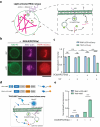
Figure 1.. TEVp-mediated cargo release from GUVs.
a. The left panel shows a schematic representation of fascin-bundled actin (F-actin) and TEVp-mediated releasable actin-binding protein (TRAP). The right panel illustrates the constructs for TRAP design: TRAP contains a cargo protein flanked by a cell-penetrating peptide (CPP) at the N-terminus and an actin-binding domain Lifeact at the C-terminus separated from the cargo by a TEVp cleavage site (TCS). TRAP-ΔCPP contains the cargo and the C-terminal TCS-actin-binding domain but lacks the N-terminal CPP domain. b. Schematic representation of cargo release from a GUV through unbinding of TRAP from the actin bundle via TEVp. TRAP is released from the GUV through its CPP domain while TRAP-ΔCPP remains inside the GUV.
Figure 2.
Figure 2.. TEVp-mediated unbinding of TRAP-mCherry from actin bundle.
a. Schematics of constructs of TRAP-mCherry and TRAP-△CPP-mCherry (left) and their corresponding protein depictions (right). b. Flow chart of the F-actin and TRAP-△CPP-mCherry co-pelleting assay. c. Representative SDS-PAGE gel image of the pellet and supernatant fractions from the co-pelleting assay of 1 μM TRAP-△CPP-mCherry and 5.5 μM of preformed actin incubated for 30 min followed by treatment of 10 units of TEVp. BSA was used as a control instead of F-actin. Lane M indicates the protein ladder. d. Bar graph of the normalized band intensities quantified from the SDS-PAGE presented in panel c. For each protein, band intensities were normalized by the sum of intensities in all lanes for the corresponding protein. e. Illustrations of fluorescence microscopy-based unbinding assay of TRAP-mCherry from F-actin in the presence of TEVp. f. Representative confocal fluorescence microscopy images of TRAP-mCherry or TRAP-△CPP-mCherry (red) and F-actin (magenta) with or without TEVp treatment for 30 min. Scale bar: 10 μm.
Figure 3
Figure 3. TEVp-mediated TRAP-mCherry release from GUVs.
a. Schematic illustrations show the binding of TRAP-mCherry and TRAP-ΔCPP-mCherry to F-actin in GUVs in the absence of TEVp. Introduction of TEVp dissociates TRAP and TRAP-ΔCPP from F-actin, leading to the release of TRAP-mCherry, but not TRAP-ΔCPP-mCherry. b. Confocal fluorescence microscopy images of GUVs (green) encapsulating TRAP-mCherry or TRAP- ΔCPP-mCherry (red) and F-actin (magenta) in the absence of TEVp. c. Normalized fluorescence intensity profiles of NBD-PE (green), actin (red), TRAP-mCherry (magenta) along the white line in the merged channel in panel b. The legend shows the fluorescence signal each profile color represents in panels c and e. d. Representative confocal fluorescence microscopy images of GUVs (green) showing the unbinding of TRAP-mCherry/TRAP-ΔCPP-mCherry (red) from F-actin (magenta) in the presence of TEVp. e. Normalized fluorescence intensity profiles of NBD-PE (green), actin (red), TRAP-mCherry (magenta) along the white line in the merged channel in panel e. f. Time course fluorescence intensity of TRAP-ΔCPP-mCherry in GUVs in the presence of TEVp. ns = non-significant. Error bars represent the standard error of the means. (n = 50 vesicles, three experiments). g. Time course fluorescence intensity change of TRAP-mCherry in GUVs in the presence of TEVp. *** represents p < 0.001. Error bars represent the standard error of the means. (n = 50 vesicles, three experiments). Scale bar: 10 μm.
Figure 4.
Figure 4.. Light-activated TEVp-mediated cargo release from GUVs.
a. Schematic representation of UV( λ= 254 nm)-activated TEVp release from DC(8,9)PC liposomes (depicted as DC(8,9)PC-{TEVp}) encapsulated in GUVs which induces cargo release from the GUVs through cleavage of TCS on TRAP. Inset shows detailed steps of UV-triggered release of TEVp, cleaving TRAP and allowing the CPP-cargo to traverse the GUV membrane b. Representative confocal fluorescence microscopy images of GUVs (green) encapsulating F-actin (magenta) and TRAP-mCherry (red) with or without 10 min exposure to UV light. Scale bar: 10 μm. c. Bar graphs comparing the mCherry fluorescence intensity in GUVs encapsulating TRAP-mCherry in the presence or absence of DC(8,9)PC{TEV} liposomes and UV illumination. Error bars represent standard error of the means, n > 50 from 3 independent trials. d. Top schematic shows the constructs of TRAP-HiBiT and TRAP-△CPP-HiBiT and their corresponding protein depictions. Bottom shows the schematic illustration of TRAP-HiBiT luminescence assay based on Promega Nano-Glo assay. e. Normalized luminescence readout from NanoBiT formed by LgBiT dimerization with TRAP-HiBiT or TRAP-△CPP-HiBiT released from GUVs in the absence of presence of DC(8,9)PC{TEVp} liposomes. Error bars represent the standard error of the means (n= 50 vesicles per condition from 3 independent experiments). *** represents p < 0.001. n.s. stands for not significant.

Similar articles

References

    1. Guindani C., da Silva L. C., Cao S., Ivanov T., Landfester K., Angewandte Chemie 2022, 134, e202110855. - PMC - PubMed
    1. Sharma B., Moghimianavval H., Hwang S.-W., Liu A. P., Membranes 2021, 11, 912. - PMC - PubMed
    1. Groaz A., Moghimianavval H., Tavella F., Giessen T. W., Vecchiarelli A. G., Yang Q., Liu A. P., WIREs Nanomedicine and Nanobiotechnology 2021, 13, e1685. - PMC - PubMed
    1. Boyd M. A., Kamat N. P., Trends in Biotechnology 2021, 39, 927. - PubMed
    1. Boyd M. A., Thavarajah W., Lucks J. B., Kamat N. P., Science Advances 2023, 9, eadd6605. - PMC - PubMed

Publication types

LinkOut - more resources